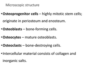
Iqra Saeed
Roll No 72
BS(Hons.)Physiology
The Skeletal System
Bones, tendons, ligaments and cartilages
Introduction
• Bones are very active and tough organ
• Tendons are collagen fibrous tissue that connect muscle to bone.
• Ligaments join one bone to another bone.
• Cartilage is a flexible connective tissue
• Support- framework that supports body soft organs
• Protection- for delicate organs, heart, lungs, brain
• Movement- bones act as levers for muscles
• Mineral storage- calcium & phosphate
• Blood cell formation- hematopoiesis
• Energy Storage- Yellow marrow serve as an
important chemical energy reserve.
• Long Bones- metacarples, metatarsals,
phelangies, humerus, ulna, radius, tibia,
fibula
• Short Bones- carpals, tarsals
• Flat Bones- rib, scapula, skull, sternum
• Irregular Bones- vertebrae, some facial bones
• Sesamoid- patella
• Wormian bones- tiny bones within the
skull that lie between major skull bones.
Distal
epiphysis
Proximal
epiphysis
diaphysis
yellow marrow
epiphyseal line
periosteum
compact bone
spongy bone
Endosteum
hyaline cartilage
Sharpey’s fibers
Bone Structure
• Expanded ends of bones that form joints with adjacent bones are called
epiphyses.
• Articular cartilages (hyaline cartilage) cover the epiphyses.
• The shaft of the bone is the diaphysis.
• A tough layer of vascular connective tissue, called the periosteum,
covers the bone and is continuous with ligaments and tendons.
•Compact bone makes up the wall of the diaphysis; the
epiphyses are filled with spongy bone to reduce the
weight of the skeleton.
•The diaphysis contains a hollow medullary cavity that
is lined with endosteum and filled with marrow.
•Epiphyseal line is the remnant of epiphyseal disk/plate
•Nutrient Foramen is a perforating canal allowing
blood vessels to enter and leave bone.
Microscopic structure
•Osteoprogenitor cells – highly mitotic stem cells;
originate in periosteum and enosteum.
•Osteoblasts – bone-forming cells.
•Osteocytes – mature osteoblasts.
•Osteoclasts – bone-destroying cells.
•Intercellular material consists of collagen and
inorganic salts.
Compact bone
solid, dense, and smooth
• Osteon – structural unit of a compact bone. cylindrical structure oriented parallel
to bone's long axis.
• Osteon consists of hollow tubes, one inside the other, called lamella.
• In the center of each osteon there is a Haverisan (also, central) canal that contains
small blood vessels, nerves and lymphatic vessels.
• Haversian canals are connected to each other by Volkmann's canals.
• Lacunae – small hollow spaces between lamellae. Each neighboring lacuna is
connected by canaliculi – hairlike passages that form a network of tiny canals.
Spongy bone
• Spongy bone is the structure located
deep in compact bone. It is made up
of thin trabeculae. This helps the bone
to resist stress while maintaining its
light weight.
• There are no osteons present in
spongy bone; however, its trabeculae
contain lamellae, lacunae
interconnected by canaliculi, and
osteocytes.
CHEMICAL COMPOSITION OF BONE
• Matrix consists of solid materials rich in minerals and salts
• 67% inorganic material; provides strength & hardness [Hydroxapatite
(Ca3(PO4)2)3 Ca(OH)2]
• 33% organic; collagenous proteins that provide reinforcement &
flexibility
BONE DEVELOPMENT AND GROWTH
• The "skeleton" of an embryo is composed of fibrous CT membranes
(formed from mesenchyme and hyaline cartilage) that are loosely
shaped like bones.
• At about 6-7 weeks gestation, ossification of this skeleton begins and
continues throughout adulthood.
• Ossification follows one of two patterns: Both mechanisms involve the
replacement of preexisting CT with bone.
A. Intramembranous Ossification is
when a bone forms on or within a
fibrous CT membrane. Flat bones are
formed in this manner
B. Endochondral Ossification occurs
when a bone is formed from a hyaline
cartilage model
• Longitudinal growth: During infancy and childhood, long bones lengthen
entirely by growth at the epiphyseal plates
• Appositional growth: bones grow in thickness
Growth at the Epiphyseal Plate
• Zone of resting cartilage
 near epiphysis, small, scattered chondrocytes,
• Zone of proliferation
 Chondrocytes undergo rapid mitosis under influence of growth
hormone.
• Zone of maturation and Hypertrophic
•  Chondrocytes stop mitosis, and begin to hypertrophy by accumulating
glycogen, lipids, and alkaline phosphatase
• Zone of calcification:
 Chondrocytes undergo apoptosis. Cartilagenous matrix begins to calcify.
Growth at the Epiphyseal Plate
• Zone of ossification: Osteoclasts and osteoblasts from the diaphyseal
side break down the calcified cartilage and replace with mineralized
bone tissue.
• a. Cartilage cells are produced by mitosis on the epiphyseal side of the
plate,
• b. They are then destroyed and replaced by bone on the diaphyseal
side of the plate.
• Therefore, the thickness of the plate remains almost constant, while the
bone on the diaphyseal side increases in length.
• The epiphyseal plate allows for bone lengthening until adulthood.
• Ossification of most bones is completed by age 25.
SKELETAL ORGANIZATION – Axial and appendicular
•The axial skeleton
consists of the skull,
hyoid bone, vertebral
column (vertebrae and
intervertebral disks),
and thorax (ribs and
sternum).
•The appendicular skeleton
consists of the pectoral girdle
(scapulae and clavicles), upper
limbs (humerus, radius, ulna,
carpals, metacarpals, and
phalanges), pelvic girdle (coxal
bones articulating with the
sacrum), and lower limbs (femur,
tibia, fibula, patella, tarsals,
metatarsals, phalanges).
7 the skeletal system

7 the skeletal system

  • 1.
    Iqra Saeed Roll No72 BS(Hons.)Physiology
  • 2.
    The Skeletal System Bones,tendons, ligaments and cartilages
  • 3.
    Introduction • Bones arevery active and tough organ • Tendons are collagen fibrous tissue that connect muscle to bone. • Ligaments join one bone to another bone. • Cartilage is a flexible connective tissue
  • 4.
    • Support- frameworkthat supports body soft organs • Protection- for delicate organs, heart, lungs, brain • Movement- bones act as levers for muscles • Mineral storage- calcium & phosphate • Blood cell formation- hematopoiesis • Energy Storage- Yellow marrow serve as an important chemical energy reserve.
  • 5.
    • Long Bones-metacarples, metatarsals, phelangies, humerus, ulna, radius, tibia, fibula • Short Bones- carpals, tarsals • Flat Bones- rib, scapula, skull, sternum • Irregular Bones- vertebrae, some facial bones • Sesamoid- patella • Wormian bones- tiny bones within the skull that lie between major skull bones.
  • 6.
    Distal epiphysis Proximal epiphysis diaphysis yellow marrow epiphyseal line periosteum compactbone spongy bone Endosteum hyaline cartilage Sharpey’s fibers
  • 7.
    Bone Structure • Expandedends of bones that form joints with adjacent bones are called epiphyses. • Articular cartilages (hyaline cartilage) cover the epiphyses. • The shaft of the bone is the diaphysis. • A tough layer of vascular connective tissue, called the periosteum, covers the bone and is continuous with ligaments and tendons.
  • 8.
    •Compact bone makesup the wall of the diaphysis; the epiphyses are filled with spongy bone to reduce the weight of the skeleton. •The diaphysis contains a hollow medullary cavity that is lined with endosteum and filled with marrow. •Epiphyseal line is the remnant of epiphyseal disk/plate •Nutrient Foramen is a perforating canal allowing blood vessels to enter and leave bone.
  • 9.
    Microscopic structure •Osteoprogenitor cells– highly mitotic stem cells; originate in periosteum and enosteum. •Osteoblasts – bone-forming cells. •Osteocytes – mature osteoblasts. •Osteoclasts – bone-destroying cells. •Intercellular material consists of collagen and inorganic salts.
  • 12.
    Compact bone solid, dense,and smooth • Osteon – structural unit of a compact bone. cylindrical structure oriented parallel to bone's long axis. • Osteon consists of hollow tubes, one inside the other, called lamella. • In the center of each osteon there is a Haverisan (also, central) canal that contains small blood vessels, nerves and lymphatic vessels. • Haversian canals are connected to each other by Volkmann's canals. • Lacunae – small hollow spaces between lamellae. Each neighboring lacuna is connected by canaliculi – hairlike passages that form a network of tiny canals.
  • 14.
    Spongy bone • Spongybone is the structure located deep in compact bone. It is made up of thin trabeculae. This helps the bone to resist stress while maintaining its light weight. • There are no osteons present in spongy bone; however, its trabeculae contain lamellae, lacunae interconnected by canaliculi, and osteocytes.
  • 16.
    CHEMICAL COMPOSITION OFBONE • Matrix consists of solid materials rich in minerals and salts • 67% inorganic material; provides strength & hardness [Hydroxapatite (Ca3(PO4)2)3 Ca(OH)2] • 33% organic; collagenous proteins that provide reinforcement & flexibility
  • 17.
    BONE DEVELOPMENT ANDGROWTH • The "skeleton" of an embryo is composed of fibrous CT membranes (formed from mesenchyme and hyaline cartilage) that are loosely shaped like bones. • At about 6-7 weeks gestation, ossification of this skeleton begins and continues throughout adulthood. • Ossification follows one of two patterns: Both mechanisms involve the replacement of preexisting CT with bone.
  • 18.
    A. Intramembranous Ossificationis when a bone forms on or within a fibrous CT membrane. Flat bones are formed in this manner B. Endochondral Ossification occurs when a bone is formed from a hyaline cartilage model
  • 19.
    • Longitudinal growth:During infancy and childhood, long bones lengthen entirely by growth at the epiphyseal plates • Appositional growth: bones grow in thickness
  • 20.
    Growth at theEpiphyseal Plate • Zone of resting cartilage  near epiphysis, small, scattered chondrocytes, • Zone of proliferation  Chondrocytes undergo rapid mitosis under influence of growth hormone. • Zone of maturation and Hypertrophic •  Chondrocytes stop mitosis, and begin to hypertrophy by accumulating glycogen, lipids, and alkaline phosphatase • Zone of calcification:  Chondrocytes undergo apoptosis. Cartilagenous matrix begins to calcify.
  • 21.
    Growth at theEpiphyseal Plate • Zone of ossification: Osteoclasts and osteoblasts from the diaphyseal side break down the calcified cartilage and replace with mineralized bone tissue.
  • 22.
    • a. Cartilagecells are produced by mitosis on the epiphyseal side of the plate, • b. They are then destroyed and replaced by bone on the diaphyseal side of the plate. • Therefore, the thickness of the plate remains almost constant, while the bone on the diaphyseal side increases in length. • The epiphyseal plate allows for bone lengthening until adulthood. • Ossification of most bones is completed by age 25.
  • 23.
    SKELETAL ORGANIZATION –Axial and appendicular •The axial skeleton consists of the skull, hyoid bone, vertebral column (vertebrae and intervertebral disks), and thorax (ribs and sternum).
  • 24.
    •The appendicular skeleton consistsof the pectoral girdle (scapulae and clavicles), upper limbs (humerus, radius, ulna, carpals, metacarpals, and phalanges), pelvic girdle (coxal bones articulating with the sacrum), and lower limbs (femur, tibia, fibula, patella, tarsals, metatarsals, phalanges).