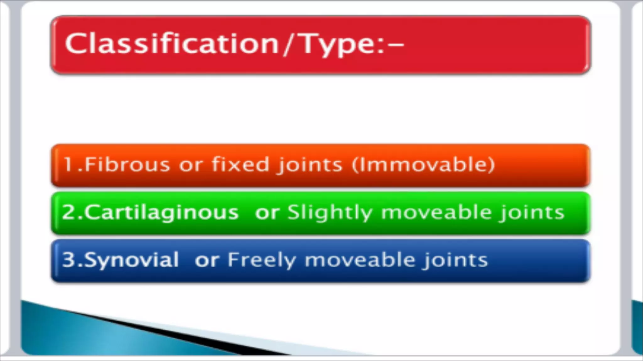
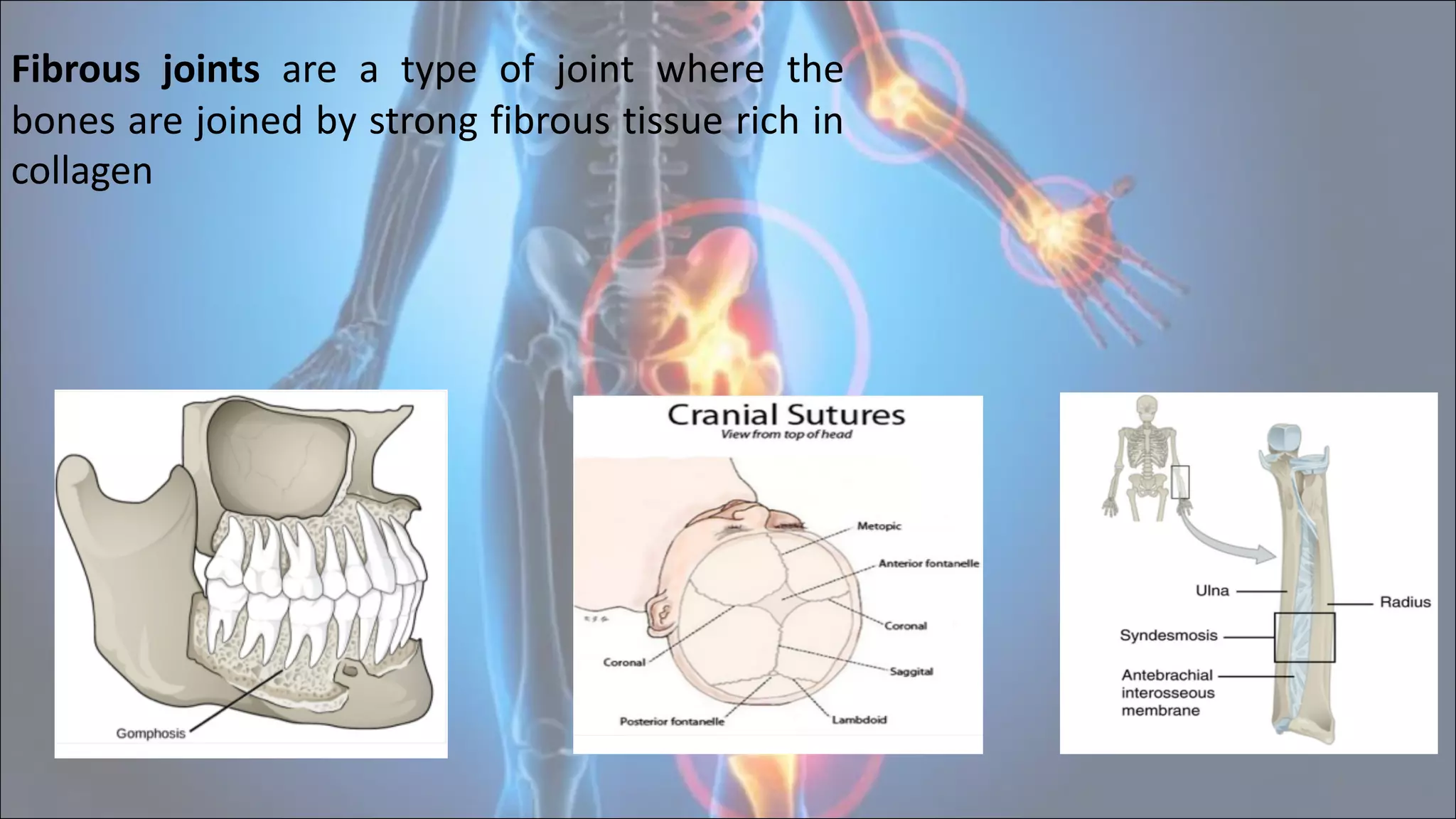
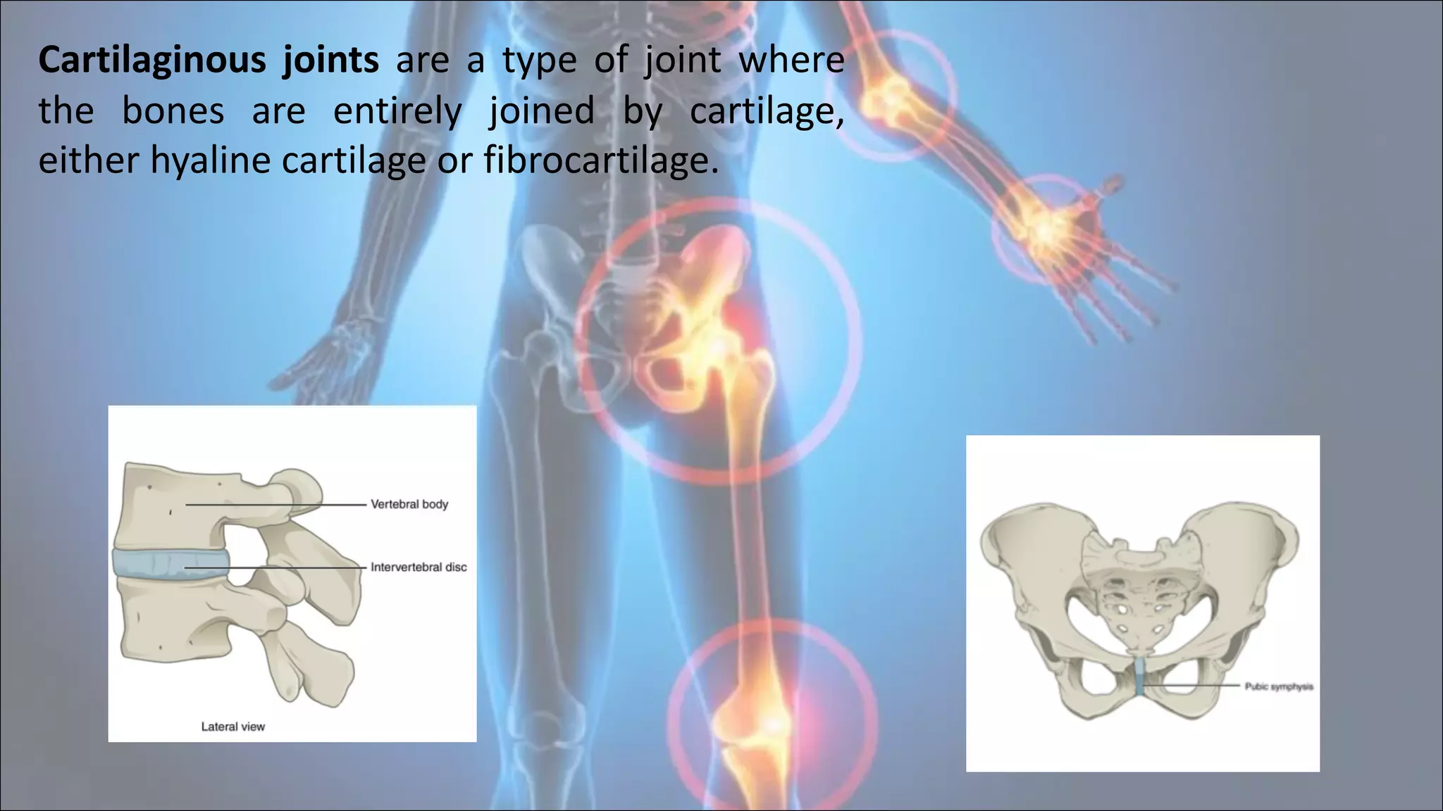
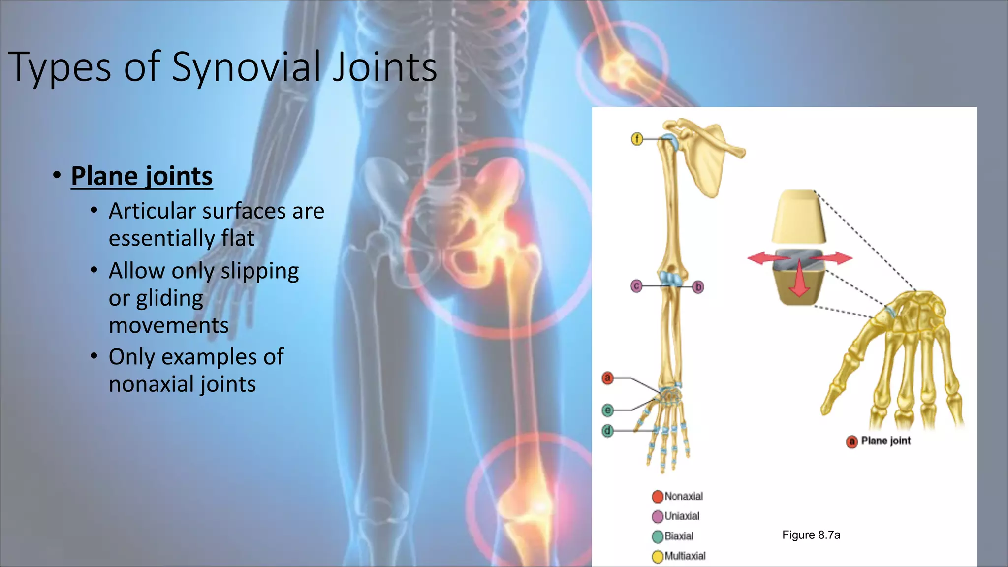
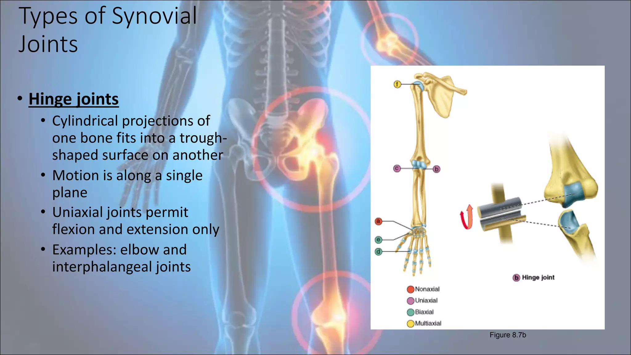
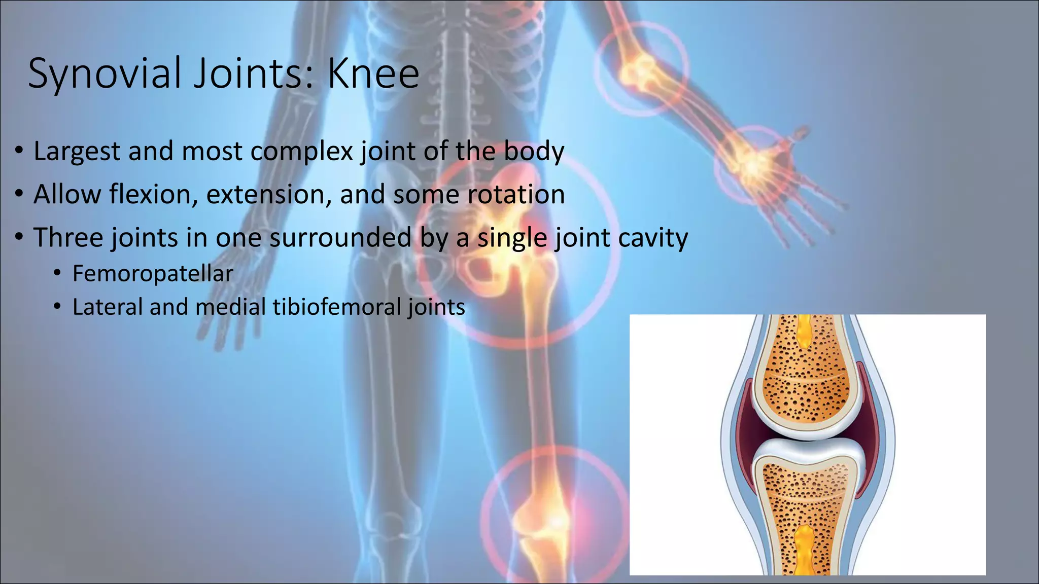
Duhok polytechnic University
Bardarash technical institute
Nursing department
Synovial Joints
Presentation By :-
• Fatima yeasin
Muhammed ghaib
•Hals hazm
•Ismail bahre
• Hogr askander
• Fazil kakl
Supervise by:-
Dr Zahir
2022
-
2023
Group G
2
Joint: is the part of the body where two or more bones
meet to allow movement also called articulation is a
point of contact
Arthrology:- It is the scientific study of
joints.
Kinesiology:- It is the study of the motion of the
human body.
3
4
Fibrous joints are a type of joint where the
bones are joined by strong fibrous tissue rich in
collagen
5
Cartilaginous joints are a type of joint where
the bones are entirely joined by cartilage,
either hyaline cartilage or fibrocartilage.
6
synovial joint is the type of joint found
between bones that move against each
other
7
Function of joint
1. to allow mobility of the skeletal
system
2. to provide a protective enclosure for
vital organs
Synovial Joints: Range of Motion
• Nonaxial – slipping movements only
• Uniaxial – movement in one plane
• Biaxial – movement in two planes
• Multiaxial – movement in or around all three planes
8
Synovial Joints:
General Structure
• Synovial joints all have
the following:
• Articular cartilage
• Joint (synovial) cavity
• Articular capsule
• Synovial fluid
• Reinforcing ligaments
9
Figure 8.3a
Synovial Joints: Friction-Reducing Structures
• Bursae – flattened, fibrous sacs lined with synovial
membranes and containing synovial fluid
• Common where ligaments, muscles, skin, tendons, or bones
rub together
• Tendon sheath – elongated bursa that wraps completely
around a tendon
10
Synovial Joints: Stability
• Stability is determined by:
• Articular surfaces – shape determines what movements are
possible
• Ligaments – unite bones and prevent excessive or undesirable
motion
• Muscles
• Muscle tendons across joints are the most important stabilizing factor
• Tendons are kept tight at all times by muscle tone
11
Synovial Joints: Movement
• Muscle attachment across a joint
• Origin – attachment to the immovable bone
• Insertion – attachment to the movable bone
• Described as movement along transverse, frontal, or sagittal
planes
12
Types of Synovial Joints
• Plane joints
• Articular surfaces are
essentially flat
• Allow only slipping
or gliding
movements
• Only examples of
nonaxial joints
13
Figure 8.7a
Types of Synovial
Joints
• Hinge joints
• Cylindrical projections of
one bone fits into a trough-
shaped surface on another
• Motion is along a single
plane
• Uniaxial joints permit
flexion and extension only
• Examples: elbow and
interphalangeal joints
14
Figure 8.7b
Pivot Joints
• Rounded end of one bone
protrudes into a “sleeve,”
or ring, composed of
bone (and possibly
ligaments) of another
• Only uniaxial movement
allowed
• Examples: joint between
the axis and the dens,
and the proximal
radioulnar joint
15
Figure 8.7c
Condyloid, or Ellipsoidal, Joints
• Oval articular surface of
one bone fits into a
complementary
depression in another
• Both articular surfaces
are oval
• Biaxial joints permit all
angular motions
• Examples: radiocarpal
(wrist) joints, and
metacarpophalangeal
(knuckle) joints
16
Figure 8.7d
Saddle Joints
• Similar to condyloid
joints but with
greater movement
• Each articular surface
has both a concave
and a convex surface
• Example:
carpometacarpal
joint of the thumb
17
Figure 8.7e
Ball-and-Socket Joints
• A spherical or
hemispherical head
of one bone
articulates with a
cuplike socket of
another
• Multiaxial joints
permit the most
freely moving
synovial joints
• Examples: shoulder
and hip joints
18
Figure 8.7f
Synovial Joints: Shoulder (Glenohumeral)
• Ball-and-socket joint in which stability is sacrificed to obtain
greater freedom of movement
• Head of humerus articulates with the glenoid fossa of the
scapula
19
Synovial Joints: Hip Joint
• Hip (coxal) joint
• Ball-and-socket joint
• Head of the femur articulates with the acetabulum
• Good range of motion, but limited by the deep socket and
strong ligaments
20
Synovial Joints: Hip Stability
• Acetabular
labrum
• Iliofemoral
ligament
• Pubofemoral
ligament
• Ischiofemoral
ligament
• Ligamentum
teres
21
Figure 8.9a
Stability:
• Annular ligament
• Ulnar collateral
ligament
• Radial collateral
ligament 22
Figure 8.10a
Synovial Joints: Elbow
• Hinge joint that
allows flexion and
extension only
• Radius and ulna
articulate with the
humerus
Synovial Joints: Knee
• Largest and most complex joint of the body
• Allow flexion, extension, and some rotation
• Three joints in one surrounded by a single joint cavity
• Femoropatellar
• Lateral and medial tibiofemoral joints
23
Reference
• https://mydr.com.au/sports-fitness/joint-synovial/
• https://www.physio-pedia.com/Synovial_Joints
• https://teachmeanatomy.info/the-basics/synovial-joint
24
25

Synovial joint .ppt

  • 1.
    Duhok polytechnic University Bardarashtechnical institute Nursing department Synovial Joints Presentation By :- • Fatima yeasin Muhammed ghaib •Hals hazm •Ismail bahre • Hogr askander • Fazil kakl Supervise by:- Dr Zahir 2022 - 2023 Group G
  • 2.
    2 Joint: is thepart of the body where two or more bones meet to allow movement also called articulation is a point of contact Arthrology:- It is the scientific study of joints. Kinesiology:- It is the study of the motion of the human body.
  • 3.
  • 4.
    4 Fibrous joints area type of joint where the bones are joined by strong fibrous tissue rich in collagen
  • 5.
    5 Cartilaginous joints area type of joint where the bones are entirely joined by cartilage, either hyaline cartilage or fibrocartilage.
  • 6.
    6 synovial joint isthe type of joint found between bones that move against each other
  • 7.
    7 Function of joint 1.to allow mobility of the skeletal system 2. to provide a protective enclosure for vital organs
  • 8.
    Synovial Joints: Rangeof Motion • Nonaxial – slipping movements only • Uniaxial – movement in one plane • Biaxial – movement in two planes • Multiaxial – movement in or around all three planes 8
  • 9.
    Synovial Joints: General Structure •Synovial joints all have the following: • Articular cartilage • Joint (synovial) cavity • Articular capsule • Synovial fluid • Reinforcing ligaments 9 Figure 8.3a
  • 10.
    Synovial Joints: Friction-ReducingStructures • Bursae – flattened, fibrous sacs lined with synovial membranes and containing synovial fluid • Common where ligaments, muscles, skin, tendons, or bones rub together • Tendon sheath – elongated bursa that wraps completely around a tendon 10
  • 11.
    Synovial Joints: Stability •Stability is determined by: • Articular surfaces – shape determines what movements are possible • Ligaments – unite bones and prevent excessive or undesirable motion • Muscles • Muscle tendons across joints are the most important stabilizing factor • Tendons are kept tight at all times by muscle tone 11
  • 12.
    Synovial Joints: Movement •Muscle attachment across a joint • Origin – attachment to the immovable bone • Insertion – attachment to the movable bone • Described as movement along transverse, frontal, or sagittal planes 12
  • 13.
    Types of SynovialJoints • Plane joints • Articular surfaces are essentially flat • Allow only slipping or gliding movements • Only examples of nonaxial joints 13 Figure 8.7a
  • 14.
    Types of Synovial Joints •Hinge joints • Cylindrical projections of one bone fits into a trough- shaped surface on another • Motion is along a single plane • Uniaxial joints permit flexion and extension only • Examples: elbow and interphalangeal joints 14 Figure 8.7b
  • 15.
    Pivot Joints • Roundedend of one bone protrudes into a “sleeve,” or ring, composed of bone (and possibly ligaments) of another • Only uniaxial movement allowed • Examples: joint between the axis and the dens, and the proximal radioulnar joint 15 Figure 8.7c
  • 16.
    Condyloid, or Ellipsoidal,Joints • Oval articular surface of one bone fits into a complementary depression in another • Both articular surfaces are oval • Biaxial joints permit all angular motions • Examples: radiocarpal (wrist) joints, and metacarpophalangeal (knuckle) joints 16 Figure 8.7d
  • 17.
    Saddle Joints • Similarto condyloid joints but with greater movement • Each articular surface has both a concave and a convex surface • Example: carpometacarpal joint of the thumb 17 Figure 8.7e
  • 18.
    Ball-and-Socket Joints • Aspherical or hemispherical head of one bone articulates with a cuplike socket of another • Multiaxial joints permit the most freely moving synovial joints • Examples: shoulder and hip joints 18 Figure 8.7f
  • 19.
    Synovial Joints: Shoulder(Glenohumeral) • Ball-and-socket joint in which stability is sacrificed to obtain greater freedom of movement • Head of humerus articulates with the glenoid fossa of the scapula 19
  • 20.
    Synovial Joints: HipJoint • Hip (coxal) joint • Ball-and-socket joint • Head of the femur articulates with the acetabulum • Good range of motion, but limited by the deep socket and strong ligaments 20
  • 21.
    Synovial Joints: HipStability • Acetabular labrum • Iliofemoral ligament • Pubofemoral ligament • Ischiofemoral ligament • Ligamentum teres 21 Figure 8.9a
  • 22.
    Stability: • Annular ligament •Ulnar collateral ligament • Radial collateral ligament 22 Figure 8.10a Synovial Joints: Elbow • Hinge joint that allows flexion and extension only • Radius and ulna articulate with the humerus
  • 23.
    Synovial Joints: Knee •Largest and most complex joint of the body • Allow flexion, extension, and some rotation • Three joints in one surrounded by a single joint cavity • Femoropatellar • Lateral and medial tibiofemoral joints 23
  • 24.
  • 25.