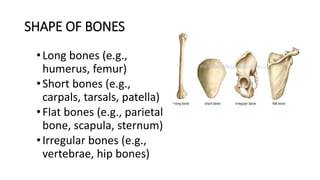
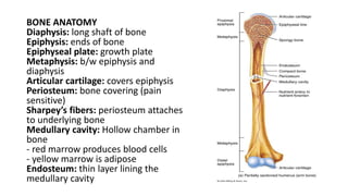
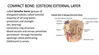
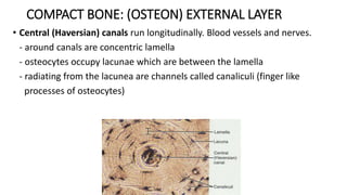
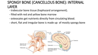
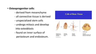
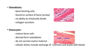
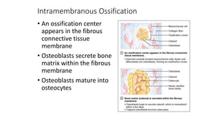
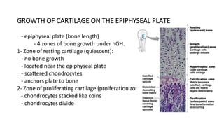
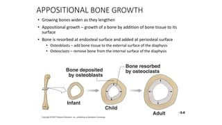
BONE PATHOLOGY
Dariusz Borys MD
dborys@lumc.edu
www.pedorthpath.com
OBJECTIVES
• Identify main functions of bone tissue
• Identify the major parts of a long bones
• Describe the cells found in bone tissue
• Describe the components of an osteon
• Compare and contrast intramembranous and endochondral
ossification.
BONE FUNCTION
• Support
• Protection (protect internal organs)
• Movement (provide leverage system for skeletal muscles,
tendons, ligaments and joints)
• Mineral homeostasis (bones act as reserves of minerals
important for the body like calcium or phosphorus)
• Hematopoiesis: blood cell formation
• Storage of adipose tissue: yellow marrow
SHAPE OF BONES
•Long bones (e.g.,
humerus, femur)
•Short bones (e.g.,
carpals, tarsals, patella)
•Flat bones (e.g., parietal
bone, scapula, sternum)
•Irregular bones (e.g.,
vertebrae, hip bones)
BONE ANATOMY
Diaphysis: long shaft of bone
Epiphysis: ends of bone
Epiphyseal plate: growth plate
Metaphysis: b/w epiphysis and
diaphysis
Articular cartilage: covers epiphysis
Periosteum: bone covering (pain
sensitive)
Sharpey’s fibers: periosteum attaches
to underlying bone
Medullary cavity: Hollow chamber in
bone
- red marrow produces blood cells
- yellow marrow is adipose
Endosteum: thin layer lining the
medullary cavity
BLOOD AND NERVE SUPPLY OF BONE
• Bone is supplied with blood by:
• Periosteal arteries accompanied by
nerves supply the periosteum and
compact bone
• Epiphyseal veins carry blood away
from long bones
• Nerves accompany the blood
vessels that supply bones
• The periosteum is rich in sensory
nerves sensitive to tearing or
tension
LONG BONES
Compact Bone – dense
outer layer
Spongy Bone – (cancellous
bone) honeycomb of
trabeculae (needle-like or
flat pieces) filled with bone
marrow
COMPACT BONE: (OSTEON) EXTERNAL LAYER
- called lamellar bone (groups of
elongated tubules called lamella)
- majority of all long bones
- protection and strength
(wt. bearing)
- concentric ring structure
- blood vessels and nerves penetrate
periosteum through horizontal
openings called perforating
(Volkmann’s) canals.
COMPACT BONE: (OSTEON) EXTERNAL LAYER
• Central (Haversian) canals run longitudinally. Blood vessels and nerves.
- around canals are concentric lamella
- osteocytes occupy lacunae which are between the lamella
- radiating from the lacunea are channels called canaliculi (finger like
processes of osteocytes)
COMPACT BONE
- Lacunae are connected to one another by canaliculi
- Osteon contains: - central canal
- surrounding lamellae
- lacunae
- osteocytes
- canaliculi
SPONGY BONE (CANCELLOUS BONE): INTERNAL
LAYER
- trabecular bone tissue (haphazard arrangement).
- filled with red and yellow bone marrow
- osteocytes get nutrients directly from circulating blood.
- short, flat and irregular bone is made up of mostly spongy bone
HISTOLOGY OF BONE
• Histology of bone tissue
Cells are surrounded by matrix.
- 25% water
- 25% protein
- 50% mineral salts
4 cell types make up osseous tissue
Osteoprogenitor cells
Osteoblasts
Osteocytes
Osteoclasts
Cells of Bone Tissue
• Osteoprogenitor cells:
- derived from mesenchyme
- all connective tissue is derived
- unspecialized stem cells
- undergo mitosis and develop
into osteoblasts
- found on inner surface of
periosteum and endosteum.
Cells of Bone Tissue
• Osteoblasts:
- bone forming cells
- found on surface of bone (arrow)
- no ability to mitotically divide
- collagen secretors
• Osteocytes:
- mature bone cells
- derived form osteoblasts
- do not secrete matrix material
- cellular duties include exchange of nutrients and waste with blood.
• Osteoclasts
- bone resorbing cells
- bone surface
- growth, maintenance and bone repair
Abundant inorganic mineral salts:
- Tricalcium phosphate in crystalline
form called hydroxyapatite
Ca3(PO4)2(OH)2
- Calcium Carbonate: CaCO3
- Magnesium Hydroxide: Mg(OH)2
- Fluoride and Sulfate
SKELETAL CARTILAGE
• Chondrocytes: cartilage producing cells.
• Lacunae: small cavities where the
chondrocytes are encased.
• Extracellular matrix: jellylike ground
substance.
• Perichondrium: layer of dense irregular
connective tissue that surrounds the
cartilage.
• No blood vessels or nerves
Types of Cartilage
• Hyaline cartilage – (glassy)
• Most abundant cartilage
• Provides support through flexibility
• Articular cartilages and costal
cartilage, larynx, trachea, and nose
• Elastic cartilage – contains many elastic
fibers
• Able to tolerate repeated bending
• Ear and epiglottis
• Fibrocartilage – resists strong
compression and strong tension
• An intermediate between hyaline and
elastic cartilage
• Intervertebral discs and pubic
symphysis
BONE FORMATION
• The process of bone formation is called ossification
• Bone formation occurs in four situations:
• 1) Formation of bone in an embryo
• 2) Growth of bones until adulthood
• 3) Remodeling of bone
• 4) Repair of fractures
Bone Formation
• Formation of Bone in an Embryo
• cartilage formation and ossification occurs during
the sixth week of embryonic development
• two patterns
• Intramembranous ossification
• Flat bones of the skull and mandible
are formed in this way
• “Soft spots” that help the fetal skull
pass through the birth canal later
become ossified forming the skull
• Endochondral ossification
• The replacement of cartilage by bone
• Most bones of the body are formed in
this way including long bones
Intramembranous Ossification
• An ossification center
appears in the fibrous
connective tissue
membrane
• Osteoblasts secrete bone
matrix within the fibrous
membrane
• Osteoblasts mature into
osteocytes
Enlarging
chondrocytes within
calcifying matrix
Chondrocytes at
the center of the
growing cartilage
model enlarge
and then die as
the matrix
calicifies.
Newly derived
osteoblasts cover
the shaft of the
cartilage in a thin
layer of bone.
Blood vessels
penetrate the
cartilage. New
osteoblasts form a
primary ossification
center.
The bone of the
shaft thickens,
and the cartilage
near each
epiphysis is
replaced by shafts
of bone.
Blood vessels invade the
epiphyses and osteo-
blasts form secondary
centers of ossification.
Cartilage
model
Bone
formation
Epiphysis
Diaphysis Marrow
cavity
Primary
ossification
center
Blood
vessel
Marrow
cavity
Blood
vessel
Secondary
ossification
center
Epiphyseal
cartilage
Articular
cartilage
Replacement of hyaline cartilage with bone
Most bones are formed this way (i.e. long bones).
Endochondral Ossification
Longitudinal Bone Growth
• Longitudinal Growth (interstitial) – cartilage continually grows and is
replaced by bone
• Bones lengthen entirely by growth of the epiphyseal plates
• Cartilage is replaced with bone as quickly as it grows
• Epiphyseal plate maintains constant thickness
GROWTH IN LENGTH
• The growth in length of long bones involves
two major events:
• Growth of cartilage on the epiphyseal
plate
• Replacement of cartilage by bone tissue
in the epiphyseal plate
- osteoblasts located beneath the
periosteum secrete bone matrix and
build bone on the surface
- osteoclasts located in the endosteum
resorbs (breakdown) bone.
GROWTH OF CARTILAGE ON THE EPIPHYSEAL PLATE
- epiphyseal plate (bone length)
- 4 zones of bone growth under hGH.
1- Zone of resting cartilage (quiescent):
- no bone growth
- located near the epiphyseal plate
- scattered chondrocytes
- anchors plate to bone
2- Zone of proliferating cartilage (prolferation zone)
- chondrocytes stacked like coins
- chondrocytes divide
3- Zone of hypertrophic (maturing) cartilage
- large chondrocytes arranged in columns
- lengthwise expansion of epiphyseal plate
4- Zone of calcified cartilage
- few cell layers thick
- occupied by osteoblasts and osteoclasts
and capillaries from the diaphysis
- cells lay down bone
- dead chondrocytes surrounded by a calcified
matrix.
Matrix resembles long spicules of calcified
cartilage.
Spicules are partly eroded by osteoclasts and then
covered in bone matrix from osteoblasts: spongy
bone is formed.
APPOSITIONAL BONE GROWTH
• Growing bones widen as they lengthen
• Appositional growth – growth of a bone by addition of bone tissue to its
surface
• Bone is resorbed at endosteal surface and added at periosteal surface
• Osteoblasts – add bone tissue to the external surface of the diaphysis
• Osteoclasts – remove bone from the internal surface of the diaphysis
Figure 6-6
BONE REMODELING
- bone continually renews itself
- never metabolically at rest
- enables Ca to be pulled from bone when blood levels are low
- osteoclasts are responsible for matrix destruction
- produce lysosomal enzymes and acids
- spongy bone replaced every 3-4 years
- compact bone every 10 years
• Fractures: Any bone break.
- blood clot will form around break
- fracture hematoma
- inflammatory process begins
- blood capillaries grow into clot
- phagocytes and osteoclasts
remove damaged tissue
- procallus forms and is invaded by
osteoprogenitor cells and
fibroblasts
- collagen and fibrocartilage turns
procallus to fibrocartilagenous
(soft) callus
- broken ends of bone are bridged by
callus
- osteoprogenitor cells are replaced by
osteoblasts and form spongy bone
- bony (hard) callus is formed
- callus is resorbed by osteoclasts and
compact bone replaces spongy bone.
Remodeling : the shaft is
reconstructed to resemble original
unbroken bone.
Closed reduction - bone ends coaxed
back into place by manipulation
Open reduction - surgery, bone ends
secured together with pins or wires
•Thank you

Bonehistology

  • 1.
    BONE PATHOLOGY Dariusz BorysMD dborys@lumc.edu www.pedorthpath.com
  • 2.
    OBJECTIVES • Identify mainfunctions of bone tissue • Identify the major parts of a long bones • Describe the cells found in bone tissue • Describe the components of an osteon • Compare and contrast intramembranous and endochondral ossification.
  • 3.
    BONE FUNCTION • Support •Protection (protect internal organs) • Movement (provide leverage system for skeletal muscles, tendons, ligaments and joints) • Mineral homeostasis (bones act as reserves of minerals important for the body like calcium or phosphorus) • Hematopoiesis: blood cell formation • Storage of adipose tissue: yellow marrow
  • 4.
    SHAPE OF BONES •Longbones (e.g., humerus, femur) •Short bones (e.g., carpals, tarsals, patella) •Flat bones (e.g., parietal bone, scapula, sternum) •Irregular bones (e.g., vertebrae, hip bones)
  • 5.
    BONE ANATOMY Diaphysis: longshaft of bone Epiphysis: ends of bone Epiphyseal plate: growth plate Metaphysis: b/w epiphysis and diaphysis Articular cartilage: covers epiphysis Periosteum: bone covering (pain sensitive) Sharpey’s fibers: periosteum attaches to underlying bone Medullary cavity: Hollow chamber in bone - red marrow produces blood cells - yellow marrow is adipose Endosteum: thin layer lining the medullary cavity
  • 6.
    BLOOD AND NERVESUPPLY OF BONE • Bone is supplied with blood by: • Periosteal arteries accompanied by nerves supply the periosteum and compact bone • Epiphyseal veins carry blood away from long bones • Nerves accompany the blood vessels that supply bones • The periosteum is rich in sensory nerves sensitive to tearing or tension
  • 7.
    LONG BONES Compact Bone– dense outer layer Spongy Bone – (cancellous bone) honeycomb of trabeculae (needle-like or flat pieces) filled with bone marrow
  • 8.
    COMPACT BONE: (OSTEON)EXTERNAL LAYER - called lamellar bone (groups of elongated tubules called lamella) - majority of all long bones - protection and strength (wt. bearing) - concentric ring structure - blood vessels and nerves penetrate periosteum through horizontal openings called perforating (Volkmann’s) canals.
  • 9.
    COMPACT BONE: (OSTEON)EXTERNAL LAYER • Central (Haversian) canals run longitudinally. Blood vessels and nerves. - around canals are concentric lamella - osteocytes occupy lacunae which are between the lamella - radiating from the lacunea are channels called canaliculi (finger like processes of osteocytes)
  • 10.
    COMPACT BONE - Lacunaeare connected to one another by canaliculi - Osteon contains: - central canal - surrounding lamellae - lacunae - osteocytes - canaliculi
  • 11.
    SPONGY BONE (CANCELLOUSBONE): INTERNAL LAYER - trabecular bone tissue (haphazard arrangement). - filled with red and yellow bone marrow - osteocytes get nutrients directly from circulating blood. - short, flat and irregular bone is made up of mostly spongy bone
  • 12.
    HISTOLOGY OF BONE •Histology of bone tissue Cells are surrounded by matrix. - 25% water - 25% protein - 50% mineral salts 4 cell types make up osseous tissue Osteoprogenitor cells Osteoblasts Osteocytes Osteoclasts Cells of Bone Tissue
  • 13.
    • Osteoprogenitor cells: -derived from mesenchyme - all connective tissue is derived - unspecialized stem cells - undergo mitosis and develop into osteoblasts - found on inner surface of periosteum and endosteum. Cells of Bone Tissue
  • 14.
    • Osteoblasts: - boneforming cells - found on surface of bone (arrow) - no ability to mitotically divide - collagen secretors • Osteocytes: - mature bone cells - derived form osteoblasts - do not secrete matrix material - cellular duties include exchange of nutrients and waste with blood.
  • 15.
    • Osteoclasts - boneresorbing cells - bone surface - growth, maintenance and bone repair Abundant inorganic mineral salts: - Tricalcium phosphate in crystalline form called hydroxyapatite Ca3(PO4)2(OH)2 - Calcium Carbonate: CaCO3 - Magnesium Hydroxide: Mg(OH)2 - Fluoride and Sulfate
  • 16.
    SKELETAL CARTILAGE • Chondrocytes:cartilage producing cells. • Lacunae: small cavities where the chondrocytes are encased. • Extracellular matrix: jellylike ground substance. • Perichondrium: layer of dense irregular connective tissue that surrounds the cartilage. • No blood vessels or nerves
  • 17.
    Types of Cartilage •Hyaline cartilage – (glassy) • Most abundant cartilage • Provides support through flexibility • Articular cartilages and costal cartilage, larynx, trachea, and nose • Elastic cartilage – contains many elastic fibers • Able to tolerate repeated bending • Ear and epiglottis • Fibrocartilage – resists strong compression and strong tension • An intermediate between hyaline and elastic cartilage • Intervertebral discs and pubic symphysis
  • 18.
    BONE FORMATION • Theprocess of bone formation is called ossification • Bone formation occurs in four situations: • 1) Formation of bone in an embryo • 2) Growth of bones until adulthood • 3) Remodeling of bone • 4) Repair of fractures
  • 19.
    Bone Formation • Formationof Bone in an Embryo • cartilage formation and ossification occurs during the sixth week of embryonic development • two patterns • Intramembranous ossification • Flat bones of the skull and mandible are formed in this way • “Soft spots” that help the fetal skull pass through the birth canal later become ossified forming the skull • Endochondral ossification • The replacement of cartilage by bone • Most bones of the body are formed in this way including long bones
  • 20.
    Intramembranous Ossification • Anossification center appears in the fibrous connective tissue membrane • Osteoblasts secrete bone matrix within the fibrous membrane • Osteoblasts mature into osteocytes
  • 21.
    Enlarging chondrocytes within calcifying matrix Chondrocytesat the center of the growing cartilage model enlarge and then die as the matrix calicifies. Newly derived osteoblasts cover the shaft of the cartilage in a thin layer of bone. Blood vessels penetrate the cartilage. New osteoblasts form a primary ossification center. The bone of the shaft thickens, and the cartilage near each epiphysis is replaced by shafts of bone. Blood vessels invade the epiphyses and osteo- blasts form secondary centers of ossification. Cartilage model Bone formation Epiphysis Diaphysis Marrow cavity Primary ossification center Blood vessel Marrow cavity Blood vessel Secondary ossification center Epiphyseal cartilage Articular cartilage Replacement of hyaline cartilage with bone Most bones are formed this way (i.e. long bones). Endochondral Ossification
  • 22.
    Longitudinal Bone Growth •Longitudinal Growth (interstitial) – cartilage continually grows and is replaced by bone • Bones lengthen entirely by growth of the epiphyseal plates • Cartilage is replaced with bone as quickly as it grows • Epiphyseal plate maintains constant thickness
  • 23.
    GROWTH IN LENGTH •The growth in length of long bones involves two major events: • Growth of cartilage on the epiphyseal plate • Replacement of cartilage by bone tissue in the epiphyseal plate - osteoblasts located beneath the periosteum secrete bone matrix and build bone on the surface - osteoclasts located in the endosteum resorbs (breakdown) bone.
  • 24.
    GROWTH OF CARTILAGEON THE EPIPHYSEAL PLATE - epiphyseal plate (bone length) - 4 zones of bone growth under hGH. 1- Zone of resting cartilage (quiescent): - no bone growth - located near the epiphyseal plate - scattered chondrocytes - anchors plate to bone 2- Zone of proliferating cartilage (prolferation zone) - chondrocytes stacked like coins - chondrocytes divide
  • 25.
    3- Zone ofhypertrophic (maturing) cartilage - large chondrocytes arranged in columns - lengthwise expansion of epiphyseal plate 4- Zone of calcified cartilage - few cell layers thick - occupied by osteoblasts and osteoclasts and capillaries from the diaphysis - cells lay down bone - dead chondrocytes surrounded by a calcified matrix. Matrix resembles long spicules of calcified cartilage. Spicules are partly eroded by osteoclasts and then covered in bone matrix from osteoblasts: spongy bone is formed.
  • 26.
    APPOSITIONAL BONE GROWTH •Growing bones widen as they lengthen • Appositional growth – growth of a bone by addition of bone tissue to its surface • Bone is resorbed at endosteal surface and added at periosteal surface • Osteoblasts – add bone tissue to the external surface of the diaphysis • Osteoclasts – remove bone from the internal surface of the diaphysis Figure 6-6
  • 27.
    BONE REMODELING - bonecontinually renews itself - never metabolically at rest - enables Ca to be pulled from bone when blood levels are low - osteoclasts are responsible for matrix destruction - produce lysosomal enzymes and acids - spongy bone replaced every 3-4 years - compact bone every 10 years
  • 28.
    • Fractures: Anybone break. - blood clot will form around break - fracture hematoma - inflammatory process begins - blood capillaries grow into clot - phagocytes and osteoclasts remove damaged tissue - procallus forms and is invaded by osteoprogenitor cells and fibroblasts - collagen and fibrocartilage turns procallus to fibrocartilagenous (soft) callus
  • 29.
    - broken endsof bone are bridged by callus - osteoprogenitor cells are replaced by osteoblasts and form spongy bone - bony (hard) callus is formed - callus is resorbed by osteoclasts and compact bone replaces spongy bone. Remodeling : the shaft is reconstructed to resemble original unbroken bone. Closed reduction - bone ends coaxed back into place by manipulation Open reduction - surgery, bone ends secured together with pins or wires
  • 30.