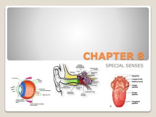
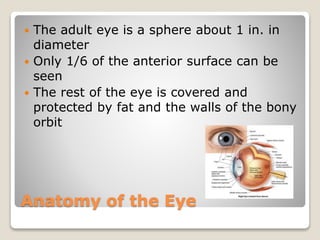
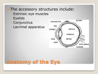
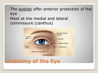
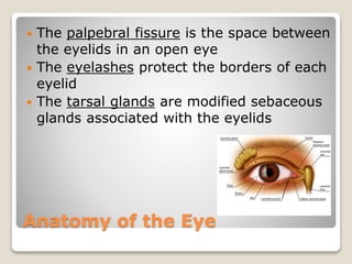
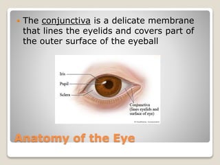
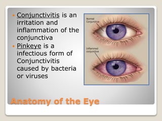
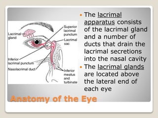
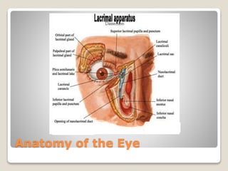
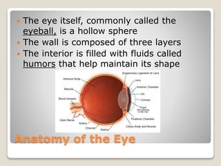
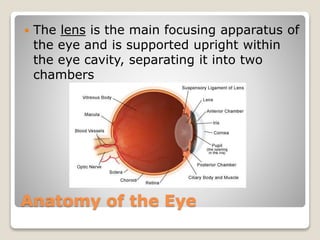
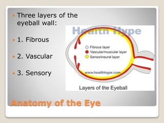
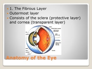
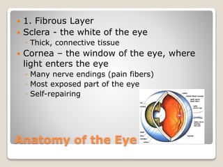
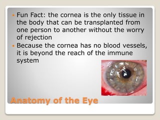
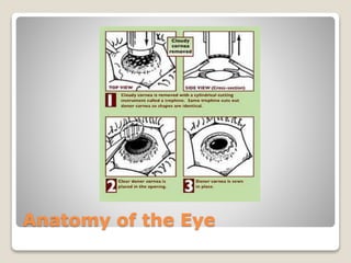
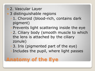
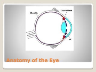
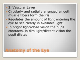
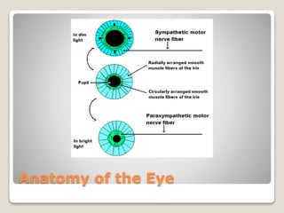
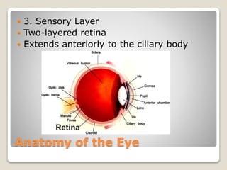
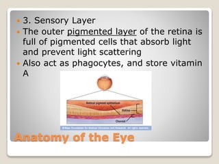
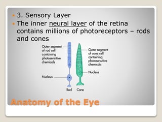
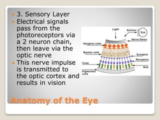
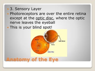
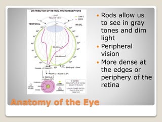
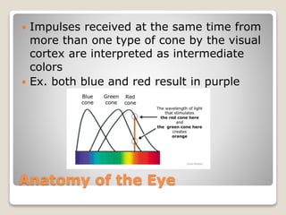
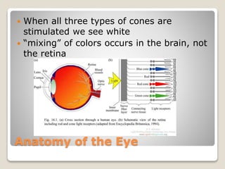
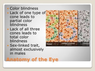
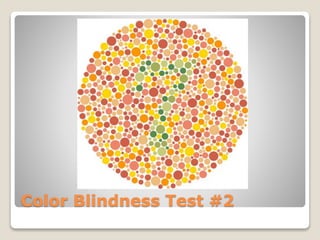
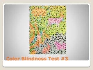
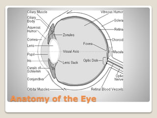
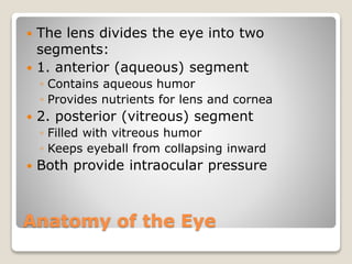
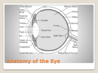
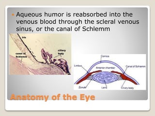
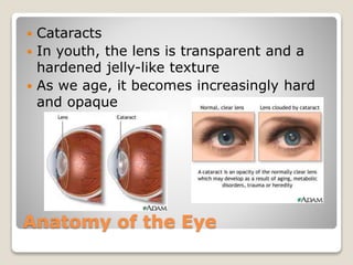
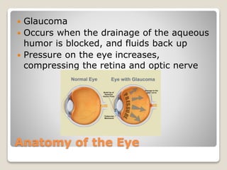
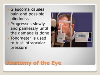
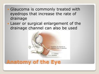
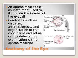
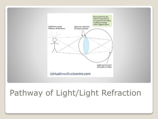
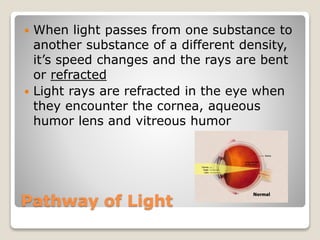
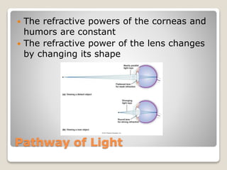
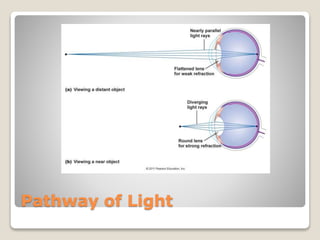
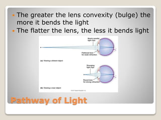
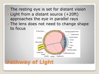
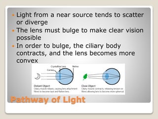
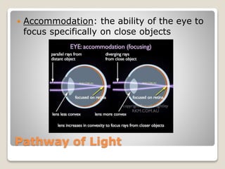
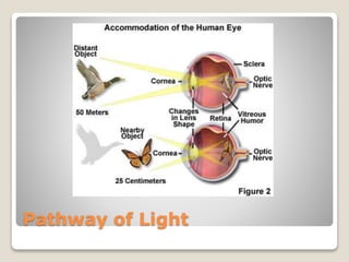
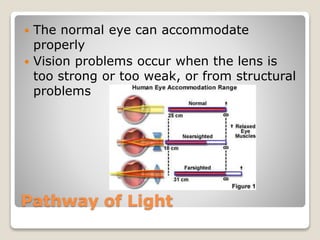
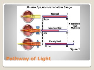
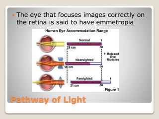
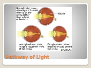
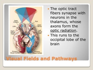
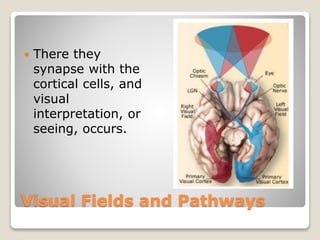
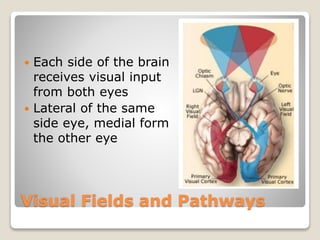
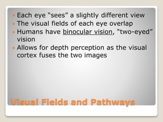
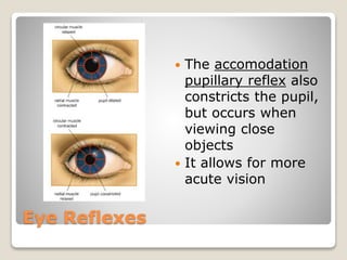
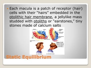
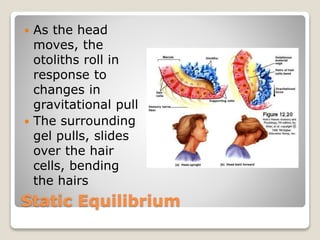
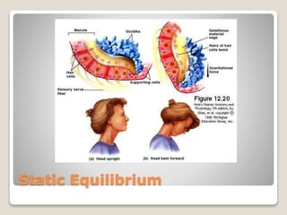
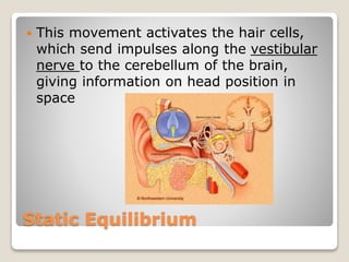
SlideShare a Scribd company logo
1 of 180
CHAPTER 8
SPECIAL SENSES
Special Senses
 Special Senses: smell, taste, sight,
hearing and equilibrium
 The special sense receptors are either
large, complex sensory organs
(eyes/ears) or localized clusters of
receptors (tastebuds/olfactory epithelium)
Special Senses
 Remember, these senses are overlapping!
 What you sense of “feel” is a blending of
stimulus effects
THE EYE AND VISION
Anatomy of the Eye
 The adult eye is a sphere about 1 in. in
diameter
 Only 1/6 of the anterior surface can be
seen
 The rest of the eye is covered and
protected by fat and the walls of the bony
orbit
Anatomy of the Eye
 The accessory structures include:
◦ Extrinsic eye muscles
◦ Eyelids
◦ Conjunctiva
◦ Lacrimal apparatus
Anatomy of the Eye
 The eyelids offer anterior protection of the
eye
 Meet at the medial and lateral
commissure (canthus)
Anatomy of the Eye
 The palpebral fissure is the space between
the eyelids in an open eye
 The eyelashes protect the borders of each
eyelid
 The tarsal glands are modified sebaceous
glands associated with the eyelids
Anatomy of the Eye
 The conjunctiva is a delicate membrane
that lines the eyelids and covers part of
the outer surface of the eyeball
Anatomy of the Eye
 Conjunctivitis is an
irritation and
inflammation of the
conjunctiva
 Pinkeye is a
infectious form of
Conjunctivitis
caused by bacteria
or viruses
Anatomy of the Eye
 The lacrimal
apparatus consists
of the lacrimal gland
and a number of
ducts that drain the
lacrimal secretions
into the nasal cavity
 The lacrimal glands
are located above
the lateral end of
each eye
Anatomy of the Eye
 The lacrimal glands continually release a
salt solution (your tears!) onto the
anterior surface of the eye
 Tears flush across the eyeball into the
lacrimal canaliculi medially, then into the
lacrimal sac and finally into the
nasolacrimal duct which empties into the
nasal cavity
Anatomy of the Eye
Anatomy of the Eye
 Lacrimal secretions are high in antibodies
and lysozyme to help cleanse and protect
the eye from foreign substances
Anatomy of the Eye
 There are six extrinsic or external eye
muscles
 Attached to the outer surface to each eye
 Produce gross eye movement
 Make it possible to follow objects
 Controlled by the cranial nerves
(abducens, oculomotor, trochlear)
Anatomy of the Eye
Anatomy of the Eye
 The eye itself, commonly called the
eyeball, is a hollow sphere
 The wall is composed of three layers
 The interior is filled with fluids called
humors that help maintain its shape
Anatomy of the Eye
 The lens is the main focusing apparatus of
the eye and is supported upright within
the eye cavity, separating it into two
chambers
Anatomy of the Eye
 Three layers of the
eyeball wall:
 1. Fibrous
 2. Vascular
 3. Sensory
Anatomy of the Eye
 1. The Fibrous Layer
 Outermost layer
 Consists of the sclera (protective layer)
and cornea (transparent layer)
Anatomy of the Eye
 1. Fibrous Layer
 Sclera - the white of the eye
◦ Thick, connective tissue
 Cornea – the window of the eye, where
light enters the eye
◦ Many nerve endings (pain fibers)
◦ Most exposed part of the eye
◦ Self-repairing
Anatomy of the Eye
 Fun Fact: the cornea is the only tissue in
the body that can be transplanted from
one person to another without the worry
of rejection
 Because the cornea has no blood vessels,
it is beyond the reach of the immune
system
Anatomy of the Eye
Anatomy of the Eye
 2. Vascular Layer
 3 distinguishable regions
◦ 1. Choroid (blood-rich, contains dark
pigment)
◦ Prevents light scattering inside the eye
◦ 2. Ciliary body (smooth muscle to which
the lens is attached by the ciliary
zonule)
◦ 3. Iris (pigmented part of the eye)
◦ Includes the pupil, where light passes
Anatomy of the Eye
Anatomy of the Eye
 2. Vascular Layer
 Circularly and radially arranged smooth
muscle fibers form the iris
 Regulates the amount of light entering the
eye to see clearly in available light
 In bright light/close vision the pupil
contracts, in dim light/distant vision the
pupil dilates
Anatomy of the Eye
Anatomy of the Eye
 3. Sensory Layer
 Two-layered retina
 Extends anteriorly to the ciliary body
Anatomy of the Eye
 3. Sensory Layer
 The outer pigmented layer of the retina is
full of pigmented cells that absorb light
and prevent light scattering
 Also act as phagocytes, and store vitamin
A
Anatomy of the Eye
 3. Sensory Layer
 The inner neural layer of the retina
contains millions of photoreceptors – rods
and cones
Anatomy of the Eye
 3. Sensory Layer
 Electrical signals
pass from the
photoreceptors via
a 2 neuron chain,
then leave via the
optic nerve
 This nerve impulse
is transmitted to
the optic cortex and
results in vision
Anatomy of the Eye
 3. Sensory Layer
 Photoreceptors are over the entire retina
except at the optic disc, where the optic
nerve leaves the eyeball
 This is your blind spot!
Anatomy of the Eye
 Rods allow us
to see in gray
tones and dim
light
 Peripheral
vision
 More dense at
the edges or
periphery of the
retina
Anatomy of the Eye
 Night Blindness
(Nyctalopia)
 Interferes with rod
function and limits the
ability to see at night
 Usually results from
prolonged vitamin A
deficiency
◦ Causes neural retina
deterioration
Anatomy of the Eye
 Cones allow for details and color to be
seen in bright light
 Densest in the center of the retina
Anatomy of the Eye
 Lateral to each blind spot is the fovea
centralis, a tiny pit that contains only
cones
 Greatest visual acuity (sharpest vision)
Anatomy of the Eye
 There are three varieties of cones:
 “blue”
 “green”
 “red” (actually responds to green and red)
Anatomy of the Eye
 Impulses received at the same time from
more than one type of cone by the visual
cortex are interpreted as intermediate
colors
 Ex. both blue and red result in purple
Anatomy of the Eye
 When all three types of cones are
stimulated we see white
 “mixing” of colors occurs in the brain, not
the retina
Anatomy of the Eye
 Color blindness
 Lack of one type of
cone leads to
partial color
blindness
 Lack of all three
cones leads to
total color
blindness
 Sex-linked trait,
almost exclusively
in males
Color Blindness Test #1
Color Blindness Test #2
Color Blindness Test #3
Anatomy of the Eye
 Lens: a flexible biconvex crystal-like
structure
 Light entering the eye is focused on the
retina by the lens
 Lens is held upright by the ciliary zonule
Anatomy of the Eye
Anatomy of the Eye
 The lens divides the eye into two
segments:
 1. anterior (aqueous) segment
◦ Contains aqueous humor
◦ Provides nutrients for lens and cornea
 2. posterior (vitreous) segment
◦ Filled with vitreous humor
◦ Keeps eyeball from collapsing inward
 Both provide intraocular pressure
Anatomy of the Eye
Anatomy of the Eye
 Aqueous humor is reabsorbed into the
venous blood through the scleral venous
sinus, or the canal of Schlemm
Anatomy of the Eye
 Cataracts
 In youth, the lens is transparent and a
hardened jelly-like texture
 As we age, it becomes increasingly hard
and opaque
Anatomy of the Eye
 Cataracts cause vision to become hazy
and distorted, and eventually cause
blindness
 Risk factors: Type II diabetes, exposure to
intense sunlight, heavy smoking
 Current treatments: surgical removal,
replacement lens implants or specialized
glasses
Cataract Surgery
Anatomy of the Eye
 Glaucoma
 Occurs when the drainage of the aqueous
humor is blocked, and fluids back up
 Pressure on the eye increases,
compressing the retina and optic nerve
Anatomy of the Eye
 Glaucoma causes
pain and possible
blindness
 Progresses slowly
and painlessly until
the damage is done
 Tonometer is used
to test intraocular
pressure
Anatomy of the Eye
 Glaucoma is commonly treated with
eyedrops that increase the rate of
drainage
 Laser or surgical enlargement of the
drainage channel can also be used
Anatomy of the Eye
 An ophthalmoscope is
an instrument used to
illuminate the interior of
the eyeball
 Conditions such as
diabetes,
arteriosclerosis, and
degeneration of the
optic nerve and retina,
can be detected by
examination with an
ophthalmoscope
Anatomy of the Eye
 Ophthalmascope
Anatomy of the Eye
 Ophthalmascope
Anatomy of the Eye
Pathway of Light/Light Refraction
Pathway of Light
 When light passes from one substance to
another substance of a different density,
it’s speed changes and the rays are bent
or refracted
 Light rays are refracted in the eye when
they encounter the cornea, aqueous
humor lens and vitreous humor
Pathway of Light
 The refractive powers of the corneas and
humors are constant
 The refractive power of the lens changes
by changing its shape
Pathway of Light
Pathway of Light
 The greater the lens convexity (bulge) the
more it bends the light
 The flatter the lens, the less it bends light
Pathway of Light
 The resting eye is set for distant vision
 Light from a distant source (+20ft)
approaches the eye in parallel rays
 The lens does not need to change shape
to focus
Pathway of Light
 Light from a near source tends to scatter
or diverge
 The lens must bulge to make clear vision
possible
 In order to bulge, the ciliary body
contracts, and the lens becomes more
convex
Pathway of Light
 Accommodation: the ability of the eye to
focus specifically on close objects
Pathway of Light
Pathway of Light
 The image formed on the retina as a
result of the light-bending activity of the
lens is a real image – it is reversed from
left to right, upside down, and smaller
than the object
 The farther away the object is, the smaller
its image on the retina
Pathway of Light
Pathway of Light
 The normal eye can accommodate
properly
 Vision problems occur when the lens is
too strong or too weak, or from structural
problems
Pathway of Light
Pathway of Light
 The eye that focuses images correctly on
the retina is said to have emmetropia
Pathway of Light
 Myopia is nearsightedness
 It occurs when the parallel light rays from
distant objects fail to reach the retina and
instead are focused in front of it
 Results from eyeball that is too long, a
lens that is too strong, or a cornea that is
too curved
 Requires concave corrective lenses
Pathway of Light
Pathway of Light
 Hyperopia is farsightedness
 It occurs when the parallel light rays from
distant objects are focused behind the
retina
 Lens is usually short or “lazy”
 Often subject to eyestrain
 Requires convex corrective lenses
Pathway of Light
Pathway of Light
 Unequal curvatures in
different parts of the
cornea or lens cause
astigmatism
 In this condition, images
occur because points of
light are focused not on
points on the retina but as
lines
 Special cylindrically ground
lenses or contacts are used
to correct this problem
Pathway of Light
Visual Fields and Visual Pathways
to the Brain
Visual Fields and Pathways
 The optic nerve is
located in back of
the eye, and
carries impulses
form the retina to
the brain
Visual Fields and Pathways
 At the optic chiasma the fibers from the
medial side of each eye cross over to the
opposite site of the brain
Visual Fields and Pathways
 The fiber tracts
that result are
called the optic
tracts
 Each optic tract
contains fibers
from the lateral
side of the eye on
the same side and
the medial side of
the opposite eye
Visual Fields and Pathways
 The optic tract
fibers synapse with
neurons in the
thalamus, whose
axons form the
optic radiation.
 This runs to the
occipital lobe of the
brain
Visual Fields and Pathways
 There they
synapse with the
cortical cells, and
visual
interpretation, or
seeing, occurs.
Visual Fields and Pathways
 Each side of the brain
receives visual input
from both eyes
 Lateral of the same
side eye, medial form
the other eye
Visual Fields and Pathways
 Each eye “sees” a slightly different view
 The visual fields of each eye overlap
 Humans have binocular vision, “two-eyed”
vision
 Allows for depth perception as the visual
cortex fuses the two images
Visual Fields and Pathways
 Hemianopia: the loss of the same side of
the visual field in both eyes
 Results from damage to the visual cortex
Visual Fields and Pathways
 People who suffer from
hemianopia are not
able to see things past
the middle of the
visual field on either
the left or right side
Visual Fields and Pathways
Eye Reflexes
Eye Reflexes
 Both the internal and external eye
muscles are needed for proper eye
function
 The external muscles, as mentioned
earlier, are responsible for following
moving objects
 They are also responsible for convergence
Eye Reflexes
 Convergence: the
reflexive movement of
the eyes medially
when viewing close
objects
 Both eyes are aimed
toward the near
object being viewed
Eye Reflexes
 When the eyes are suddenly exposed to
bright light, the pupils instantly constrict
 This is called the photopupillary reflex
Eye Reflexes
 The photopupillary reflex prevents
excessively bright light from damaging
the photoreceptors
Eye Reflexes
 The accomodation
pupillary reflex also
constricts the pupil,
but occurs when
viewing close
objects
 It allows for more
acute vision
Eye Reflexes
 Reading requires almost continuous work
by both sets of muscles
 Ciliary fibers cause the lens to bulge, the
iris constricts the pupil, convergence
occurs
 This is why eyestrain often occurs
Ears
Hearing and Balance
The Ear
 Ears are key to the senses of hearing and
balance
 Sound vibrations move fluids to stimulate
hearing receptors
 Gross movements of the head move fluids
in the balance organs
The Ear
 Receptors that respond to physical forces
are called mechanoreceptors
 These two sense organs are housed in the
ear
 They respond to different stimuli and are
activated independently of one another
Anatomy of the Ear
Anatomy of the Ear
 Anatomically, the ear is divided into 3
major sections:
 1. external (outer) ear - hearing
 2. middle ear - hearing
 3. internal (inner) ear – hearing and
equilibrium
Anatomy of the Ear
External Ear
 The external (outer) ear is composed of
the auricle and the external acoustic
meatus
External Ear
 The auricle (pinna) is
“the ear,” the shell-
shaped structure
surrounding the
auditory canal opening
 It collects and directs
sound waves into the
auditory canal
 Function is weak in
humans
External Ear
 The external acoustic meatus (auditory
canal) is a short, narrow chamber (1x.25
in) carved into the temporal bone of the
skull
External Ear
 The auditory
external acoustic
meatus has skin-
lined walls with
ceruminous glands
which secrete
cerumen (earwax)
 It provides a sticky
trap for foreign
bodies and repels
insects
External Ear
 Sound waves entering the auditory canal
eventually hit the tympanic membrane
(eardrum) and cause it to vibrate
 The canal ends at the eardrum, which
separate the external from the middle ear
Middle Ear
 The tympanic cavity or middle ear is a
small, air-filled, mucosa-lined cavity
within the temporal bone
Middle Ear
Middle Ear
 It is flanked laterally
by the eardrum and
medially by a bony
wall with two
openings, the oval
window and the
inferior, membrane-
covered round
window
Middle Ear
 The pharyngotympanic (auditory) tube
also called the eustachian tube runs
obliquely downward to link the middle ear
cavity with the throat, and the mucosae
lining the two regions are continuous
Middle Ear
 Normally the auditory tube is flattened
and closed
 Swallowing and yawning opens this tube
briefly to equalize the pressure in the
middle ear cavity
 The eardrum does not vibrate freely
unless the pressure on both surfaces is
the same
Middle Ear
 When the pressures are
unequal, the eardrum
bulges inward or
outward making hearing
difficult or causes
earaches
 The sensation of ear-
popping is the equalizing
of pressures
Middle Ear
 An inflammation of the otitis media is
common in children with sore throats
 The eardrum becomes inflamed, bulges
and often the cavity fills with fluid or pus
 A myringotomy is sometimes required to
relieve the pressure
 A tiny tube can also be used for drainage
Middle Ear
Middle Ear
 The tympanic membrane is spanned by
the three smallest bones in the body, the
ossicles
 The ossicles transmit the vibratory motion
of the eardrum to the fluids of the inner
ear
Middle Ear
 The ossicles are named for their shape:
 Hammer (malleus)
 Anvil (incus)
 Stirrup (stapes)
Middle Ear
 When the eardrum moves, the hammer
moves as well, and transfers the vibration
to the anvil
 The anvil passes the vibration to the
stirrup
Middle Ear
 The stirrup which presses on the oval
window of the inner ear
 The movement at the oval window sets
the fluids of the inner ear into motion,
eventually exciting the hearing receptors
 How Hearing Works
Inner Ear
 The internal ear is a
maze of bony
chambers called the
bony, or osseous,
labyrinth, located
deep within the
temporal bone
behind the eye
socket
Inner Ear
 The three subdivisions of the bony
labyrinth are the:
 1. cochlea (spiral, pea size)
 2. vestibule
 3. semicircular canals
Inner Ear
 The inner ear is a
cavity filled with a
plasmalike fluid called
perilymph
 Suspended in the
perilymph is a
membraneous
labyrinth, a system of
membrane sacs that
more or less follow the
shape of the bony
labyrinth
Inner Ear
 Suspended in the
perilymph is a
membranous
labyrinth, a system of
membrane sacs that
more or less follow
the shape of the bony
labyrinth
 The membranous
labyrinth itself
contains a thicker
fluid called endolymph
 How Hearing Works
Mechanisms of Equilibrium
 Equilibrium is not an easy sense to
describe
 It responds to various head movements
 The equilibrium receptors of the inner ear,
are collectively called the vestibular
apparatus
Mechanisms of Equilibrium
 The vestibular apparatus can be divided
into two functional aspects:
 1. static equilibrium
 2. dynamic equilibrium
Static Equilibrium
Static Equilibrium
 Within the membrane
sacs of the vestibule
are receptors called
maculae
 The maculae are
essential to the sense
of static equilibrium:
balance concerned
with changes in the
position of the head
Static Equilibrium
 The maculae report on changes in the
position of the head in space with respect
to the pull of gravity when the body is not
moving
 Give information on which way is up or
down
 Help keep the head erect
Static Equilibrium
Static Equilibrium
 Each macula is a patch of receptor (hair)
cells with their “hairs” embedded in the
otolithic hair membrane, a jellylike mass
studded with otoliths or “earstones,” tiny
stones made of calcium salts
Static Equilibrium
 As the head
moves, the
otoliths roll in
response to
changes in
gravitational pull
 The surrounding
gel pulls, slides
over the hair
cells, bending
the hairs
Static Equilibrium
Static Equilibrium
 This movement activates the hair cells,
which send impulses along the vestibular
nerve to the cerebellum of the brain,
giving information on head position in
space
Dynamic Equilibrium
Dynamic Equilibrium
 Dynamic equilibrium receptors, found in
the semicircular canals, respond to
angular or rotary movements of the head
Dynamic Equilibrium
 Within the ampulla, a swollen region at
the base of each membranous
semicircular canal is a receptor region
called a crista ampulliaris or crista
Dynamic Equilibrium
 The crista consists of a tuft of hair cells
covered with a gelatinous cap called
cupula
 When your head moves in arclike/angular
motion, the endolymph lags behind
Dynamic Equilibrium
 As the cupula drags against the stationary
endolymph the cupula bends with the
body’s motion
Dynamic Equilibrium
 This motion
stimulates the hair
cells, and impulses
are transmitted up
the vestibular
nerve to the
cerebellum
 Bending the
cupula in the
opposite direction
reduces impulse
generation
Dynamic Equilibrium
 When you are
moving at a
constant rate, the
receptors gradually
stop sending
impulses, and you
no longer have the
sensation of
motion until your
speed or direction
of movement
changes
Dynamic Equilibrium
 The receptors of the semicircular canal
and vestibule are responsible for dynamic
and static equilibrium separately, they
usually act together
Dynamic Equilibrium
 Besides these equilibrium senses, sight
and proprioceptors of the muscles and
tendons are also important in providing
information used to control balance to the
cerebellum
Mechanism of Hearing
 Within the cochlear duct, the endolymph-
containing membranous labyrinth of the
cochlea is the spiral organ of Corti
 This contains the hearing receptors, or
hair cells
Mechanism of Hearing
 The scalae, chambers, above and below
the cochlear duct contain perilymph
 Sound waves that reach the cochlea
through vibrations of the eardrum,
ossicles and oval window set the cochlear
fluids in motion
Mechanism of Hearing
 As the sound waves are transmitted by
the ossicles, their force is increased by
the lever activity of the ossicles
 The total force exerted in the large
eardrum reaches the oval window, which
sets the inner ear fluids in motion
Mechanism of Hearing
 The pressure waves set up vibrations in
the basilar membrane
 The receptor cells are stimulated when
the hairs are bent or tweaked by the
movement of the tectorial membrane
Mechanism of Hearing
 The length of the fibers spanning the
basilar membrane “tunes” specific regions
to vibrate at specific frequencies
 Shorter fibers are disturbed by high-
pitched sounds
 Longer fibers are disturbed by low-pitched
sounds
Mechanism of Hearing
 Once stimulated, the hair cells transmit
impulses along the cochlear nerve to the
auditory cortex
Mechanism of Hearing
 Sound usually reaches the two ears at
different times, or “in stereo”
 This helps us determine the location of
sounds
Mechanism of Hearing
 We are able to habituate, or adapt, to
constant, repetitive sounds
Mechanism of Hearing
 Hearing is also the last sense to leave us
when we fall asleep and the first to return
when we wake up
Hearing and Equilibrium Deficits
 Deafness: hearing loss of any degree
 Two main kinds:
 1. conduction
 2. sensorineural
Hearing and Equilibrium Deficits
 Temporary or
permanent
conduction
deafness results
when something
interferes with
the conduction
of sound
vibrations to the
fluids of the
inner ear
Hearing and Equilibrium Deficits
 Conduction
deafness can be
caused by
 Earwax
 Otosclerosis, the
fusion of the
ossicles
 Ruptured eardrum
Hearing and Equilibrium Deficits
 Sensorineural deafness occurs when there
is degeneration or damage to the receptor
cells in the spiral organ of Corti, cochlear
nerve or neurons in the auditory cortex
Hearing and Equilibrium Deficits
 Hearing aids use the
skull bones to conduct
sound vibrations to
the inner ear
 They are generally
very successful in
helping people with
conduction deafness to
hear
 They are less helpful
for people with
sensorineural deafness
Hearing and Equilibrium Deficits
 Equilibrium problems are usually obvious;
nausea, dizziness, and problems in
maintaining balance
 There are often strange, jerky eye
movements
Hearing and Equilibrium Deficits
 Meniere’s Syndrome
causes progressive
deafness of the inner
ear
 Sufferers become
nauseated and often
have howling or
ringing sounds in
their ears and vertigo
Hearing and Equilibrium Deficits
 Vertigo: a sense of spinning so severe
that they cannot stand up without
extreme discomfort
Chemical Senses - Taste and Smell
Taste and Smell
 Both senses use chemoreceptors
Stimulated by chemicals in solutions
Taste has four types of receptors
Smell can differentiate a large range of
chemicals
 Both senses complement each other and
respond to many of the same stimuli
Olfaction - Smell
 Olfactory
receptors are in
the roof of the
nasal cavity
Neurons with
long cilia
Chemicals must
be dissolved in
mucus for
detection
Olfaction - Smell
Olfaction - Smell
 Impulses are transmitted via the olfactory
nerve
 Interpretation of smells is made in the cortex
(olfactory area of temporal lobe)
http://asb.aecom.yu.edu/histology/labs/images/slides/A74_OlfactoryEpith_40X.jpg
Taste
 Taste buds house the receptor
organs
 Location of taste buds
Most on the tongue
Soft palate
Cheeks
Tongue and Taste
 The tongue is covered
with projections called
papillae
 Filiform papillae –
sharp with no taste
buds
 Fungiform papillae –
rounded with taste
buds
 Circumvallate papillae
– large papillae with
taste buds
 Taste buds are found on
the sides of papillae
http://neuromedia.neurobio.ucla.edu/campbell/oral_cavity/wp_images/96_fungiform.gif
http://www.esg.montana.edu/esg/kla/ta/vallate.jpg
Structure of Taste Buds
 Gustatory cells
are the
receptors
Have gustatory
hairs (long
microvilli)
Hairs are
stimulated by
chemicals
dissolved in
saliva
Structure of taste buds
 Impulses are carried to the gustatory
complex (parietal lobe) by several
cranial nerves because taste buds are
found in different areas
Facial nerve
Glossopharyngeal nerve
Vagus nerve
http://www.biosci.ohiou.edu/introbioslab/Bios171/images/lab6/Tastebuds.JPG
Taste Sensations
 Sweet receptors
Sugars
Saccharine
Some amino acids
 Sour receptors
Acids
 Bitter receptors
Alkaloids
 Salty receptors
Metal ions
 Umami
Glutamate, aspartate (MSG, meats)
Development of the Special
Senses
 Formed early in embryonic development
 Eyes are outgrowths of the brain
 All special senses are functional at birth

More Related Content

What's hot

What's hot (20)

Body cavities
Body cavitiesBody cavities
Body cavities
 
cartilage
cartilage cartilage
cartilage
 
Anatomy of Lymphatic system
Anatomy of Lymphatic systemAnatomy of Lymphatic system
Anatomy of Lymphatic system
 
connective tissue.
 connective tissue. connective tissue.
connective tissue.
 
Lungs
LungsLungs
Lungs
 
Skeletal system. anatomy and physiology of skeletal system. appendicular skel...
Skeletal system. anatomy and physiology of skeletal system. appendicular skel...Skeletal system. anatomy and physiology of skeletal system. appendicular skel...
Skeletal system. anatomy and physiology of skeletal system. appendicular skel...
 
Sensory receptor.pptx
Sensory receptor.pptxSensory receptor.pptx
Sensory receptor.pptx
 
Nervous system notes
Nervous system notesNervous system notes
Nervous system notes
 
Cardiovascular system
Cardiovascular systemCardiovascular system
Cardiovascular system
 
Anatomy and physiology of the Eye-Nursing
Anatomy and physiology of the Eye-NursingAnatomy and physiology of the Eye-Nursing
Anatomy and physiology of the Eye-Nursing
 
Special Senses
Special SensesSpecial Senses
Special Senses
 
Anatomy of Nephron
Anatomy of NephronAnatomy of Nephron
Anatomy of Nephron
 
SPECIAL SENSES
SPECIAL SENSESSPECIAL SENSES
SPECIAL SENSES
 
Sense organs
Sense organsSense organs
Sense organs
 
Anatomy of nervous system
Anatomy of nervous systemAnatomy of nervous system
Anatomy of nervous system
 
The cranial bones
The cranial bonesThe cranial bones
The cranial bones
 
Appendicular skeleton
Appendicular skeletonAppendicular skeleton
Appendicular skeleton
 
Structure of the Cardiac muscles
Structure of the Cardiac muscles Structure of the Cardiac muscles
Structure of the Cardiac muscles
 
Skull, neck and muscle
Skull, neck and muscleSkull, neck and muscle
Skull, neck and muscle
 
anatomy of central nervous system
anatomy of central nervous systemanatomy of central nervous system
anatomy of central nervous system
 

Similar to Chapter 8 special senses

Eye Conditions presentation for medical students
Eye Conditions presentation for medical studentsEye Conditions presentation for medical students
Eye Conditions presentation for medical studentsIbrahimKargbo13
 
Eye Conditions portable display format for medical students
Eye Conditions portable display format for medical studentsEye Conditions portable display format for medical students
Eye Conditions portable display format for medical studentsIbrahimKargbo13
 
Ocular Anatomy
Ocular AnatomyOcular Anatomy
Ocular Anatomywcbvi
 
Chapter 8 ppt-senses.ppt
Chapter 8 ppt-senses.pptChapter 8 ppt-senses.ppt
Chapter 8 ppt-senses.pptNehaPandey199
 
Biology 161 the_eye_and_the_ear_without_
Biology 161 the_eye_and_the_ear_without_Biology 161 the_eye_and_the_ear_without_
Biology 161 the_eye_and_the_ear_without_jydyck
 
Eye structure.pptx
Eye structure.pptxEye structure.pptx
Eye structure.pptxij1009
 
INTRODUCTION TO OPHTHALMOLOGY.pdf
INTRODUCTION TO OPHTHALMOLOGY.pdfINTRODUCTION TO OPHTHALMOLOGY.pdf
INTRODUCTION TO OPHTHALMOLOGY.pdfSimon Maina
 
VISION IN ANIMALS VETERINARY PHYSIOLOGY.pdf
VISION IN ANIMALS VETERINARY PHYSIOLOGY.pdfVISION IN ANIMALS VETERINARY PHYSIOLOGY.pdf
VISION IN ANIMALS VETERINARY PHYSIOLOGY.pdfTatendaMageja
 
Anatomy of the eye and Layers of eyeball
Anatomy of the eye and Layers of eyeball Anatomy of the eye and Layers of eyeball
Anatomy of the eye and Layers of eyeball AnamSehreen
 

Similar to Chapter 8 special senses (20)

Ocular anatomy
Ocular anatomyOcular anatomy
Ocular anatomy
 
anatomy of eye
anatomy of eyeanatomy of eye
anatomy of eye
 
Eye Conditions presentation for medical students
Eye Conditions presentation for medical studentsEye Conditions presentation for medical students
Eye Conditions presentation for medical students
 
Eye Conditions portable display format for medical students
Eye Conditions portable display format for medical studentsEye Conditions portable display format for medical students
Eye Conditions portable display format for medical students
 
Ocular Anatomy
Ocular AnatomyOcular Anatomy
Ocular Anatomy
 
Ocular Anatomy
Ocular AnatomyOcular Anatomy
Ocular Anatomy
 
Chapter 8 ppt-senses.ppt
Chapter 8 ppt-senses.pptChapter 8 ppt-senses.ppt
Chapter 8 ppt-senses.ppt
 
Biology 161 the_eye_and_the_ear_without_
Biology 161 the_eye_and_the_ear_without_Biology 161 the_eye_and_the_ear_without_
Biology 161 the_eye_and_the_ear_without_
 
Eye anatomy
Eye anatomyEye anatomy
Eye anatomy
 
diagram of eye
diagram of eyediagram of eye
diagram of eye
 
the eye.ppsx
the eye.ppsxthe eye.ppsx
the eye.ppsx
 
Ophthalmology 5th year, 1st 2 lectures (Dr. Bakhtyar)
Ophthalmology 5th year, 1st 2 lectures (Dr. Bakhtyar)Ophthalmology 5th year, 1st 2 lectures (Dr. Bakhtyar)
Ophthalmology 5th year, 1st 2 lectures (Dr. Bakhtyar)
 
Sss5
Sss5Sss5
Sss5
 
Eye structure.pptx
Eye structure.pptxEye structure.pptx
Eye structure.pptx
 
INTRODUCTION TO OPHTHALMOLOGY.pdf
INTRODUCTION TO OPHTHALMOLOGY.pdfINTRODUCTION TO OPHTHALMOLOGY.pdf
INTRODUCTION TO OPHTHALMOLOGY.pdf
 
VISION IN ANIMALS VETERINARY PHYSIOLOGY.pdf
VISION IN ANIMALS VETERINARY PHYSIOLOGY.pdfVISION IN ANIMALS VETERINARY PHYSIOLOGY.pdf
VISION IN ANIMALS VETERINARY PHYSIOLOGY.pdf
 
Anatomy of the eye and Layers of eyeball
Anatomy of the eye and Layers of eyeball Anatomy of the eye and Layers of eyeball
Anatomy of the eye and Layers of eyeball
 
Sense organs
Sense organsSense organs
Sense organs
 
Sense organs
Sense organsSense organs
Sense organs
 
Eye presentation1
Eye presentation1Eye presentation1
Eye presentation1
 

More from watsonma12

Special topic genomics and personalized medicine
Special topic genomics and personalized medicineSpecial topic genomics and personalized medicine
Special topic genomics and personalized medicinewatsonma12
 
Chapter 4 skin power point
Chapter 4 skin power pointChapter 4 skin power point
Chapter 4 skin power pointwatsonma12
 
Ch. 4 Cell Structures
Ch. 4 Cell StructuresCh. 4 Cell Structures
Ch. 4 Cell Structureswatsonma12
 
Chapter 5 Cell Transport
Chapter 5 Cell Transport Chapter 5 Cell Transport
Chapter 5 Cell Transport watsonma12
 
Chapter 7 Dna fingerprinting
Chapter 7   Dna fingerprintingChapter 7   Dna fingerprinting
Chapter 7 Dna fingerprintingwatsonma12
 
Chapter 8 power point
Chapter 8 power pointChapter 8 power point
Chapter 8 power pointwatsonma12
 
Chapter 7 cells power point student version
Chapter 7 cells power point student versionChapter 7 cells power point student version
Chapter 7 cells power point student versionwatsonma12
 
Blood splatter informational power point
Blood splatter informational power pointBlood splatter informational power point
Blood splatter informational power pointwatsonma12
 
Chapter 4 the six ma jor biomes
Chapter 4 the six ma jor biomesChapter 4 the six ma jor biomes
Chapter 4 the six ma jor biomeswatsonma12
 
Chapter 3 biosphere and ecology
Chapter 3 biosphere and ecologyChapter 3 biosphere and ecology
Chapter 3 biosphere and ecologywatsonma12
 
Chapter 3 good buddies! final
Chapter 3 good buddies! finalChapter 3 good buddies! final
Chapter 3 good buddies! finalwatsonma12
 
Chapter 8 blood typing
Chapter 8 blood typingChapter 8 blood typing
Chapter 8 blood typingwatsonma12
 
Chapter 6 fingerprints power point
Chapter 6 fingerprints power pointChapter 6 fingerprints power point
Chapter 6 fingerprints power pointwatsonma12
 
Chapter 3 newtons first law of motion
Chapter 3 newtons first law of motion Chapter 3 newtons first law of motion
Chapter 3 newtons first law of motion watsonma12
 
Chapter 2 chemistry power point
Chapter 2 chemistry power pointChapter 2 chemistry power point
Chapter 2 chemistry power pointwatsonma12
 
Chapter 2 Crime Scene Investigation
Chapter 2 Crime Scene InvestigationChapter 2 Crime Scene Investigation
Chapter 2 Crime Scene Investigationwatsonma12
 
Chapter 2 mechanical equilibrium watson
Chapter 2 mechanical equilibrium watsonChapter 2 mechanical equilibrium watson
Chapter 2 mechanical equilibrium watsonwatsonma12
 
Chapter 1 power point from book
Chapter 1 power point from bookChapter 1 power point from book
Chapter 1 power point from bookwatsonma12
 
Chapter 1 introduction to science and biology
Chapter 1  introduction to science and biologyChapter 1  introduction to science and biology
Chapter 1 introduction to science and biologywatsonma12
 
Chapter 1 Observation Skills
Chapter 1 Observation SkillsChapter 1 Observation Skills
Chapter 1 Observation Skillswatsonma12
 

More from watsonma12 (20)

Special topic genomics and personalized medicine
Special topic genomics and personalized medicineSpecial topic genomics and personalized medicine
Special topic genomics and personalized medicine
 
Chapter 4 skin power point
Chapter 4 skin power pointChapter 4 skin power point
Chapter 4 skin power point
 
Ch. 4 Cell Structures
Ch. 4 Cell StructuresCh. 4 Cell Structures
Ch. 4 Cell Structures
 
Chapter 5 Cell Transport
Chapter 5 Cell Transport Chapter 5 Cell Transport
Chapter 5 Cell Transport
 
Chapter 7 Dna fingerprinting
Chapter 7   Dna fingerprintingChapter 7   Dna fingerprinting
Chapter 7 Dna fingerprinting
 
Chapter 8 power point
Chapter 8 power pointChapter 8 power point
Chapter 8 power point
 
Chapter 7 cells power point student version
Chapter 7 cells power point student versionChapter 7 cells power point student version
Chapter 7 cells power point student version
 
Blood splatter informational power point
Blood splatter informational power pointBlood splatter informational power point
Blood splatter informational power point
 
Chapter 4 the six ma jor biomes
Chapter 4 the six ma jor biomesChapter 4 the six ma jor biomes
Chapter 4 the six ma jor biomes
 
Chapter 3 biosphere and ecology
Chapter 3 biosphere and ecologyChapter 3 biosphere and ecology
Chapter 3 biosphere and ecology
 
Chapter 3 good buddies! final
Chapter 3 good buddies! finalChapter 3 good buddies! final
Chapter 3 good buddies! final
 
Chapter 8 blood typing
Chapter 8 blood typingChapter 8 blood typing
Chapter 8 blood typing
 
Chapter 6 fingerprints power point
Chapter 6 fingerprints power pointChapter 6 fingerprints power point
Chapter 6 fingerprints power point
 
Chapter 3 newtons first law of motion
Chapter 3 newtons first law of motion Chapter 3 newtons first law of motion
Chapter 3 newtons first law of motion
 
Chapter 2 chemistry power point
Chapter 2 chemistry power pointChapter 2 chemistry power point
Chapter 2 chemistry power point
 
Chapter 2 Crime Scene Investigation
Chapter 2 Crime Scene InvestigationChapter 2 Crime Scene Investigation
Chapter 2 Crime Scene Investigation
 
Chapter 2 mechanical equilibrium watson
Chapter 2 mechanical equilibrium watsonChapter 2 mechanical equilibrium watson
Chapter 2 mechanical equilibrium watson
 
Chapter 1 power point from book
Chapter 1 power point from bookChapter 1 power point from book
Chapter 1 power point from book
 
Chapter 1 introduction to science and biology
Chapter 1  introduction to science and biologyChapter 1  introduction to science and biology
Chapter 1 introduction to science and biology
 
Chapter 1 Observation Skills
Chapter 1 Observation SkillsChapter 1 Observation Skills
Chapter 1 Observation Skills
 

Recently uploaded

Call Girls in Munirka Delhi 💯Call Us 🔝9953322196🔝 💯Escort.
Call Girls in Munirka Delhi 💯Call Us 🔝9953322196🔝 💯Escort.Call Girls in Munirka Delhi 💯Call Us 🔝9953322196🔝 💯Escort.
Call Girls in Munirka Delhi 💯Call Us 🔝9953322196🔝 💯Escort.aasikanpl
 
Hubble Asteroid Hunter III. Physical properties of newly found asteroids
Hubble Asteroid Hunter III. Physical properties of newly found asteroidsHubble Asteroid Hunter III. Physical properties of newly found asteroids
Hubble Asteroid Hunter III. Physical properties of newly found asteroidsSérgio Sacani
 
SOLUBLE PATTERN RECOGNITION RECEPTORS.pptx
SOLUBLE PATTERN RECOGNITION RECEPTORS.pptxSOLUBLE PATTERN RECOGNITION RECEPTORS.pptx
SOLUBLE PATTERN RECOGNITION RECEPTORS.pptxkessiyaTpeter
 
Analytical Profile of Coleus Forskohlii | Forskolin .pdf
Analytical Profile of Coleus Forskohlii | Forskolin .pdfAnalytical Profile of Coleus Forskohlii | Forskolin .pdf
Analytical Profile of Coleus Forskohlii | Forskolin .pdfSwapnil Therkar
 
Stunning ➥8448380779▻ Call Girls In Panchshil Enclave Delhi NCR
Stunning ➥8448380779▻ Call Girls In Panchshil Enclave Delhi NCRStunning ➥8448380779▻ Call Girls In Panchshil Enclave Delhi NCR
Stunning ➥8448380779▻ Call Girls In Panchshil Enclave Delhi NCRDelhi Call girls
 
Is RISC-V ready for HPC workload? Maybe?
Is RISC-V ready for HPC workload? Maybe?Is RISC-V ready for HPC workload? Maybe?
Is RISC-V ready for HPC workload? Maybe?Patrick Diehl
 
Isotopic evidence of long-lived volcanism on Io
Isotopic evidence of long-lived volcanism on IoIsotopic evidence of long-lived volcanism on Io
Isotopic evidence of long-lived volcanism on IoSérgio Sacani
 
Physiochemical properties of nanomaterials and its nanotoxicity.pptx
Physiochemical properties of nanomaterials and its nanotoxicity.pptxPhysiochemical properties of nanomaterials and its nanotoxicity.pptx
Physiochemical properties of nanomaterials and its nanotoxicity.pptxAArockiyaNisha
 
Recombination DNA Technology (Nucleic Acid Hybridization )
Recombination DNA Technology (Nucleic Acid Hybridization )Recombination DNA Technology (Nucleic Acid Hybridization )
Recombination DNA Technology (Nucleic Acid Hybridization )aarthirajkumar25
 
Animal Communication- Auditory and Visual.pptx
Animal Communication- Auditory and Visual.pptxAnimal Communication- Auditory and Visual.pptx
Animal Communication- Auditory and Visual.pptxUmerFayaz5
 
Labelling Requirements and Label Claims for Dietary Supplements and Recommend...
Labelling Requirements and Label Claims for Dietary Supplements and Recommend...Labelling Requirements and Label Claims for Dietary Supplements and Recommend...
Labelling Requirements and Label Claims for Dietary Supplements and Recommend...Lokesh Kothari
 
NAVSEA PEO USC - Unmanned & Small Combatants 26Oct23.pdf
NAVSEA PEO USC - Unmanned & Small Combatants 26Oct23.pdfNAVSEA PEO USC - Unmanned & Small Combatants 26Oct23.pdf
NAVSEA PEO USC - Unmanned & Small Combatants 26Oct23.pdfWadeK3
 
Scheme-of-Work-Science-Stage-4 cambridge science.docx
Scheme-of-Work-Science-Stage-4 cambridge science.docxScheme-of-Work-Science-Stage-4 cambridge science.docx
Scheme-of-Work-Science-Stage-4 cambridge science.docxyaramohamed343013
 
Artificial Intelligence In Microbiology by Dr. Prince C P
Artificial Intelligence In Microbiology by Dr. Prince C PArtificial Intelligence In Microbiology by Dr. Prince C P
Artificial Intelligence In Microbiology by Dr. Prince C PPRINCE C P
 
Call Us ≽ 9953322196 ≼ Call Girls In Mukherjee Nagar(Delhi) |
Call Us ≽ 9953322196 ≼ Call Girls In Mukherjee Nagar(Delhi) |Call Us ≽ 9953322196 ≼ Call Girls In Mukherjee Nagar(Delhi) |
Call Us ≽ 9953322196 ≼ Call Girls In Mukherjee Nagar(Delhi) |aasikanpl
 
Luciferase in rDNA technology (biotechnology).pptx
Luciferase in rDNA technology (biotechnology).pptxLuciferase in rDNA technology (biotechnology).pptx
Luciferase in rDNA technology (biotechnology).pptxAleenaTreesaSaji
 
Grafana in space: Monitoring Japan's SLIM moon lander in real time
Grafana in space: Monitoring Japan's SLIM moon lander  in real timeGrafana in space: Monitoring Japan's SLIM moon lander  in real time
Grafana in space: Monitoring Japan's SLIM moon lander in real timeSatoshi NAKAHIRA
 
PossibleEoarcheanRecordsoftheGeomagneticFieldPreservedintheIsuaSupracrustalBe...
PossibleEoarcheanRecordsoftheGeomagneticFieldPreservedintheIsuaSupracrustalBe...PossibleEoarcheanRecordsoftheGeomagneticFieldPreservedintheIsuaSupracrustalBe...
PossibleEoarcheanRecordsoftheGeomagneticFieldPreservedintheIsuaSupracrustalBe...Sérgio Sacani
 

Recently uploaded (20)

Call Girls in Munirka Delhi 💯Call Us 🔝9953322196🔝 💯Escort.
Call Girls in Munirka Delhi 💯Call Us 🔝9953322196🔝 💯Escort.Call Girls in Munirka Delhi 💯Call Us 🔝9953322196🔝 💯Escort.
Call Girls in Munirka Delhi 💯Call Us 🔝9953322196🔝 💯Escort.
 
Hubble Asteroid Hunter III. Physical properties of newly found asteroids
Hubble Asteroid Hunter III. Physical properties of newly found asteroidsHubble Asteroid Hunter III. Physical properties of newly found asteroids
Hubble Asteroid Hunter III. Physical properties of newly found asteroids
 
Engler and Prantl system of classification in plant taxonomy
Engler and Prantl system of classification in plant taxonomyEngler and Prantl system of classification in plant taxonomy
Engler and Prantl system of classification in plant taxonomy
 
SOLUBLE PATTERN RECOGNITION RECEPTORS.pptx
SOLUBLE PATTERN RECOGNITION RECEPTORS.pptxSOLUBLE PATTERN RECOGNITION RECEPTORS.pptx
SOLUBLE PATTERN RECOGNITION RECEPTORS.pptx
 
Analytical Profile of Coleus Forskohlii | Forskolin .pdf
Analytical Profile of Coleus Forskohlii | Forskolin .pdfAnalytical Profile of Coleus Forskohlii | Forskolin .pdf
Analytical Profile of Coleus Forskohlii | Forskolin .pdf
 
Stunning ➥8448380779▻ Call Girls In Panchshil Enclave Delhi NCR
Stunning ➥8448380779▻ Call Girls In Panchshil Enclave Delhi NCRStunning ➥8448380779▻ Call Girls In Panchshil Enclave Delhi NCR
Stunning ➥8448380779▻ Call Girls In Panchshil Enclave Delhi NCR
 
Is RISC-V ready for HPC workload? Maybe?
Is RISC-V ready for HPC workload? Maybe?Is RISC-V ready for HPC workload? Maybe?
Is RISC-V ready for HPC workload? Maybe?
 
Isotopic evidence of long-lived volcanism on Io
Isotopic evidence of long-lived volcanism on IoIsotopic evidence of long-lived volcanism on Io
Isotopic evidence of long-lived volcanism on Io
 
Physiochemical properties of nanomaterials and its nanotoxicity.pptx
Physiochemical properties of nanomaterials and its nanotoxicity.pptxPhysiochemical properties of nanomaterials and its nanotoxicity.pptx
Physiochemical properties of nanomaterials and its nanotoxicity.pptx
 
Recombination DNA Technology (Nucleic Acid Hybridization )
Recombination DNA Technology (Nucleic Acid Hybridization )Recombination DNA Technology (Nucleic Acid Hybridization )
Recombination DNA Technology (Nucleic Acid Hybridization )
 
Animal Communication- Auditory and Visual.pptx
Animal Communication- Auditory and Visual.pptxAnimal Communication- Auditory and Visual.pptx
Animal Communication- Auditory and Visual.pptx
 
Labelling Requirements and Label Claims for Dietary Supplements and Recommend...
Labelling Requirements and Label Claims for Dietary Supplements and Recommend...Labelling Requirements and Label Claims for Dietary Supplements and Recommend...
Labelling Requirements and Label Claims for Dietary Supplements and Recommend...
 
NAVSEA PEO USC - Unmanned & Small Combatants 26Oct23.pdf
NAVSEA PEO USC - Unmanned & Small Combatants 26Oct23.pdfNAVSEA PEO USC - Unmanned & Small Combatants 26Oct23.pdf
NAVSEA PEO USC - Unmanned & Small Combatants 26Oct23.pdf
 
Scheme-of-Work-Science-Stage-4 cambridge science.docx
Scheme-of-Work-Science-Stage-4 cambridge science.docxScheme-of-Work-Science-Stage-4 cambridge science.docx
Scheme-of-Work-Science-Stage-4 cambridge science.docx
 
Artificial Intelligence In Microbiology by Dr. Prince C P
Artificial Intelligence In Microbiology by Dr. Prince C PArtificial Intelligence In Microbiology by Dr. Prince C P
Artificial Intelligence In Microbiology by Dr. Prince C P
 
9953056974 Young Call Girls In Mahavir enclave Indian Quality Escort service
9953056974 Young Call Girls In Mahavir enclave Indian Quality Escort service9953056974 Young Call Girls In Mahavir enclave Indian Quality Escort service
9953056974 Young Call Girls In Mahavir enclave Indian Quality Escort service
 
Call Us ≽ 9953322196 ≼ Call Girls In Mukherjee Nagar(Delhi) |
Call Us ≽ 9953322196 ≼ Call Girls In Mukherjee Nagar(Delhi) |Call Us ≽ 9953322196 ≼ Call Girls In Mukherjee Nagar(Delhi) |
Call Us ≽ 9953322196 ≼ Call Girls In Mukherjee Nagar(Delhi) |
 
Luciferase in rDNA technology (biotechnology).pptx
Luciferase in rDNA technology (biotechnology).pptxLuciferase in rDNA technology (biotechnology).pptx
Luciferase in rDNA technology (biotechnology).pptx
 
Grafana in space: Monitoring Japan's SLIM moon lander in real time
Grafana in space: Monitoring Japan's SLIM moon lander  in real timeGrafana in space: Monitoring Japan's SLIM moon lander  in real time
Grafana in space: Monitoring Japan's SLIM moon lander in real time
 
PossibleEoarcheanRecordsoftheGeomagneticFieldPreservedintheIsuaSupracrustalBe...
PossibleEoarcheanRecordsoftheGeomagneticFieldPreservedintheIsuaSupracrustalBe...PossibleEoarcheanRecordsoftheGeomagneticFieldPreservedintheIsuaSupracrustalBe...
PossibleEoarcheanRecordsoftheGeomagneticFieldPreservedintheIsuaSupracrustalBe...
 

Chapter 8 special senses

  • 2. Special Senses  Special Senses: smell, taste, sight, hearing and equilibrium  The special sense receptors are either large, complex sensory organs (eyes/ears) or localized clusters of receptors (tastebuds/olfactory epithelium)
  • 3. Special Senses  Remember, these senses are overlapping!  What you sense of “feel” is a blending of stimulus effects
  • 4. THE EYE AND VISION
  • 5. Anatomy of the Eye  The adult eye is a sphere about 1 in. in diameter  Only 1/6 of the anterior surface can be seen  The rest of the eye is covered and protected by fat and the walls of the bony orbit
  • 6. Anatomy of the Eye  The accessory structures include: ◦ Extrinsic eye muscles ◦ Eyelids ◦ Conjunctiva ◦ Lacrimal apparatus
  • 7. Anatomy of the Eye  The eyelids offer anterior protection of the eye  Meet at the medial and lateral commissure (canthus)
  • 8. Anatomy of the Eye  The palpebral fissure is the space between the eyelids in an open eye  The eyelashes protect the borders of each eyelid  The tarsal glands are modified sebaceous glands associated with the eyelids
  • 9. Anatomy of the Eye  The conjunctiva is a delicate membrane that lines the eyelids and covers part of the outer surface of the eyeball
  • 10. Anatomy of the Eye  Conjunctivitis is an irritation and inflammation of the conjunctiva  Pinkeye is a infectious form of Conjunctivitis caused by bacteria or viruses
  • 11. Anatomy of the Eye  The lacrimal apparatus consists of the lacrimal gland and a number of ducts that drain the lacrimal secretions into the nasal cavity  The lacrimal glands are located above the lateral end of each eye
  • 12. Anatomy of the Eye  The lacrimal glands continually release a salt solution (your tears!) onto the anterior surface of the eye  Tears flush across the eyeball into the lacrimal canaliculi medially, then into the lacrimal sac and finally into the nasolacrimal duct which empties into the nasal cavity
  • 14. Anatomy of the Eye  Lacrimal secretions are high in antibodies and lysozyme to help cleanse and protect the eye from foreign substances
  • 15. Anatomy of the Eye  There are six extrinsic or external eye muscles  Attached to the outer surface to each eye  Produce gross eye movement  Make it possible to follow objects  Controlled by the cranial nerves (abducens, oculomotor, trochlear)
  • 17. Anatomy of the Eye  The eye itself, commonly called the eyeball, is a hollow sphere  The wall is composed of three layers  The interior is filled with fluids called humors that help maintain its shape
  • 18. Anatomy of the Eye  The lens is the main focusing apparatus of the eye and is supported upright within the eye cavity, separating it into two chambers
  • 19. Anatomy of the Eye  Three layers of the eyeball wall:  1. Fibrous  2. Vascular  3. Sensory
  • 20. Anatomy of the Eye  1. The Fibrous Layer  Outermost layer  Consists of the sclera (protective layer) and cornea (transparent layer)
  • 21. Anatomy of the Eye  1. Fibrous Layer  Sclera - the white of the eye ◦ Thick, connective tissue  Cornea – the window of the eye, where light enters the eye ◦ Many nerve endings (pain fibers) ◦ Most exposed part of the eye ◦ Self-repairing
  • 22. Anatomy of the Eye  Fun Fact: the cornea is the only tissue in the body that can be transplanted from one person to another without the worry of rejection  Because the cornea has no blood vessels, it is beyond the reach of the immune system
  • 24. Anatomy of the Eye  2. Vascular Layer  3 distinguishable regions ◦ 1. Choroid (blood-rich, contains dark pigment) ◦ Prevents light scattering inside the eye ◦ 2. Ciliary body (smooth muscle to which the lens is attached by the ciliary zonule) ◦ 3. Iris (pigmented part of the eye) ◦ Includes the pupil, where light passes
  • 26. Anatomy of the Eye  2. Vascular Layer  Circularly and radially arranged smooth muscle fibers form the iris  Regulates the amount of light entering the eye to see clearly in available light  In bright light/close vision the pupil contracts, in dim light/distant vision the pupil dilates
  • 28. Anatomy of the Eye  3. Sensory Layer  Two-layered retina  Extends anteriorly to the ciliary body
  • 29. Anatomy of the Eye  3. Sensory Layer  The outer pigmented layer of the retina is full of pigmented cells that absorb light and prevent light scattering  Also act as phagocytes, and store vitamin A
  • 30. Anatomy of the Eye  3. Sensory Layer  The inner neural layer of the retina contains millions of photoreceptors – rods and cones
  • 31. Anatomy of the Eye  3. Sensory Layer  Electrical signals pass from the photoreceptors via a 2 neuron chain, then leave via the optic nerve  This nerve impulse is transmitted to the optic cortex and results in vision
  • 32. Anatomy of the Eye  3. Sensory Layer  Photoreceptors are over the entire retina except at the optic disc, where the optic nerve leaves the eyeball  This is your blind spot!
  • 33. Anatomy of the Eye  Rods allow us to see in gray tones and dim light  Peripheral vision  More dense at the edges or periphery of the retina
  • 34. Anatomy of the Eye  Night Blindness (Nyctalopia)  Interferes with rod function and limits the ability to see at night  Usually results from prolonged vitamin A deficiency ◦ Causes neural retina deterioration
  • 35. Anatomy of the Eye  Cones allow for details and color to be seen in bright light  Densest in the center of the retina
  • 36. Anatomy of the Eye  Lateral to each blind spot is the fovea centralis, a tiny pit that contains only cones  Greatest visual acuity (sharpest vision)
  • 37. Anatomy of the Eye  There are three varieties of cones:  “blue”  “green”  “red” (actually responds to green and red)
  • 38. Anatomy of the Eye  Impulses received at the same time from more than one type of cone by the visual cortex are interpreted as intermediate colors  Ex. both blue and red result in purple
  • 39. Anatomy of the Eye  When all three types of cones are stimulated we see white  “mixing” of colors occurs in the brain, not the retina
  • 40. Anatomy of the Eye  Color blindness  Lack of one type of cone leads to partial color blindness  Lack of all three cones leads to total color blindness  Sex-linked trait, almost exclusively in males
  • 44. Anatomy of the Eye  Lens: a flexible biconvex crystal-like structure  Light entering the eye is focused on the retina by the lens  Lens is held upright by the ciliary zonule
  • 46. Anatomy of the Eye  The lens divides the eye into two segments:  1. anterior (aqueous) segment ◦ Contains aqueous humor ◦ Provides nutrients for lens and cornea  2. posterior (vitreous) segment ◦ Filled with vitreous humor ◦ Keeps eyeball from collapsing inward  Both provide intraocular pressure
  • 48. Anatomy of the Eye  Aqueous humor is reabsorbed into the venous blood through the scleral venous sinus, or the canal of Schlemm
  • 49. Anatomy of the Eye  Cataracts  In youth, the lens is transparent and a hardened jelly-like texture  As we age, it becomes increasingly hard and opaque
  • 50. Anatomy of the Eye  Cataracts cause vision to become hazy and distorted, and eventually cause blindness  Risk factors: Type II diabetes, exposure to intense sunlight, heavy smoking  Current treatments: surgical removal, replacement lens implants or specialized glasses
  • 52. Anatomy of the Eye  Glaucoma  Occurs when the drainage of the aqueous humor is blocked, and fluids back up  Pressure on the eye increases, compressing the retina and optic nerve
  • 53. Anatomy of the Eye  Glaucoma causes pain and possible blindness  Progresses slowly and painlessly until the damage is done  Tonometer is used to test intraocular pressure
  • 54. Anatomy of the Eye  Glaucoma is commonly treated with eyedrops that increase the rate of drainage  Laser or surgical enlargement of the drainage channel can also be used
  • 55. Anatomy of the Eye  An ophthalmoscope is an instrument used to illuminate the interior of the eyeball  Conditions such as diabetes, arteriosclerosis, and degeneration of the optic nerve and retina, can be detected by examination with an ophthalmoscope
  • 56. Anatomy of the Eye  Ophthalmascope
  • 57. Anatomy of the Eye  Ophthalmascope
  • 60. Pathway of Light  When light passes from one substance to another substance of a different density, it’s speed changes and the rays are bent or refracted  Light rays are refracted in the eye when they encounter the cornea, aqueous humor lens and vitreous humor
  • 61. Pathway of Light  The refractive powers of the corneas and humors are constant  The refractive power of the lens changes by changing its shape
  • 63. Pathway of Light  The greater the lens convexity (bulge) the more it bends the light  The flatter the lens, the less it bends light
  • 64. Pathway of Light  The resting eye is set for distant vision  Light from a distant source (+20ft) approaches the eye in parallel rays  The lens does not need to change shape to focus
  • 65. Pathway of Light  Light from a near source tends to scatter or diverge  The lens must bulge to make clear vision possible  In order to bulge, the ciliary body contracts, and the lens becomes more convex
  • 66. Pathway of Light  Accommodation: the ability of the eye to focus specifically on close objects
  • 68. Pathway of Light  The image formed on the retina as a result of the light-bending activity of the lens is a real image – it is reversed from left to right, upside down, and smaller than the object  The farther away the object is, the smaller its image on the retina
  • 70. Pathway of Light  The normal eye can accommodate properly  Vision problems occur when the lens is too strong or too weak, or from structural problems
  • 72. Pathway of Light  The eye that focuses images correctly on the retina is said to have emmetropia
  • 73. Pathway of Light  Myopia is nearsightedness  It occurs when the parallel light rays from distant objects fail to reach the retina and instead are focused in front of it  Results from eyeball that is too long, a lens that is too strong, or a cornea that is too curved  Requires concave corrective lenses
  • 75. Pathway of Light  Hyperopia is farsightedness  It occurs when the parallel light rays from distant objects are focused behind the retina  Lens is usually short or “lazy”  Often subject to eyestrain  Requires convex corrective lenses
  • 77. Pathway of Light  Unequal curvatures in different parts of the cornea or lens cause astigmatism  In this condition, images occur because points of light are focused not on points on the retina but as lines  Special cylindrically ground lenses or contacts are used to correct this problem
  • 79. Visual Fields and Visual Pathways to the Brain
  • 80. Visual Fields and Pathways  The optic nerve is located in back of the eye, and carries impulses form the retina to the brain
  • 81. Visual Fields and Pathways  At the optic chiasma the fibers from the medial side of each eye cross over to the opposite site of the brain
  • 82. Visual Fields and Pathways  The fiber tracts that result are called the optic tracts  Each optic tract contains fibers from the lateral side of the eye on the same side and the medial side of the opposite eye
  • 83. Visual Fields and Pathways  The optic tract fibers synapse with neurons in the thalamus, whose axons form the optic radiation.  This runs to the occipital lobe of the brain
  • 84. Visual Fields and Pathways  There they synapse with the cortical cells, and visual interpretation, or seeing, occurs.
  • 85. Visual Fields and Pathways  Each side of the brain receives visual input from both eyes  Lateral of the same side eye, medial form the other eye
  • 86. Visual Fields and Pathways  Each eye “sees” a slightly different view  The visual fields of each eye overlap  Humans have binocular vision, “two-eyed” vision  Allows for depth perception as the visual cortex fuses the two images
  • 87. Visual Fields and Pathways  Hemianopia: the loss of the same side of the visual field in both eyes  Results from damage to the visual cortex
  • 88. Visual Fields and Pathways  People who suffer from hemianopia are not able to see things past the middle of the visual field on either the left or right side
  • 89. Visual Fields and Pathways
  • 91. Eye Reflexes  Both the internal and external eye muscles are needed for proper eye function  The external muscles, as mentioned earlier, are responsible for following moving objects  They are also responsible for convergence
  • 92. Eye Reflexes  Convergence: the reflexive movement of the eyes medially when viewing close objects  Both eyes are aimed toward the near object being viewed
  • 93. Eye Reflexes  When the eyes are suddenly exposed to bright light, the pupils instantly constrict  This is called the photopupillary reflex
  • 94. Eye Reflexes  The photopupillary reflex prevents excessively bright light from damaging the photoreceptors
  • 95. Eye Reflexes  The accomodation pupillary reflex also constricts the pupil, but occurs when viewing close objects  It allows for more acute vision
  • 96. Eye Reflexes  Reading requires almost continuous work by both sets of muscles  Ciliary fibers cause the lens to bulge, the iris constricts the pupil, convergence occurs  This is why eyestrain often occurs
  • 98. The Ear  Ears are key to the senses of hearing and balance  Sound vibrations move fluids to stimulate hearing receptors  Gross movements of the head move fluids in the balance organs
  • 99. The Ear  Receptors that respond to physical forces are called mechanoreceptors  These two sense organs are housed in the ear  They respond to different stimuli and are activated independently of one another
  • 101. Anatomy of the Ear  Anatomically, the ear is divided into 3 major sections:  1. external (outer) ear - hearing  2. middle ear - hearing  3. internal (inner) ear – hearing and equilibrium
  • 103. External Ear  The external (outer) ear is composed of the auricle and the external acoustic meatus
  • 104. External Ear  The auricle (pinna) is “the ear,” the shell- shaped structure surrounding the auditory canal opening  It collects and directs sound waves into the auditory canal  Function is weak in humans
  • 105. External Ear  The external acoustic meatus (auditory canal) is a short, narrow chamber (1x.25 in) carved into the temporal bone of the skull
  • 106. External Ear  The auditory external acoustic meatus has skin- lined walls with ceruminous glands which secrete cerumen (earwax)  It provides a sticky trap for foreign bodies and repels insects
  • 107. External Ear  Sound waves entering the auditory canal eventually hit the tympanic membrane (eardrum) and cause it to vibrate  The canal ends at the eardrum, which separate the external from the middle ear
  • 108. Middle Ear  The tympanic cavity or middle ear is a small, air-filled, mucosa-lined cavity within the temporal bone
  • 110. Middle Ear  It is flanked laterally by the eardrum and medially by a bony wall with two openings, the oval window and the inferior, membrane- covered round window
  • 111. Middle Ear  The pharyngotympanic (auditory) tube also called the eustachian tube runs obliquely downward to link the middle ear cavity with the throat, and the mucosae lining the two regions are continuous
  • 112. Middle Ear  Normally the auditory tube is flattened and closed  Swallowing and yawning opens this tube briefly to equalize the pressure in the middle ear cavity  The eardrum does not vibrate freely unless the pressure on both surfaces is the same
  • 113. Middle Ear  When the pressures are unequal, the eardrum bulges inward or outward making hearing difficult or causes earaches  The sensation of ear- popping is the equalizing of pressures
  • 114. Middle Ear  An inflammation of the otitis media is common in children with sore throats  The eardrum becomes inflamed, bulges and often the cavity fills with fluid or pus  A myringotomy is sometimes required to relieve the pressure  A tiny tube can also be used for drainage
  • 116. Middle Ear  The tympanic membrane is spanned by the three smallest bones in the body, the ossicles  The ossicles transmit the vibratory motion of the eardrum to the fluids of the inner ear
  • 117. Middle Ear  The ossicles are named for their shape:  Hammer (malleus)  Anvil (incus)  Stirrup (stapes)
  • 118. Middle Ear  When the eardrum moves, the hammer moves as well, and transfers the vibration to the anvil  The anvil passes the vibration to the stirrup
  • 119. Middle Ear  The stirrup which presses on the oval window of the inner ear  The movement at the oval window sets the fluids of the inner ear into motion, eventually exciting the hearing receptors  How Hearing Works
  • 120. Inner Ear  The internal ear is a maze of bony chambers called the bony, or osseous, labyrinth, located deep within the temporal bone behind the eye socket
  • 121. Inner Ear  The three subdivisions of the bony labyrinth are the:  1. cochlea (spiral, pea size)  2. vestibule  3. semicircular canals
  • 122. Inner Ear  The inner ear is a cavity filled with a plasmalike fluid called perilymph  Suspended in the perilymph is a membraneous labyrinth, a system of membrane sacs that more or less follow the shape of the bony labyrinth
  • 123. Inner Ear  Suspended in the perilymph is a membranous labyrinth, a system of membrane sacs that more or less follow the shape of the bony labyrinth  The membranous labyrinth itself contains a thicker fluid called endolymph  How Hearing Works
  • 125. Mechanisms of Equilibrium  Equilibrium is not an easy sense to describe  It responds to various head movements  The equilibrium receptors of the inner ear, are collectively called the vestibular apparatus
  • 126. Mechanisms of Equilibrium  The vestibular apparatus can be divided into two functional aspects:  1. static equilibrium  2. dynamic equilibrium
  • 128. Static Equilibrium  Within the membrane sacs of the vestibule are receptors called maculae  The maculae are essential to the sense of static equilibrium: balance concerned with changes in the position of the head
  • 129. Static Equilibrium  The maculae report on changes in the position of the head in space with respect to the pull of gravity when the body is not moving  Give information on which way is up or down  Help keep the head erect
  • 131. Static Equilibrium  Each macula is a patch of receptor (hair) cells with their “hairs” embedded in the otolithic hair membrane, a jellylike mass studded with otoliths or “earstones,” tiny stones made of calcium salts
  • 132. Static Equilibrium  As the head moves, the otoliths roll in response to changes in gravitational pull  The surrounding gel pulls, slides over the hair cells, bending the hairs
  • 134. Static Equilibrium  This movement activates the hair cells, which send impulses along the vestibular nerve to the cerebellum of the brain, giving information on head position in space
  • 136. Dynamic Equilibrium  Dynamic equilibrium receptors, found in the semicircular canals, respond to angular or rotary movements of the head
  • 137. Dynamic Equilibrium  Within the ampulla, a swollen region at the base of each membranous semicircular canal is a receptor region called a crista ampulliaris or crista
  • 138. Dynamic Equilibrium  The crista consists of a tuft of hair cells covered with a gelatinous cap called cupula  When your head moves in arclike/angular motion, the endolymph lags behind
  • 139. Dynamic Equilibrium  As the cupula drags against the stationary endolymph the cupula bends with the body’s motion
  • 140. Dynamic Equilibrium  This motion stimulates the hair cells, and impulses are transmitted up the vestibular nerve to the cerebellum  Bending the cupula in the opposite direction reduces impulse generation
  • 141. Dynamic Equilibrium  When you are moving at a constant rate, the receptors gradually stop sending impulses, and you no longer have the sensation of motion until your speed or direction of movement changes
  • 142. Dynamic Equilibrium  The receptors of the semicircular canal and vestibule are responsible for dynamic and static equilibrium separately, they usually act together
  • 143. Dynamic Equilibrium  Besides these equilibrium senses, sight and proprioceptors of the muscles and tendons are also important in providing information used to control balance to the cerebellum
  • 145. Mechanism of Hearing  Within the cochlear duct, the endolymph- containing membranous labyrinth of the cochlea is the spiral organ of Corti  This contains the hearing receptors, or hair cells
  • 146. Mechanism of Hearing  The scalae, chambers, above and below the cochlear duct contain perilymph  Sound waves that reach the cochlea through vibrations of the eardrum, ossicles and oval window set the cochlear fluids in motion
  • 147. Mechanism of Hearing  As the sound waves are transmitted by the ossicles, their force is increased by the lever activity of the ossicles  The total force exerted in the large eardrum reaches the oval window, which sets the inner ear fluids in motion
  • 148. Mechanism of Hearing  The pressure waves set up vibrations in the basilar membrane  The receptor cells are stimulated when the hairs are bent or tweaked by the movement of the tectorial membrane
  • 150. Mechanism of Hearing  The length of the fibers spanning the basilar membrane “tunes” specific regions to vibrate at specific frequencies  Shorter fibers are disturbed by high- pitched sounds  Longer fibers are disturbed by low-pitched sounds
  • 152. Mechanism of Hearing  Once stimulated, the hair cells transmit impulses along the cochlear nerve to the auditory cortex
  • 153. Mechanism of Hearing  Sound usually reaches the two ears at different times, or “in stereo”  This helps us determine the location of sounds
  • 154. Mechanism of Hearing  We are able to habituate, or adapt, to constant, repetitive sounds
  • 155. Mechanism of Hearing  Hearing is also the last sense to leave us when we fall asleep and the first to return when we wake up
  • 157. Hearing and Equilibrium Deficits  Deafness: hearing loss of any degree  Two main kinds:  1. conduction  2. sensorineural
  • 158. Hearing and Equilibrium Deficits  Temporary or permanent conduction deafness results when something interferes with the conduction of sound vibrations to the fluids of the inner ear
  • 159. Hearing and Equilibrium Deficits  Conduction deafness can be caused by  Earwax  Otosclerosis, the fusion of the ossicles  Ruptured eardrum
  • 160. Hearing and Equilibrium Deficits  Sensorineural deafness occurs when there is degeneration or damage to the receptor cells in the spiral organ of Corti, cochlear nerve or neurons in the auditory cortex
  • 161. Hearing and Equilibrium Deficits  Hearing aids use the skull bones to conduct sound vibrations to the inner ear  They are generally very successful in helping people with conduction deafness to hear  They are less helpful for people with sensorineural deafness
  • 162. Hearing and Equilibrium Deficits  Equilibrium problems are usually obvious; nausea, dizziness, and problems in maintaining balance  There are often strange, jerky eye movements
  • 163. Hearing and Equilibrium Deficits  Meniere’s Syndrome causes progressive deafness of the inner ear  Sufferers become nauseated and often have howling or ringing sounds in their ears and vertigo
  • 164. Hearing and Equilibrium Deficits  Vertigo: a sense of spinning so severe that they cannot stand up without extreme discomfort
  • 165. Chemical Senses - Taste and Smell
  • 166. Taste and Smell  Both senses use chemoreceptors Stimulated by chemicals in solutions Taste has four types of receptors Smell can differentiate a large range of chemicals  Both senses complement each other and respond to many of the same stimuli
  • 167. Olfaction - Smell  Olfactory receptors are in the roof of the nasal cavity Neurons with long cilia Chemicals must be dissolved in mucus for detection
  • 169. Olfaction - Smell  Impulses are transmitted via the olfactory nerve  Interpretation of smells is made in the cortex (olfactory area of temporal lobe)
  • 171. Taste  Taste buds house the receptor organs  Location of taste buds Most on the tongue Soft palate Cheeks
  • 172. Tongue and Taste  The tongue is covered with projections called papillae  Filiform papillae – sharp with no taste buds  Fungiform papillae – rounded with taste buds  Circumvallate papillae – large papillae with taste buds  Taste buds are found on the sides of papillae
  • 175. Structure of Taste Buds  Gustatory cells are the receptors Have gustatory hairs (long microvilli) Hairs are stimulated by chemicals dissolved in saliva
  • 177. Structure of taste buds  Impulses are carried to the gustatory complex (parietal lobe) by several cranial nerves because taste buds are found in different areas Facial nerve Glossopharyngeal nerve Vagus nerve
  • 179. Taste Sensations  Sweet receptors Sugars Saccharine Some amino acids  Sour receptors Acids  Bitter receptors Alkaloids  Salty receptors Metal ions  Umami Glutamate, aspartate (MSG, meats)
  • 180. Development of the Special Senses  Formed early in embryonic development  Eyes are outgrowths of the brain  All special senses are functional at birth