CARDIAC TAMPONADE
Mrs. D.Melba Sahaya
Sweety.D
M.Sc Nursing
GIMSAR
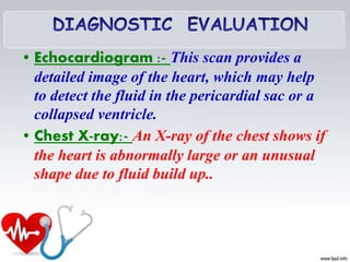
Cardiac tamponade is a serious
medical condition in which
blood or fluids fill the space
between the sac that encases
the heart and the heart
muscle. This places extreme
pressure on your heart. The
pressure prevents the heart’s
ventricles from expanding
fully and keeps your heart
from functioning properly. So
the heart can’t pump enough
• Cardiac tamponade is a clinical syndrome
caused by the accumulation of fluid in the
pericardial space, resulting in reduced
ventricular filling and subsequent
hemodynamic compromise. Cardiac
tamponade is a medical emergency.
• Cardiac tamponade is cardiac
dysfunction caused by external
compression of heart by the accumulation
of excessive contents in the pericardial
space
• Cardiac tamponade is the
accumulation of excess fluid within
the pericardial space, resulting in
impaired cardiac filling, reduction in
stroke volume, and epicardial
coronary artery compression with
resultant myocardial ischemia. This
fluid, which can be blood , pus, or air
in the pericardial sac. Accumulates
fast enough and in sufficient quantity
to compress the heart and restrict
blood flow in & out of the ventricles.
Incidence : 2-5 cases per 10000
• 2% penetrating injuries result
into cardiac tamponade •
• In adults male: female = 1.25:1
• Cardiac tamponade develops due to pericardial
effusion
• Pericardial effusion: accumulation of excess fluid in
pericardial space It may occur rapidly or insidiously
 Rapid pericardial effusion
• Trauma : both stabbing and blunt
• Post myocardial infarction
• Heart surgery (open heart surgery, CABG)
• Aortic dissection
• Drugs and medications ( antihypertensives e.g.
minoxidil, hydralazine , procainamide)
Insidious pericardial effusion
 • Cancers ( lung or breast cancer) 30-60%
 • Infections (viral, bacterial, fungal) 5-10%
 • Uremic pericarditis (10-15%)
 • Hypothyroidism
 • Chronic inflammation of connective tissue (SLE,
rheumatoid)
 high levels of radiation to the chest
 kidney failure
Pleural effusion due to various causes exert pressure in heart walls
Impairs relaxation and filling of the ventricles Chambers do not fill
properly
Less cardiac output (too little
oxygen reaches the tissues)
Increase venous pressure
Hypotension,
shock ,
Reflex
tachycardia
↑JVP hepatomegaly ascites
peripheral edema
Rales
Systemic Congestion Pulmonary congestion
• Chest discomfort
• Pleuritic pain
• Tachypnea and dyspnea on exertion that
progresses to air hunger at rest are the key
symptoms
• Convulsions, unconscious
• Most patients are weak and faint at
presentation and can have vague symptoms
such as anorexia, dysphagia, and cough
• The initial symptom may also be one of the
complications of tamponade, such as renal
failure
• Elevated venous pressure , Distended
neck veins
• Kussmaul’s sign{distended neck
veins}
• Hypotension
• Narrow pulse pressure
• Dyspnoea
• Cyanosis of lips and nails
• Restlessness and anxiety
• Pain in the right upper abdomen,
Upset stomach
• Fever, for any infection
• Restlessness and anxiety
• Diaphoresis
• Muffled heart sounds
• Pulsus paradoxus
• Beck's triad (cardiology)
• Beck's triad. :- Classical cardiac tamponade
presents three signs, known as Beck's triad-
Hypotension occurs because of decreased
stroke volume, - jugular venous distension
due to impaired venous return to the heart –
muffled heart sounds due to fluid inside the
pericardium
• History Collection :- Collect history regarding the
etiological factors and symptoms
• Physical Examination :- Beck's triad, Pulsus
paradoxus (a drop of at least 10 mmHg in arterial
blood pressure on inspiration) ,There may also be
general signs & symptoms of shock (such as
tachycardia , more than 90 beats) per minute
breathlessness and decreasing level of
consciousness) , Can be bradycardia? (uremia and
patients with hypothyroidism) ,Rub is a frequent
finding in patients with inflammatory effusions
Electrocardiographic Findings :- May be associated
with ST segment , low voltage QRS complexes , In some
cases, electrical alternans will be present in which case the
height of the QRS varies from beat to beat , Tachycardia
will likely be present as well , Combined P and QRS
alternation is virtually specific for tamponade
• Echocardiogram :- This scan provides a
detailed image of the heart, which may help
to detect the fluid in the pericardial sac or a
collapsed ventricle.
• Chest X-ray:- An X-ray of the chest shows if
the heart is abnormally large or an unusual
shape due to fluid build up..
• Computerized tomography (CT) scan:-
A CT scan of the chest can confirm the
presence of extra fluid in the pericardium.
• Magnetic resonance angiogram (MRA):-
An MRA uses a magnetic field and radio
waves to detect any abnormalities in how the
blood flows through the blood vessels of the
heart.
Heart failure
Pulmonary edema
Bleeding
Shock
Death
• The main aim of client with cardiac Tamponade is :
• 1. Save the patient life
• 2. improve the heart functions
• 3. Relive from symptoms
• Treatment that are administered for cardiac
tamponade include:
• 1. IV fluids to maintain normal BP
• 2. Antibiotics
• 3. Supplemental oxygen to reduce work load on the
heart
4.Bed rest and leg elevation
5. Inotropic drugs ( Dobutamine)
Mechanical ventilation with positive
airway pressure should be avoided in
patients with tamponade, because this
further decreases cardiac output
• PERICARDIOCENTESIS •
Pericardiocentesis, also called a
pericardial tap, is a surgical invasive
procedure ( use both diagnostic and
therapeutic purpose) in which
abnormal or excessive fluid is
removed from the pericardium sac the
sac around your heart. Or
Pericardiocentesis is the removal by
needle of pericardial fluid from the
sac surrounding the heart for
diagnostic or therapeutic purposes.
• Monitor strictly vital signs, especially respiratory frequency.
• Give the semi-Fowler position if not contraindicated.
• Give oxygen as indicated
• Monitor urine output hourly; a drop in urine output may
indicate decreased renal perfusion as a result of decreased
stroke volume secondary to cardiac compression.
• Continuously monitor ECG for dysrhythmia formation,
which may result of myocardial ischemia secondary to
epicardial coronary artery compression.
• Monitor the BP every 5 to 15 minutes during the acute
phase.
• Auscultation of breathsounds and heart sounds. Listen to
the murmur.
• Maintain bed rest in a comfortable position during the
acuteperiod.
• Educatethe patient about the disease condition and
treatment
• Educate the patient about the pericardiocentesis
• Encourage the patient to ask questions
• Ineffective Breathing Pattern related to
hyperventilation as evidenced by Dyspnea.
• Decreased cardiac output related to reduced
ventricularfilling secondary to increased
intrapericardial pressure as manifested by
tachycardia.
• Activity intolerance related to shortness of
breath and chest discomfort as manifested
restlessness and fatigue
Cardiac tamponade

Cardiac tamponade

  • 1.
    CARDIAC TAMPONADE Mrs. D.MelbaSahaya Sweety.D M.Sc Nursing GIMSAR
  • 2.
    Cardiac tamponade isa serious medical condition in which blood or fluids fill the space between the sac that encases the heart and the heart muscle. This places extreme pressure on your heart. The pressure prevents the heart’s ventricles from expanding fully and keeps your heart from functioning properly. So the heart can’t pump enough
  • 3.
    • Cardiac tamponadeis a clinical syndrome caused by the accumulation of fluid in the pericardial space, resulting in reduced ventricular filling and subsequent hemodynamic compromise. Cardiac tamponade is a medical emergency. • Cardiac tamponade is cardiac dysfunction caused by external compression of heart by the accumulation of excessive contents in the pericardial space
  • 4.
    • Cardiac tamponadeis the accumulation of excess fluid within the pericardial space, resulting in impaired cardiac filling, reduction in stroke volume, and epicardial coronary artery compression with resultant myocardial ischemia. This fluid, which can be blood , pus, or air in the pericardial sac. Accumulates fast enough and in sufficient quantity to compress the heart and restrict blood flow in & out of the ventricles.
  • 5.
    Incidence : 2-5cases per 10000 • 2% penetrating injuries result into cardiac tamponade • • In adults male: female = 1.25:1
  • 6.
    • Cardiac tamponadedevelops due to pericardial effusion • Pericardial effusion: accumulation of excess fluid in pericardial space It may occur rapidly or insidiously  Rapid pericardial effusion • Trauma : both stabbing and blunt • Post myocardial infarction • Heart surgery (open heart surgery, CABG) • Aortic dissection • Drugs and medications ( antihypertensives e.g. minoxidil, hydralazine , procainamide)
  • 7.
    Insidious pericardial effusion • Cancers ( lung or breast cancer) 30-60%  • Infections (viral, bacterial, fungal) 5-10%  • Uremic pericarditis (10-15%)  • Hypothyroidism  • Chronic inflammation of connective tissue (SLE, rheumatoid)  high levels of radiation to the chest  kidney failure
  • 8.
    Pleural effusion dueto various causes exert pressure in heart walls Impairs relaxation and filling of the ventricles Chambers do not fill properly Less cardiac output (too little oxygen reaches the tissues) Increase venous pressure Hypotension, shock , Reflex tachycardia ↑JVP hepatomegaly ascites peripheral edema Rales Systemic Congestion Pulmonary congestion
  • 9.
    • Chest discomfort •Pleuritic pain • Tachypnea and dyspnea on exertion that progresses to air hunger at rest are the key symptoms • Convulsions, unconscious • Most patients are weak and faint at presentation and can have vague symptoms such as anorexia, dysphagia, and cough • The initial symptom may also be one of the complications of tamponade, such as renal failure
  • 10.
    • Elevated venouspressure , Distended neck veins • Kussmaul’s sign{distended neck veins} • Hypotension • Narrow pulse pressure • Dyspnoea • Cyanosis of lips and nails • Restlessness and anxiety • Pain in the right upper abdomen, Upset stomach • Fever, for any infection
  • 11.
    • Restlessness andanxiety • Diaphoresis • Muffled heart sounds • Pulsus paradoxus • Beck's triad (cardiology) • Beck's triad. :- Classical cardiac tamponade presents three signs, known as Beck's triad- Hypotension occurs because of decreased stroke volume, - jugular venous distension due to impaired venous return to the heart – muffled heart sounds due to fluid inside the pericardium
  • 12.
    • History Collection:- Collect history regarding the etiological factors and symptoms • Physical Examination :- Beck's triad, Pulsus paradoxus (a drop of at least 10 mmHg in arterial blood pressure on inspiration) ,There may also be general signs & symptoms of shock (such as tachycardia , more than 90 beats) per minute breathlessness and decreasing level of consciousness) , Can be bradycardia? (uremia and patients with hypothyroidism) ,Rub is a frequent finding in patients with inflammatory effusions
  • 13.
    Electrocardiographic Findings :-May be associated with ST segment , low voltage QRS complexes , In some cases, electrical alternans will be present in which case the height of the QRS varies from beat to beat , Tachycardia will likely be present as well , Combined P and QRS alternation is virtually specific for tamponade
  • 14.
    • Echocardiogram :-This scan provides a detailed image of the heart, which may help to detect the fluid in the pericardial sac or a collapsed ventricle. • Chest X-ray:- An X-ray of the chest shows if the heart is abnormally large or an unusual shape due to fluid build up..
  • 15.
    • Computerized tomography(CT) scan:- A CT scan of the chest can confirm the presence of extra fluid in the pericardium. • Magnetic resonance angiogram (MRA):- An MRA uses a magnetic field and radio waves to detect any abnormalities in how the blood flows through the blood vessels of the heart.
  • 16.
  • 17.
    • The mainaim of client with cardiac Tamponade is : • 1. Save the patient life • 2. improve the heart functions • 3. Relive from symptoms • Treatment that are administered for cardiac tamponade include: • 1. IV fluids to maintain normal BP • 2. Antibiotics • 3. Supplemental oxygen to reduce work load on the heart
  • 18.
    4.Bed rest andleg elevation 5. Inotropic drugs ( Dobutamine) Mechanical ventilation with positive airway pressure should be avoided in patients with tamponade, because this further decreases cardiac output
  • 19.
    • PERICARDIOCENTESIS • Pericardiocentesis,also called a pericardial tap, is a surgical invasive procedure ( use both diagnostic and therapeutic purpose) in which abnormal or excessive fluid is removed from the pericardium sac the sac around your heart. Or Pericardiocentesis is the removal by needle of pericardial fluid from the sac surrounding the heart for diagnostic or therapeutic purposes.
  • 20.
    • Monitor strictlyvital signs, especially respiratory frequency. • Give the semi-Fowler position if not contraindicated. • Give oxygen as indicated • Monitor urine output hourly; a drop in urine output may indicate decreased renal perfusion as a result of decreased stroke volume secondary to cardiac compression. • Continuously monitor ECG for dysrhythmia formation, which may result of myocardial ischemia secondary to epicardial coronary artery compression.
  • 21.
    • Monitor theBP every 5 to 15 minutes during the acute phase. • Auscultation of breathsounds and heart sounds. Listen to the murmur. • Maintain bed rest in a comfortable position during the acuteperiod. • Educatethe patient about the disease condition and treatment • Educate the patient about the pericardiocentesis • Encourage the patient to ask questions
  • 22.
    • Ineffective BreathingPattern related to hyperventilation as evidenced by Dyspnea. • Decreased cardiac output related to reduced ventricularfilling secondary to increased intrapericardial pressure as manifested by tachycardia. • Activity intolerance related to shortness of breath and chest discomfort as manifested restlessness and fatigue