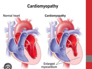
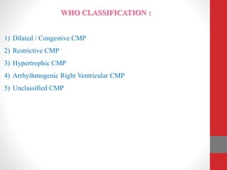
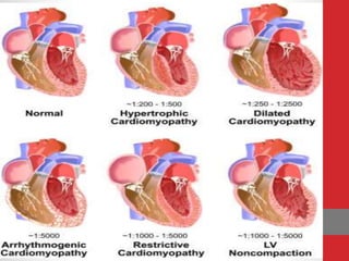
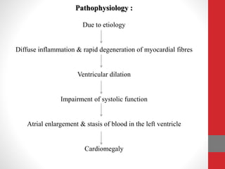
CARDIOMYOPATHY
Presented By:
Mr. Nandish.S
Asso. Professor
Mandya Institute of Nursing Sciences
DEFINITION :
• It is a disease of the heart muscle that is associated with cardiac
dysfunction.
• It is a term used to describe a group of heart muscle diseases that
primarily affect the structural or functional ability of the
myocardium.
• It is a disease of heart muscle in which heart loses its ability to pump
blood effectively.
INCIDENCE :
 Dilated cardiomyopathy is the most common form affects 5 to 7 in
100,000 adults and 0.50 in 100,000 children.
 Hypertrophic cardiomyopathy is the leading cause of sudden death
with an incidence of 1 in 500 persons (Athletes) & 0.2% in general
population.
 Restrictive cardiomyopathy is the rarest form, it accounts 5% of all
cardiomyopathy cases.
GENERAL CLASSIFICATION :
1) Primary CMP : these are conditions in which the etiology of the
heart disease is unknown. Heart muscles are the only part involved in
this type and other portions remain normal.
2) Secondary CMP : here the cause of myocardial disease is known and
are due to another disease process. They are not limited to heart
muscles.
WHO CLASSIFICATION :
1) Dilated / Congestive CMP
2) Restrictive CMP
3) Hypertrophic CMP
4) Arrhythmogenic Right Ventricular CMP
5) Unclassified CMP
DILATED CMP:
• It is the most common condition characterized by ventricular
dilation, impairment of systolic function, atrial enlargement and
stasis of blood in Left Ventricle.
• It is the result of primary ventricular dilation.
Etiology :
 Idiopathic
 Cardiotoxic agents : Alcohol, cocaine, Doxorubicin
 Genetic or familial history
 Coronary artery disease (CAD)
 Metabolic disorders
 Muscular Dystrophy
 Myocarditis
 Pregnancy
 Valvular disorder
Pathophysiology :
Due to etiology
Diffuse inflammation & rapid degeneration of myocardial fibres
Ventricular dilation
Impairment of systolic function
Atrial enlargement & stasis of blood in the left ventricle
Cardiomegaly
Symptoms :
 Change in exercise tolerance
 Fatigue
 Dry cough
 Dyspnoea
 Paroxysmal Nocturnal Dyspnea
 Orthopnea
 Palpitations
 Anorexia
 Abdominal Bloating
Signs :
 S3 and S4 Sounds are heard
 Tachycardia
 Edema
 Pulmonary crackles
 Weak peripheral pulse
 Pallor
 Hepatomegaly
 Jugular venous distension
Diagnostic studies :
• History collection
• Physical examination
• Chest X – Ray
• Electro cardiogram
• Echocardiography
• Cardiac catheterization
Management :
Medical :
Nitrates : Isosorbitrate
Loop diuretics : Furosemide
ACE Inhibitors : Captopril
Beta Adrenergic blockers : Atenolol
Aldosterone agonists : Spironolactone
Cardiac glycoside : Digoxin
Anticoagulants : Warfarin
Surgical : For terminal end stage patients
- Cardiac transplantation
HYPERTROPHIC CMP
 it is an autosomal dominant condition.
 It is asymmetric myocardial hypertrophy without ventricular dilation.
 When the septum between two ventricles becomes enlarged which
reduces ventricular size and obstructs the blood flow from left
ventricle and it is known as hypertrophic obstructive
cardiomyopathy.
Etiology :
• Aortic stenosis
• Hypertension
• Genetic or family history
• It is more common in men than women and it is the most common
cause of sudden death among health young people.
Pathophysiology
Due to etiological factors
Thickened intra-ventricular septum & ventricular wall
Ventricular hypertrophy
Ventricular dysfunction
Impaired ventricular filling & decreased outflow
Decreased cardiac output
Clinical features
Clinical Features :
 Exertional dyspnea (shortness of breath during exercise) - due to
elevated left ventricular diastolic pressure.
 Fatigue - because of decrease in cardiac output.
 Angina – increased left ventricular muscle mass.
 Syncope – increase in obstruction to aortic outflow.
 Hypertension
 Arrhythmias
Diagnostic studies :
 History collection
 Physical examination
 12 lead ECG : increased duration of QRS Complex.
 Echocardiography
 Doppler imaging
 Cardiovascular MRI
 Cardiac Catheterization
Management :
Medical :
 Beta Adrenergic blockers – Atenolol
 Calcium channel blockers – Verapamil
 Antidysrythmic drugs – Amiodarone
 In case of Arrythmias, alternate treatment may be Implantable
defibrillator. It is a small instrument placed in the chest as a
pacemaker. It reduces outflow obstruction.
Surgical :
Indication for surgery include severe symptoms refractory to medications
& obstruction to Aortic outflow.
 Ventriculomyotomy & Myectomy
It involves incision of the hypertrophied septal muscle and resection of
some of the hypertrophied muscle.
 Alcohol Septal Ablation
It is a new procedure performed in the catheterization laboratory. It
consists of administering absolute alcohol into the first septal artery (Left
Anterior Descending Artery).
Restrictive CMP
 It is characterized by diastolic dysfunction caused by rigid ventricular
walls that impair diastolic filling and ventricular stretch.
 Systolic functions remain unaffected.
 It is the least common type.
Etiology :
 Idiopathic
 Pathologic processes involved are : Myocardial Fibrosis,
Hypertrophy, Infiltration
 Secondary causes : Amyloidosis (amyloid – a protein substance, is
deposited within the cells), Endocardial Fibrosis.
 Post radiation therapy
 Sarcoidosis (growth of tiny collections of inflammatory cells)
 Ventricular Thrombus
Pathophysiology :
Due to etiological factors
Stiffness of the ventricular wall with loss of ventricular compliance
Ventricles become resistant to filling
Decrease cardiac output
Clinical Features :
 Angina
 Syncope
 Fatigue
 Dyspnea on exertion
 Exercise Intolerance
 Tachycardia
 Peripheral edema & Ascites
 Palpitations
 Kussmaul’s Sign (bulging of the internal jugular neck veins on
inspiration)
Diagnostic Studies :
 History Collection & Physical Examination
 Chest X – Ray
 ECG – shows sign of arrhythmias
 Echocardiography – thickened ventricular wall
 CT Scan
 Endomyocardial Biopsy
 Nuclear Imaging
Management :
Medical :
 Beta Adrenergic Blockers : Atenolol
 Calcium Channel Blockers : Verapamil
 Steroids : Hydrocortisone
 Antidysrhythmic Drugs : Amiodarone
Surgical :
 Cardiac Transplantation
 Artificial heart implantation (made up of Titanium & patented
polyurethane)
Arrhythmogenic Right Ventricular Cardiomyopathy :
 It is an uncommon form of inherited heart muscle disease.
 It occurs when myocardium is progressively infiltrated & replaced by
fibrous scar and adipose tissue.
 As the disease progress, entire heart is affected. Right ventricle dilates
and develops poor contractility.
 Palpitations, Syncope & Ventricular tachycardia are the symptoms
commonly seen.
 12 Lead ECG, Holter Monitor, Echocardiography, Cardiac MRI are
done to rule out the disease.
Unclassified CMP :
 It is a different form, have characteristics of more than one of
previous types.
 It is caused due to Fibroelastosis, Noncompacted Myocardium,
systolic dysfunction and mitochondrial disease.
 Unclassified CMP include Left Ventricular Noncompaction & stress
induced CMP.
Nursing Management :
- Decreased Cardiac Output related to ventricular dysfunction.
- Impaired Tissue perfusion related to insufficient blood supply.
- Impaired breathing pattern related to decreased thoracic excursion.
- Activity intolerance related to shortness of breath.
- Imbalanced nutrition, less than body requirement related to restricted
diet.
- Fatigue related to cardiac dysfunction.
Cardiomyopathy.pptx

Cardiomyopathy.pptx

  • 1.
    CARDIOMYOPATHY Presented By: Mr. Nandish.S Asso.Professor Mandya Institute of Nursing Sciences
  • 3.
    DEFINITION : • Itis a disease of the heart muscle that is associated with cardiac dysfunction. • It is a term used to describe a group of heart muscle diseases that primarily affect the structural or functional ability of the myocardium. • It is a disease of heart muscle in which heart loses its ability to pump blood effectively.
  • 4.
    INCIDENCE :  Dilatedcardiomyopathy is the most common form affects 5 to 7 in 100,000 adults and 0.50 in 100,000 children.  Hypertrophic cardiomyopathy is the leading cause of sudden death with an incidence of 1 in 500 persons (Athletes) & 0.2% in general population.  Restrictive cardiomyopathy is the rarest form, it accounts 5% of all cardiomyopathy cases.
  • 5.
    GENERAL CLASSIFICATION : 1)Primary CMP : these are conditions in which the etiology of the heart disease is unknown. Heart muscles are the only part involved in this type and other portions remain normal. 2) Secondary CMP : here the cause of myocardial disease is known and are due to another disease process. They are not limited to heart muscles.
  • 6.
    WHO CLASSIFICATION : 1)Dilated / Congestive CMP 2) Restrictive CMP 3) Hypertrophic CMP 4) Arrhythmogenic Right Ventricular CMP 5) Unclassified CMP
  • 8.
    DILATED CMP: • Itis the most common condition characterized by ventricular dilation, impairment of systolic function, atrial enlargement and stasis of blood in Left Ventricle. • It is the result of primary ventricular dilation.
  • 10.
    Etiology :  Idiopathic Cardiotoxic agents : Alcohol, cocaine, Doxorubicin  Genetic or familial history  Coronary artery disease (CAD)  Metabolic disorders  Muscular Dystrophy  Myocarditis  Pregnancy  Valvular disorder
  • 11.
    Pathophysiology : Due toetiology Diffuse inflammation & rapid degeneration of myocardial fibres Ventricular dilation Impairment of systolic function Atrial enlargement & stasis of blood in the left ventricle Cardiomegaly
  • 12.
    Symptoms :  Changein exercise tolerance  Fatigue  Dry cough  Dyspnoea  Paroxysmal Nocturnal Dyspnea  Orthopnea  Palpitations  Anorexia  Abdominal Bloating
  • 13.
    Signs :  S3and S4 Sounds are heard  Tachycardia  Edema  Pulmonary crackles  Weak peripheral pulse  Pallor  Hepatomegaly  Jugular venous distension
  • 14.
    Diagnostic studies : •History collection • Physical examination • Chest X – Ray • Electro cardiogram • Echocardiography • Cardiac catheterization
  • 15.
    Management : Medical : Nitrates: Isosorbitrate Loop diuretics : Furosemide ACE Inhibitors : Captopril Beta Adrenergic blockers : Atenolol Aldosterone agonists : Spironolactone Cardiac glycoside : Digoxin Anticoagulants : Warfarin Surgical : For terminal end stage patients - Cardiac transplantation
  • 16.
    HYPERTROPHIC CMP  itis an autosomal dominant condition.  It is asymmetric myocardial hypertrophy without ventricular dilation.  When the septum between two ventricles becomes enlarged which reduces ventricular size and obstructs the blood flow from left ventricle and it is known as hypertrophic obstructive cardiomyopathy.
  • 18.
    Etiology : • Aorticstenosis • Hypertension • Genetic or family history • It is more common in men than women and it is the most common cause of sudden death among health young people.
  • 19.
    Pathophysiology Due to etiologicalfactors Thickened intra-ventricular septum & ventricular wall Ventricular hypertrophy Ventricular dysfunction Impaired ventricular filling & decreased outflow Decreased cardiac output Clinical features
  • 20.
    Clinical Features : Exertional dyspnea (shortness of breath during exercise) - due to elevated left ventricular diastolic pressure.  Fatigue - because of decrease in cardiac output.  Angina – increased left ventricular muscle mass.  Syncope – increase in obstruction to aortic outflow.  Hypertension  Arrhythmias
  • 21.
    Diagnostic studies : History collection  Physical examination  12 lead ECG : increased duration of QRS Complex.  Echocardiography  Doppler imaging  Cardiovascular MRI  Cardiac Catheterization
  • 22.
    Management : Medical : Beta Adrenergic blockers – Atenolol  Calcium channel blockers – Verapamil  Antidysrythmic drugs – Amiodarone  In case of Arrythmias, alternate treatment may be Implantable defibrillator. It is a small instrument placed in the chest as a pacemaker. It reduces outflow obstruction.
  • 23.
    Surgical : Indication forsurgery include severe symptoms refractory to medications & obstruction to Aortic outflow.  Ventriculomyotomy & Myectomy It involves incision of the hypertrophied septal muscle and resection of some of the hypertrophied muscle.  Alcohol Septal Ablation It is a new procedure performed in the catheterization laboratory. It consists of administering absolute alcohol into the first septal artery (Left Anterior Descending Artery).
  • 25.
    Restrictive CMP  Itis characterized by diastolic dysfunction caused by rigid ventricular walls that impair diastolic filling and ventricular stretch.  Systolic functions remain unaffected.  It is the least common type.
  • 27.
    Etiology :  Idiopathic Pathologic processes involved are : Myocardial Fibrosis, Hypertrophy, Infiltration  Secondary causes : Amyloidosis (amyloid – a protein substance, is deposited within the cells), Endocardial Fibrosis.  Post radiation therapy  Sarcoidosis (growth of tiny collections of inflammatory cells)  Ventricular Thrombus
  • 28.
    Pathophysiology : Due toetiological factors Stiffness of the ventricular wall with loss of ventricular compliance Ventricles become resistant to filling Decrease cardiac output
  • 29.
    Clinical Features : Angina  Syncope  Fatigue  Dyspnea on exertion  Exercise Intolerance  Tachycardia  Peripheral edema & Ascites  Palpitations  Kussmaul’s Sign (bulging of the internal jugular neck veins on inspiration)
  • 30.
    Diagnostic Studies : History Collection & Physical Examination  Chest X – Ray  ECG – shows sign of arrhythmias  Echocardiography – thickened ventricular wall  CT Scan  Endomyocardial Biopsy  Nuclear Imaging
  • 31.
    Management : Medical : Beta Adrenergic Blockers : Atenolol  Calcium Channel Blockers : Verapamil  Steroids : Hydrocortisone  Antidysrhythmic Drugs : Amiodarone Surgical :  Cardiac Transplantation  Artificial heart implantation (made up of Titanium & patented polyurethane)
  • 32.
    Arrhythmogenic Right VentricularCardiomyopathy :  It is an uncommon form of inherited heart muscle disease.  It occurs when myocardium is progressively infiltrated & replaced by fibrous scar and adipose tissue.  As the disease progress, entire heart is affected. Right ventricle dilates and develops poor contractility.  Palpitations, Syncope & Ventricular tachycardia are the symptoms commonly seen.  12 Lead ECG, Holter Monitor, Echocardiography, Cardiac MRI are done to rule out the disease.
  • 34.
    Unclassified CMP : It is a different form, have characteristics of more than one of previous types.  It is caused due to Fibroelastosis, Noncompacted Myocardium, systolic dysfunction and mitochondrial disease.  Unclassified CMP include Left Ventricular Noncompaction & stress induced CMP.
  • 35.
    Nursing Management : -Decreased Cardiac Output related to ventricular dysfunction. - Impaired Tissue perfusion related to insufficient blood supply. - Impaired breathing pattern related to decreased thoracic excursion. - Activity intolerance related to shortness of breath. - Imbalanced nutrition, less than body requirement related to restricted diet. - Fatigue related to cardiac dysfunction.