Downloaded 1,219 times


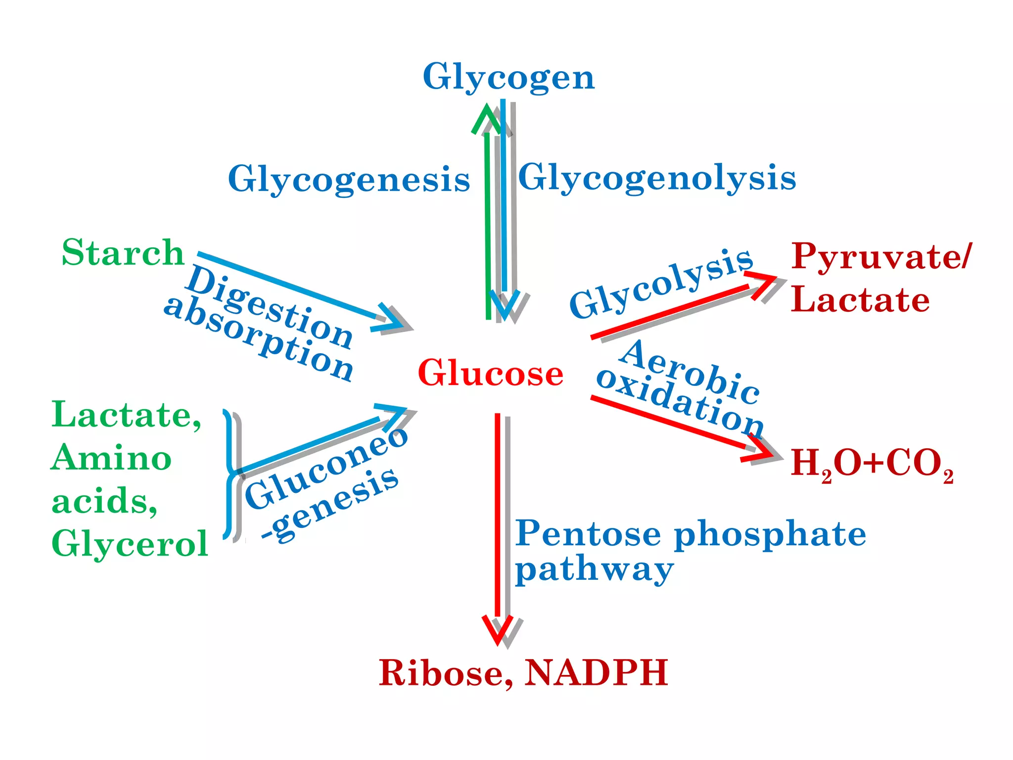


The document provides a comprehensive overview of carbohydrate metabolism, detailing the definitions, significance, digestion, and various metabolic pathways involving carbohydrates. Key processes such as glycolysis, aerobic oxidation, the tricarboxylic acid cycle, and the pentose phosphate pathway are explained along with their enzymes and regulation mechanisms. Additionally, the document discusses the significance of glycogen synthesis and catabolism.


![M.Carbohydrate Metabolism[2].pdf UNDERGRADUATE](https://cdn.slidesharecdn.com/ss_thumbnails/m-250505131013-597a5b82-thumbnail.jpg?width=640&height=640&fit=bounds)


