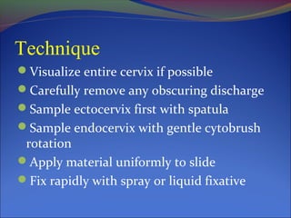
PAP smear: named after
Dr. George Papanicolaou (1883-1962)
Vaginal smears from guinea pigs (1917)
Women (1920)
Hormonal

cycles
Pathological conditions (1928)
Cytologic screening for cervical cancer
Usefulness of pap smear in the screening programme

for cancer cervix is shown by the following:

Long latent period of 10-15 years between CIN and

invasive cancer allows adequate treatment of CIN
and prevention of invasive cancer

Proved successful in reducing the incidence of

invasive cancer by 80% and the mortality by 70%
When to screen
Start within 3 years of onset of sexual activity or by age

of 21, whichever is first.
High risk factors for cervical dysplasia:
Early onset of sexual activity
Multiple sexual partners
Smoking habits
Oral contraceptives
HPV and HIV positive women
Screening frequency
Yearly until three consecutive normal pap smears, then

may decrease frequency to every 2-3 years
Annual screening for high-risk women is highly

recommend.
When to stop routine screening
Age 70 and “adequate recent screening”
Three consecutive negative pap smears
No abnormal pap smears in last 10 years
Hysterectomy for benign lesion
Original Squamous Epithelium
Vagina and outer ectocervix
4 cell layers
Well-glycogenated (pink) unless atrophic
Columnar Epithelium
Upper and middle endo-cervical canal
Single layer of columnar cells arranged in

folds
Mucin producing (not true glands)
Squamous Metaplasia
Central ectocervix and lower endocervical canal
Replacement of columnar cells by squamous epithelium
Progressive and stimulated by
Acidic environment with onset of puberty
Estrogen causing eversion of endocervix
Original Squamo-columnar Junction
Placement determined between 18-20 weeks gestation
Most often found on ectocervix
Can be found in vagina or vaginal fornices
Less apparent over time with maturation of

epithelium
“New” Squamo-columnar Junction
Border between squamous epithelium and columnar

epithelium
Found on ecto-cervix or in endo-cervical canal
Majority of cervical cancers and precursor lesions

arise in immature squamous metaplasia, i.e. the
leading edge of the squamo-columnar junction
Transformation Zone
Zone between original squamo-columnar junction and

the “new” squamo-columnar junction
Nabothian cysts visually identify the transformation

zone if present
Squamous Epithelium
Parabasal Cells
Intermediate Cells
Superficial

Cells
Endocervix
Endocervical Cells
Technique
Visualize entire cervix if possible
Carefully remove any obscuring discharge
Sample ectocervix first with spatula
Sample endocervix with gentle cytobrush

rotation
Apply material uniformly to slide
Fix rapidly with spray or liquid fixative
Classification of Pap smear
Class

Reagen(WHO)

Ruchart

Bethesda

Class 1

negative

negative

Within normal

Class 2

inflammation

Class 3

Mild dysplasia

CIN-l (HPV)

LSIL (HPV)

Class 4

Mod dysplasia
Seve dysplasia
Carcinoma in situ

CIN-ll
CIN-lll

HSIL

Class 5

Invasive cancer

Invasive cancer

Invasive cancer

------

ASCUS
“Normal” Pap Smear
Negative for intraepithelial lesion or malignancy
Other non-neoplastic findings
Reactive cellular changes
Glandular cells status post hysterectomy
Atrophy

Other
Endometrial cells (women ≥ 40 yrs)
Normal smear
Epithelial Cell Abnormalities:
Squamous
Atypical squamous cells
ASC-US: undetermined significance
ASC-H: cannot exclude HSIL

LSIL: low grade (CIN 1)
HSIL: high grade (CIN 2 - 3)
Squamous cell carcinoma
SIL and CIN
Various types of cervical lesions as seen on Pap smears:
CIN I.
Various types of cervical lesions as seen on Pap smears:
CIN Il
Various types of cervical lesions as seen on Pap smears:
CIN lll
Various types of cervical lesions as seen on Pap smears:
invasive squamous cell carcinoma.
Epithelial Cell Abnormalities:
Glandular
Atypical glandular cells,specify site of origin,if

possible
Atypical glandular cells - favor neoplastia
Endocervical adenocarcinoma in situ
Adenocarcinoma
Various types of cervical lesions as seen on Pap smears:
adenocarcinoma
Accuracy
Single pap smear-diagnostic sensitivity 60%
False negative results upto25% due to:

too scanty,too thick,too bloody,poorly stained
smear
misinterpretation by the cytologist
• In the presence of infection repeat cytology has to be
done after the infection is controlled
Abnormal cytology is an indication of colposcopic
evaluation and directed biopsy

Pap smear (2)

  • 2.
    PAP smear: namedafter Dr. George Papanicolaou (1883-1962) Vaginal smears from guinea pigs (1917) Women (1920) Hormonal cycles Pathological conditions (1928)
  • 3.
    Cytologic screening forcervical cancer Usefulness of pap smear in the screening programme for cancer cervix is shown by the following: Long latent period of 10-15 years between CIN and invasive cancer allows adequate treatment of CIN and prevention of invasive cancer Proved successful in reducing the incidence of invasive cancer by 80% and the mortality by 70%
  • 4.
    When to screen Startwithin 3 years of onset of sexual activity or by age of 21, whichever is first. High risk factors for cervical dysplasia: Early onset of sexual activity Multiple sexual partners Smoking habits Oral contraceptives HPV and HIV positive women
  • 5.
    Screening frequency Yearly untilthree consecutive normal pap smears, then may decrease frequency to every 2-3 years Annual screening for high-risk women is highly recommend.
  • 6.
    When to stoproutine screening Age 70 and “adequate recent screening” Three consecutive negative pap smears No abnormal pap smears in last 10 years Hysterectomy for benign lesion
  • 8.
    Original Squamous Epithelium Vaginaand outer ectocervix 4 cell layers Well-glycogenated (pink) unless atrophic
  • 9.
    Columnar Epithelium Upper andmiddle endo-cervical canal Single layer of columnar cells arranged in folds Mucin producing (not true glands)
  • 10.
    Squamous Metaplasia Central ectocervixand lower endocervical canal Replacement of columnar cells by squamous epithelium Progressive and stimulated by Acidic environment with onset of puberty Estrogen causing eversion of endocervix
  • 11.
    Original Squamo-columnar Junction Placementdetermined between 18-20 weeks gestation Most often found on ectocervix Can be found in vagina or vaginal fornices Less apparent over time with maturation of epithelium
  • 12.
    “New” Squamo-columnar Junction Borderbetween squamous epithelium and columnar epithelium Found on ecto-cervix or in endo-cervical canal Majority of cervical cancers and precursor lesions arise in immature squamous metaplasia, i.e. the leading edge of the squamo-columnar junction
  • 13.
    Transformation Zone Zone betweenoriginal squamo-columnar junction and the “new” squamo-columnar junction Nabothian cysts visually identify the transformation zone if present
  • 14.
  • 15.
  • 16.
  • 17.
  • 18.
  • 19.
  • 20.
    Technique Visualize entire cervixif possible Carefully remove any obscuring discharge Sample ectocervix first with spatula Sample endocervix with gentle cytobrush rotation Apply material uniformly to slide Fix rapidly with spray or liquid fixative
  • 23.
    Classification of Papsmear Class Reagen(WHO) Ruchart Bethesda Class 1 negative negative Within normal Class 2 inflammation Class 3 Mild dysplasia CIN-l (HPV) LSIL (HPV) Class 4 Mod dysplasia Seve dysplasia Carcinoma in situ CIN-ll CIN-lll HSIL Class 5 Invasive cancer Invasive cancer Invasive cancer ------ ASCUS
  • 24.
    “Normal” Pap Smear Negativefor intraepithelial lesion or malignancy Other non-neoplastic findings Reactive cellular changes Glandular cells status post hysterectomy Atrophy Other Endometrial cells (women ≥ 40 yrs)
  • 25.
  • 26.
    Epithelial Cell Abnormalities: Squamous Atypicalsquamous cells ASC-US: undetermined significance ASC-H: cannot exclude HSIL LSIL: low grade (CIN 1) HSIL: high grade (CIN 2 - 3) Squamous cell carcinoma
  • 27.
  • 28.
    Various types ofcervical lesions as seen on Pap smears: CIN I.
  • 29.
    Various types ofcervical lesions as seen on Pap smears: CIN Il
  • 30.
    Various types ofcervical lesions as seen on Pap smears: CIN lll
  • 31.
    Various types ofcervical lesions as seen on Pap smears: invasive squamous cell carcinoma.
  • 32.
    Epithelial Cell Abnormalities: Glandular Atypicalglandular cells,specify site of origin,if possible Atypical glandular cells - favor neoplastia Endocervical adenocarcinoma in situ Adenocarcinoma
  • 33.
    Various types ofcervical lesions as seen on Pap smears: adenocarcinoma
  • 34.
    Accuracy Single pap smear-diagnosticsensitivity 60% False negative results upto25% due to: too scanty,too thick,too bloody,poorly stained smear misinterpretation by the cytologist • In the presence of infection repeat cytology has to be done after the infection is controlled Abnormal cytology is an indication of colposcopic evaluation and directed biopsy