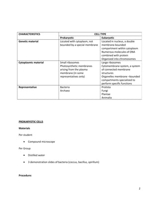
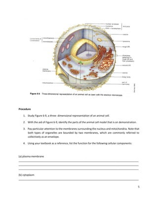
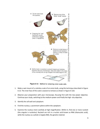
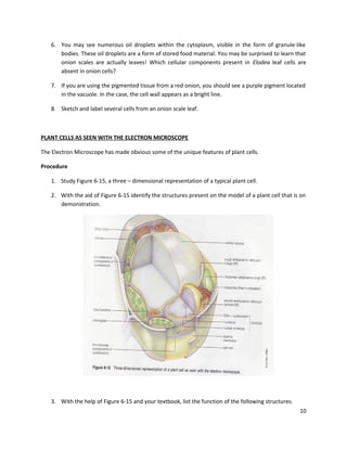
STRUCTURE AND FUNCTION OF LIVING CELLS
Objectives
After completing this exercise, you will be able to
1.

Define cell, cell theory, prokaryotic, eukaryotic, nucleus, cytomembrane system, organelle,
multinucleate, cytoplasmic streaming, envelope;

2.

List the structural features shared by all cells;

3.

Describe the similarities and differences between prokaryotic and eukaryotic cells;

4.

Identify the cell parts described in this exercise

5.

State the function for each cell part

6.

Distinguish between plant and animal cells

7.

Recognize the structures presented in boldface in the procedure sections

Introduction
Structurally and functionally, all life has one common feature: All living organisms are composed of cells.
The development of the concept began with Robert Hooke’s seventeenth-century observation that
slices of cork were made up small units. He called these units “cells” because their structure reminded
him of the small cubicles that monks lived in. Over the next 100 years, the cell theory emerged. This
theory has three principles: (1) all organisms are composed of one or more cells; (2) the cell is the basic
living unit of organization; (3) all cells arise from pre-existing cells.
Although cells vary in organization, size, and function, all share three structural features: (1) All possess
a plasma membrane defining the boundary of the living material; (2) all contain a region of DNA
(deoxyribonucleic acid), which stores genetic information; and (3) all contain cytoplasm, everything
inside the plasma membrane that is not part of the DNA region.
With respect to internal organization, there are two basic types of cells, prokaryotic and eukaryotic.
Study the table below comparing the most important differences between prokaryotic and eukaryotic
cells. The Greek word Karyon means “Kernel”, referring to the nucleus. Prokaryotic means ‘before a
nucleus’, while eukaryotic indicates the presence of a “true nucleus.” Prokaryotic cells are typical of
bacteria, cynabacteria, and archaea. Eukaryotic cells include those that comprise the bodies of protists,
fungi, plants, and animals.
This exercise will familiarize you with the basics of cell structure and the function of prokaryotes
(prokaryotic cells) and eukaryotes (eukaryotic cells).

1
CHARACTERISTICS
Genetic material

Cytoplasmic material

Representative

CELL TYPE
Prokaryotic
Eukaryotic
Located with cytoplasm, not
Located in nucleus, a double
bounded by a special membrane membrane-bounded
compartment within cytoplasm
Numerous molecules of DNA
combined with protein
Organized into chromosomes
Small ribosomes
Large ribosomes
Photosynthetic membranes
Cytomembrane system, a system
arising from the plasma
of connected membrane
membrane (in some
structures
representatives only)
Organelles membrane –bounded
compartments specialized to
perform specific functions
Bacteria
Protista
Archaea
Fungi
Plantae
Animalia

PROKARYOTIC CELLS
Materials
Per student
•

Compound microscope

Per Group
•

Distilled water

•

3 demonstration slides of bacteria (coccus, bacillus, spirillum)

Procedure:

2
1. Observe the microscopic slides of the three types of the bacteria and sketch what you see
through the microscope.

EUKARYOTIC CELLS

Materials
•

Toothpick

•

Textbook

•

Microscope slide

•

Coverslip

•

Compound microscope

•

Forceps

•

Dissecting needle

•

Methylene blue

•

Distilled water

•

Model of animal cell

•

Model of plant cell

•

Prepared slide of Elodea

ANIMAL CELLS AS OBSERVED WITH THE LIGHT MICROSCOPE
Procedure
1. Human cheek cells - Using the broad end of a clean toothpick, gently scrape the inside of your
cheek. Stir the scraping into a drop of distilled water on a clean microscope slide and add a
coverslip. Dispose of used toothpicks in the jar containing alcohol.
3
2. Because the cells are almost transparent, decrease the amount of light entering the objective
lens to increase the contrast. Find the cells using the low-power objective of your microscope,
then switch to the high-dry objective for detailed study.
3. Find the nucleus, a centrally located spherical body within the cytoplasm of each cell.
4. Now stain your cheek cells with a dilute solution of methylene blue, a dye that stains the nucleus
darker than the surrounding cytoplasm.
Without removing the coverslip, add a drop of the stain to one edge of the coverslip. Then draw the
stain under the coverslip by touching a piece of tissue paper to the opposite side of the coverslip.
5. Sketch the cheek cells, labelling the cytoplasm, nucleus, and the location of the plasma
membrane. (A light microscope cannot resolve the plasma membrane, but the boundary
between the cytoplasm and the external medium indicates its location). Many of the cells will be
folded or wrinkled due to their thin, flexible nature. Estimate and record in your sketch the size
of the cells.

ANIMAL CELLS AS OBSERVED WITH THE ELECTRON MICROSCOPE
Studies with the electron microscope have yielded a wealth of information on the structure of
eukaryotic cells. Structures too small to be seen with the light microscope have been identified.
These include many organelles, structures in the cytoplasm that have been separated
(“compartmentalized”) by enclosure in membranes. Examples of organelles are the nucleus,
mitochondrion, endoplasmic reticulum, and golgi reticulum, and golgi bodies. Although the cells in
each of the six kingdoms have some peculiarities unique to that kingdom, electron microscopy has
revealed that all cells are fundamentally similar.

4
Procedure
1. Study Figure 6-9, a three- dimensional representation of an animal cell.
2. With the aid of Figure 6-9, identify the parts of the animal cell model that is on demonstration.
3. Pay particular attention to the membranes surrounding the nucleus and mitochondria. Note that
both types of organelles are bounded by two membranes, which are commonly referred to
collectively as an envelope.
4. Using your textbook as a reference, list the function for the following cellular components:

(a) plasma membrane
_____________________________________________________________________________________
_____________________________________________________________________________________
_____________________________________________________________________________________
(b) cytoplasm
_____________________________________________________________________________________
5
_____________________________________________________________________________________
_____________________________________________________________________________________
(C) nucleus (plural: nuclei)
_____________________________________________________________________________________
_____________________________________________________________________________________
_____________________________________________________________________________________
(d) nuclear envelope
_____________________________________________________________________________________
_____________________________________________________________________________________
_____________________________________________________________________________________
(e) nuclear pores
_____________________________________________________________________________________
_____________________________________________________________________________________
_____________________________________________________________________________________
(f) chromatin
_____________________________________________________________________________________
_____________________________________________________________________________________
_____________________________________________________________________________________
(g) nucleolus (plural: nucleoli)
_____________________________________________________________________________________
_____________________________________________________________________________________
_____________________________________________________________________________________
(h) rough endoplasmic reticulum (RER)
_____________________________________________________________________________________
_____________________________________________________________________________________
_____________________________________________________________________________________
(i) smooth endoplasmic reticulum (SER)
_____________________________________________________________________________________
_____________________________________________________________________________________
_____________________________________________________________________________________
(j) golgi body
_____________________________________________________________________________________
_____________________________________________________________________________________
_____________________________________________________________________________________
(K) mitochondrion (plural mitochondria)
_____________________________________________________________________________________
6
_____________________________________________________________________________________
_____________________________________________________________________________________

PLANT CELLS SEEN WITH THE LIGHT MICROSCOPE

1. Examine the Elodea slide and observe the abundance of green bodies in the cytoplasm. These
are chloroplasts, organelles that function in photosynthesis and that are typical of green plants.
2. Locate the numerous dark lines running parallel to the long axis of the leaf. These are the air –
containing intercellular spaces.
3. Find the cell wall, a structure distinguishing plant from animal cells, visible as a clear area
surrounding the cytoplasm.
4. Remember that you are looking at a three-dimensional object. In the middle portion of the cell
is the large, clear central vacuole, which can take up from 50% to 90% of the cell interior.
Because the vacuole in Elodea is transparent, it cannot be seen with the light microscope.
5. The chloroplasts occur in the cytoplasm surrounding the vacuole, so they will appear to be in
different locations, depending on where you focus in the cell. Focus on the upper or lower
surface and observe that the chloroplasts appear to be scattered throughout the cell.
7
6. Now focus in the center of the cell (by raising or lowering the objective with the fine focus
knob), and note that the chloroplasts lie in a thin layer of cytoplasm along the wall.
7. Describe the three-dimensional shape of the Elodea leaf cell.
______________________________________________________________________________
______________________________________________________________________________
______________________________________________________________________________
8. Locate the nucleus within the cytoplasm. It will appear as a clear or slightly amber body that is
slightly larger than the chloroplasts. (You may need to examine several cells to find a clearly
defined nucleus.)
9. What are the shapes of the chloroplasts and nucleus?
______________________________________________________________________________
______________________________________________________________________________
______________________________________________________________________________
10. Now add a drop of methylene blue stain to make the cell wall more obvious.
11. Look for the mitochondria. (If you have an oil-immersion lens on your microscope, you should
use that lens.)
12. Compare the size of the mitochondria to chloroplasts.
______________________________________________________________________________
______________________________________________________________________________
______________________________________________________________________________

ONION LEAF CELLS

8
1. Make a wet mount of a colorless scale of an onion bulb, using the technique described in Figure
6-12. The inner face of the scale is easiest to remove as shown in Figure 6-12d.
2. Observe your preparation with your microscope, focusing first with the low power objective.
Continue your study, switching to the medium-power and finally the high –dry objective.
3. Identify the cell wall and cytoplasm.
4. Find the nucleus, a prominent sphere within the cytoplasm.
5. Examine the nucleus more carefully at high magnification. Within it, find one or more nucleoli
(the singular is nucleolus). Nucleoli are rich in a nucleic acid known as RNA (ribonucleic acid),
while the nucleus as a whole is largely DNA, the genetic material.
9
6. You may see numerous oil droplets within the cytoplasm, visible in the form of granule-like
bodies. These oil droplets are a form of stored food material. You may be surprised to learn that
onion scales are actually leaves! Which cellular components present in Elodea leaf cells are
absent in onion cells?
7. If you are using the pigmented tissue from a red onion, you should see a purple pigment located
in the vacuole. In the case, the cell wall appears as a bright line.
8. Sketch and label several cells from an onion scale leaf.

PLANT CELLS AS SEEN WITH THE ELECTRON MICROSCOPE
The Electron Microscope has made obvious some of the unique features of plant cells.
Procedure
1. Study Figure 6-15, a three – dimensional representation of a typical plant cell.
2. With the aid of Figure 6-15 identify the structures present on the model of a plant cell that is on
demonstration.

3. With the help of Figure 6-15 and your textbook, list the function of the following structures.
10
(a) Cell wall
______________________________________________________________________________
______________________________________________________________________________
______________________________________________________________________________
(b) Chloroplast
______________________________________________________________________________
______________________________________________________________________________
______________________________________________________________________________
(c) Vacuole
______________________________________________________________________________
______________________________________________________________________________
______________________________________________________________________________
(d) Vacuolar Membrane
______________________________________________________________________________
______________________________________________________________________________
______________________________________________________________________________
(e) Plasma membrane
______________________________________________________________________________
______________________________________________________________________________
______________________________________________________________________________
(f) Cytoplasm
______________________________________________________________________________
______________________________________________________________________________
______________________________________________________________________________
(g) Nucleus

11
______________________________________________________________________________
______________________________________________________________________________
______________________________________________________________________________
(h) Nuclear envelope
______________________________________________________________________________
______________________________________________________________________________
______________________________________________________________________________
(i) Nuclear pore
______________________________________________________________________________
______________________________________________________________________________
______________________________________________________________________________
(j) Chromatin
______________________________________________________________________________
______________________________________________________________________________
______________________________________________________________________________
(k) Nucleolus
______________________________________________________________________________
______________________________________________________________________________
______________________________________________________________________________
(l) Rough endoplasmic reticulum (RER)
______________________________________________________________________________
______________________________________________________________________________
______________________________________________________________________________
(m) Smooth endoplasmic reticulum (SER)

12
______________________________________________________________________________
______________________________________________________________________________
______________________________________________________________________________
(n) Golgi body
______________________________________________________________________________
______________________________________________________________________________
______________________________________________________________________________
(o) Mitochondrion
______________________________________________________________________________
______________________________________________________________________________
______________________________________________________________________________

POST LAB QUESTIONS
Prokaryotic Cells

1. Did all living cells that you saw in the lab contain mitochondria?

2. Below is a high-magnification photomicrograph of an organism you observed in this exercise.
Each rectangular box is a single cell. What organelle is absent from each cell that makes it
“prokaryotic?”

13
Eukaryotic Cells
1. Is it possible for a cell to contain more than one nucleus? Explain.

2. When students are asked to distinguish between an animal cell and a plant cell, they typically
answer that plant cells contain chloroplasts and animal cells do not. If you were the professor
reading that answer, what sort of credit would you give and why?

3. Describe a major distinction between most plant cells and animal cells.

14
4. Observe the electron micrograph below. Is the cell prokaryotic or eukaryotic? Identify the label
structures.

A. _______________________________
B. _______________________________
C. _______________________________

5. Look at the photomicrograph below, which was taken with a technique that gives a threedimensional impression. Identify the structures labelled A, B and C

15
A______________________

B_______________________

C_______________________

6. What are the numerous “wavy lines” within the cell (labelled C)?
__________________________________________________________________________________
__________________________________________________________________________________

7. What structural differences did you observe between prokaryotic and eukaryotic cells?

8. Are the cells in the electron micrograph below prokaryotic or eukaryotic? How do you know?

16
17

3 structure and_function_of_living_cells

  • 1.
    STRUCTURE AND FUNCTIONOF LIVING CELLS Objectives After completing this exercise, you will be able to 1. Define cell, cell theory, prokaryotic, eukaryotic, nucleus, cytomembrane system, organelle, multinucleate, cytoplasmic streaming, envelope; 2. List the structural features shared by all cells; 3. Describe the similarities and differences between prokaryotic and eukaryotic cells; 4. Identify the cell parts described in this exercise 5. State the function for each cell part 6. Distinguish between plant and animal cells 7. Recognize the structures presented in boldface in the procedure sections Introduction Structurally and functionally, all life has one common feature: All living organisms are composed of cells. The development of the concept began with Robert Hooke’s seventeenth-century observation that slices of cork were made up small units. He called these units “cells” because their structure reminded him of the small cubicles that monks lived in. Over the next 100 years, the cell theory emerged. This theory has three principles: (1) all organisms are composed of one or more cells; (2) the cell is the basic living unit of organization; (3) all cells arise from pre-existing cells. Although cells vary in organization, size, and function, all share three structural features: (1) All possess a plasma membrane defining the boundary of the living material; (2) all contain a region of DNA (deoxyribonucleic acid), which stores genetic information; and (3) all contain cytoplasm, everything inside the plasma membrane that is not part of the DNA region. With respect to internal organization, there are two basic types of cells, prokaryotic and eukaryotic. Study the table below comparing the most important differences between prokaryotic and eukaryotic cells. The Greek word Karyon means “Kernel”, referring to the nucleus. Prokaryotic means ‘before a nucleus’, while eukaryotic indicates the presence of a “true nucleus.” Prokaryotic cells are typical of bacteria, cynabacteria, and archaea. Eukaryotic cells include those that comprise the bodies of protists, fungi, plants, and animals. This exercise will familiarize you with the basics of cell structure and the function of prokaryotes (prokaryotic cells) and eukaryotes (eukaryotic cells). 1
  • 2.
    CHARACTERISTICS Genetic material Cytoplasmic material Representative CELLTYPE Prokaryotic Eukaryotic Located with cytoplasm, not Located in nucleus, a double bounded by a special membrane membrane-bounded compartment within cytoplasm Numerous molecules of DNA combined with protein Organized into chromosomes Small ribosomes Large ribosomes Photosynthetic membranes Cytomembrane system, a system arising from the plasma of connected membrane membrane (in some structures representatives only) Organelles membrane –bounded compartments specialized to perform specific functions Bacteria Protista Archaea Fungi Plantae Animalia PROKARYOTIC CELLS Materials Per student • Compound microscope Per Group • Distilled water • 3 demonstration slides of bacteria (coccus, bacillus, spirillum) Procedure: 2
  • 3.
    1. Observe themicroscopic slides of the three types of the bacteria and sketch what you see through the microscope. EUKARYOTIC CELLS Materials • Toothpick • Textbook • Microscope slide • Coverslip • Compound microscope • Forceps • Dissecting needle • Methylene blue • Distilled water • Model of animal cell • Model of plant cell • Prepared slide of Elodea ANIMAL CELLS AS OBSERVED WITH THE LIGHT MICROSCOPE Procedure 1. Human cheek cells - Using the broad end of a clean toothpick, gently scrape the inside of your cheek. Stir the scraping into a drop of distilled water on a clean microscope slide and add a coverslip. Dispose of used toothpicks in the jar containing alcohol. 3
  • 4.
    2. Because thecells are almost transparent, decrease the amount of light entering the objective lens to increase the contrast. Find the cells using the low-power objective of your microscope, then switch to the high-dry objective for detailed study. 3. Find the nucleus, a centrally located spherical body within the cytoplasm of each cell. 4. Now stain your cheek cells with a dilute solution of methylene blue, a dye that stains the nucleus darker than the surrounding cytoplasm. Without removing the coverslip, add a drop of the stain to one edge of the coverslip. Then draw the stain under the coverslip by touching a piece of tissue paper to the opposite side of the coverslip. 5. Sketch the cheek cells, labelling the cytoplasm, nucleus, and the location of the plasma membrane. (A light microscope cannot resolve the plasma membrane, but the boundary between the cytoplasm and the external medium indicates its location). Many of the cells will be folded or wrinkled due to their thin, flexible nature. Estimate and record in your sketch the size of the cells. ANIMAL CELLS AS OBSERVED WITH THE ELECTRON MICROSCOPE Studies with the electron microscope have yielded a wealth of information on the structure of eukaryotic cells. Structures too small to be seen with the light microscope have been identified. These include many organelles, structures in the cytoplasm that have been separated (“compartmentalized”) by enclosure in membranes. Examples of organelles are the nucleus, mitochondrion, endoplasmic reticulum, and golgi reticulum, and golgi bodies. Although the cells in each of the six kingdoms have some peculiarities unique to that kingdom, electron microscopy has revealed that all cells are fundamentally similar. 4
  • 5.
    Procedure 1. Study Figure6-9, a three- dimensional representation of an animal cell. 2. With the aid of Figure 6-9, identify the parts of the animal cell model that is on demonstration. 3. Pay particular attention to the membranes surrounding the nucleus and mitochondria. Note that both types of organelles are bounded by two membranes, which are commonly referred to collectively as an envelope. 4. Using your textbook as a reference, list the function for the following cellular components: (a) plasma membrane _____________________________________________________________________________________ _____________________________________________________________________________________ _____________________________________________________________________________________ (b) cytoplasm _____________________________________________________________________________________ 5
  • 6.
    _____________________________________________________________________________________ _____________________________________________________________________________________ (C) nucleus (plural:nuclei) _____________________________________________________________________________________ _____________________________________________________________________________________ _____________________________________________________________________________________ (d) nuclear envelope _____________________________________________________________________________________ _____________________________________________________________________________________ _____________________________________________________________________________________ (e) nuclear pores _____________________________________________________________________________________ _____________________________________________________________________________________ _____________________________________________________________________________________ (f) chromatin _____________________________________________________________________________________ _____________________________________________________________________________________ _____________________________________________________________________________________ (g) nucleolus (plural: nucleoli) _____________________________________________________________________________________ _____________________________________________________________________________________ _____________________________________________________________________________________ (h) rough endoplasmic reticulum (RER) _____________________________________________________________________________________ _____________________________________________________________________________________ _____________________________________________________________________________________ (i) smooth endoplasmic reticulum (SER) _____________________________________________________________________________________ _____________________________________________________________________________________ _____________________________________________________________________________________ (j) golgi body _____________________________________________________________________________________ _____________________________________________________________________________________ _____________________________________________________________________________________ (K) mitochondrion (plural mitochondria) _____________________________________________________________________________________ 6
  • 7.
    _____________________________________________________________________________________ _____________________________________________________________________________________ PLANT CELLS SEENWITH THE LIGHT MICROSCOPE 1. Examine the Elodea slide and observe the abundance of green bodies in the cytoplasm. These are chloroplasts, organelles that function in photosynthesis and that are typical of green plants. 2. Locate the numerous dark lines running parallel to the long axis of the leaf. These are the air – containing intercellular spaces. 3. Find the cell wall, a structure distinguishing plant from animal cells, visible as a clear area surrounding the cytoplasm. 4. Remember that you are looking at a three-dimensional object. In the middle portion of the cell is the large, clear central vacuole, which can take up from 50% to 90% of the cell interior. Because the vacuole in Elodea is transparent, it cannot be seen with the light microscope. 5. The chloroplasts occur in the cytoplasm surrounding the vacuole, so they will appear to be in different locations, depending on where you focus in the cell. Focus on the upper or lower surface and observe that the chloroplasts appear to be scattered throughout the cell. 7
  • 8.
    6. Now focusin the center of the cell (by raising or lowering the objective with the fine focus knob), and note that the chloroplasts lie in a thin layer of cytoplasm along the wall. 7. Describe the three-dimensional shape of the Elodea leaf cell. ______________________________________________________________________________ ______________________________________________________________________________ ______________________________________________________________________________ 8. Locate the nucleus within the cytoplasm. It will appear as a clear or slightly amber body that is slightly larger than the chloroplasts. (You may need to examine several cells to find a clearly defined nucleus.) 9. What are the shapes of the chloroplasts and nucleus? ______________________________________________________________________________ ______________________________________________________________________________ ______________________________________________________________________________ 10. Now add a drop of methylene blue stain to make the cell wall more obvious. 11. Look for the mitochondria. (If you have an oil-immersion lens on your microscope, you should use that lens.) 12. Compare the size of the mitochondria to chloroplasts. ______________________________________________________________________________ ______________________________________________________________________________ ______________________________________________________________________________ ONION LEAF CELLS 8
  • 9.
    1. Make awet mount of a colorless scale of an onion bulb, using the technique described in Figure 6-12. The inner face of the scale is easiest to remove as shown in Figure 6-12d. 2. Observe your preparation with your microscope, focusing first with the low power objective. Continue your study, switching to the medium-power and finally the high –dry objective. 3. Identify the cell wall and cytoplasm. 4. Find the nucleus, a prominent sphere within the cytoplasm. 5. Examine the nucleus more carefully at high magnification. Within it, find one or more nucleoli (the singular is nucleolus). Nucleoli are rich in a nucleic acid known as RNA (ribonucleic acid), while the nucleus as a whole is largely DNA, the genetic material. 9
  • 10.
    6. You maysee numerous oil droplets within the cytoplasm, visible in the form of granule-like bodies. These oil droplets are a form of stored food material. You may be surprised to learn that onion scales are actually leaves! Which cellular components present in Elodea leaf cells are absent in onion cells? 7. If you are using the pigmented tissue from a red onion, you should see a purple pigment located in the vacuole. In the case, the cell wall appears as a bright line. 8. Sketch and label several cells from an onion scale leaf. PLANT CELLS AS SEEN WITH THE ELECTRON MICROSCOPE The Electron Microscope has made obvious some of the unique features of plant cells. Procedure 1. Study Figure 6-15, a three – dimensional representation of a typical plant cell. 2. With the aid of Figure 6-15 identify the structures present on the model of a plant cell that is on demonstration. 3. With the help of Figure 6-15 and your textbook, list the function of the following structures. 10
  • 11.
    (a) Cell wall ______________________________________________________________________________ ______________________________________________________________________________ ______________________________________________________________________________ (b)Chloroplast ______________________________________________________________________________ ______________________________________________________________________________ ______________________________________________________________________________ (c) Vacuole ______________________________________________________________________________ ______________________________________________________________________________ ______________________________________________________________________________ (d) Vacuolar Membrane ______________________________________________________________________________ ______________________________________________________________________________ ______________________________________________________________________________ (e) Plasma membrane ______________________________________________________________________________ ______________________________________________________________________________ ______________________________________________________________________________ (f) Cytoplasm ______________________________________________________________________________ ______________________________________________________________________________ ______________________________________________________________________________ (g) Nucleus 11
  • 12.
    ______________________________________________________________________________ ______________________________________________________________________________ ______________________________________________________________________________ (h) Nuclear envelope ______________________________________________________________________________ ______________________________________________________________________________ ______________________________________________________________________________ (i)Nuclear pore ______________________________________________________________________________ ______________________________________________________________________________ ______________________________________________________________________________ (j) Chromatin ______________________________________________________________________________ ______________________________________________________________________________ ______________________________________________________________________________ (k) Nucleolus ______________________________________________________________________________ ______________________________________________________________________________ ______________________________________________________________________________ (l) Rough endoplasmic reticulum (RER) ______________________________________________________________________________ ______________________________________________________________________________ ______________________________________________________________________________ (m) Smooth endoplasmic reticulum (SER) 12
  • 13.
    ______________________________________________________________________________ ______________________________________________________________________________ ______________________________________________________________________________ (n) Golgi body ______________________________________________________________________________ ______________________________________________________________________________ ______________________________________________________________________________ (o)Mitochondrion ______________________________________________________________________________ ______________________________________________________________________________ ______________________________________________________________________________ POST LAB QUESTIONS Prokaryotic Cells 1. Did all living cells that you saw in the lab contain mitochondria? 2. Below is a high-magnification photomicrograph of an organism you observed in this exercise. Each rectangular box is a single cell. What organelle is absent from each cell that makes it “prokaryotic?” 13
  • 14.
    Eukaryotic Cells 1. Isit possible for a cell to contain more than one nucleus? Explain. 2. When students are asked to distinguish between an animal cell and a plant cell, they typically answer that plant cells contain chloroplasts and animal cells do not. If you were the professor reading that answer, what sort of credit would you give and why? 3. Describe a major distinction between most plant cells and animal cells. 14
  • 15.
    4. Observe theelectron micrograph below. Is the cell prokaryotic or eukaryotic? Identify the label structures. A. _______________________________ B. _______________________________ C. _______________________________ 5. Look at the photomicrograph below, which was taken with a technique that gives a threedimensional impression. Identify the structures labelled A, B and C 15
  • 16.
    A______________________ B_______________________ C_______________________ 6. What arethe numerous “wavy lines” within the cell (labelled C)? __________________________________________________________________________________ __________________________________________________________________________________ 7. What structural differences did you observe between prokaryotic and eukaryotic cells? 8. Are the cells in the electron micrograph below prokaryotic or eukaryotic? How do you know? 16
  • 17.