Basic Staining Techniques in
Microbiology
Mr. Vikas Lalji Gupta
Asst. Professor,
VIVA College,MH-India
Staining
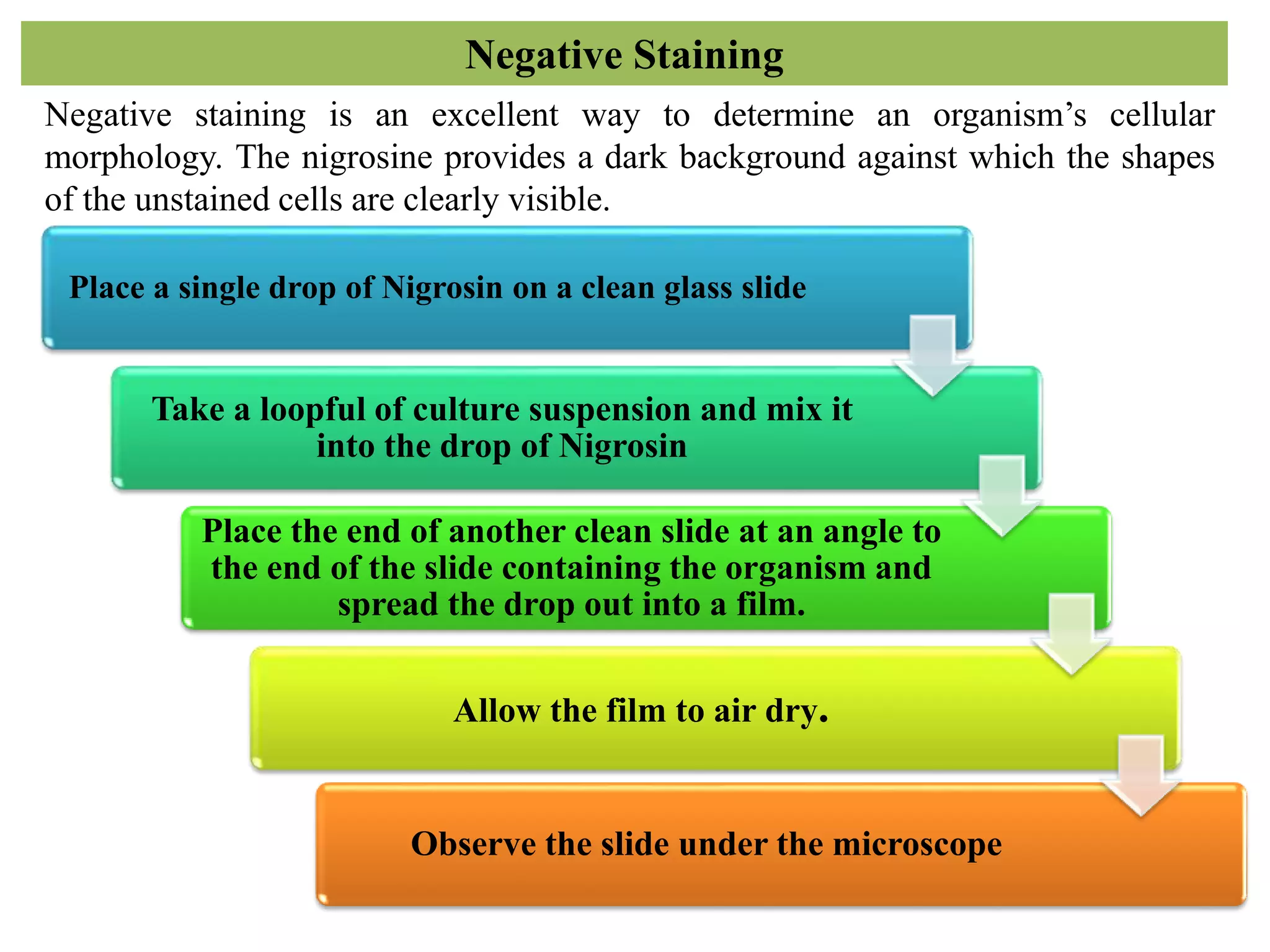
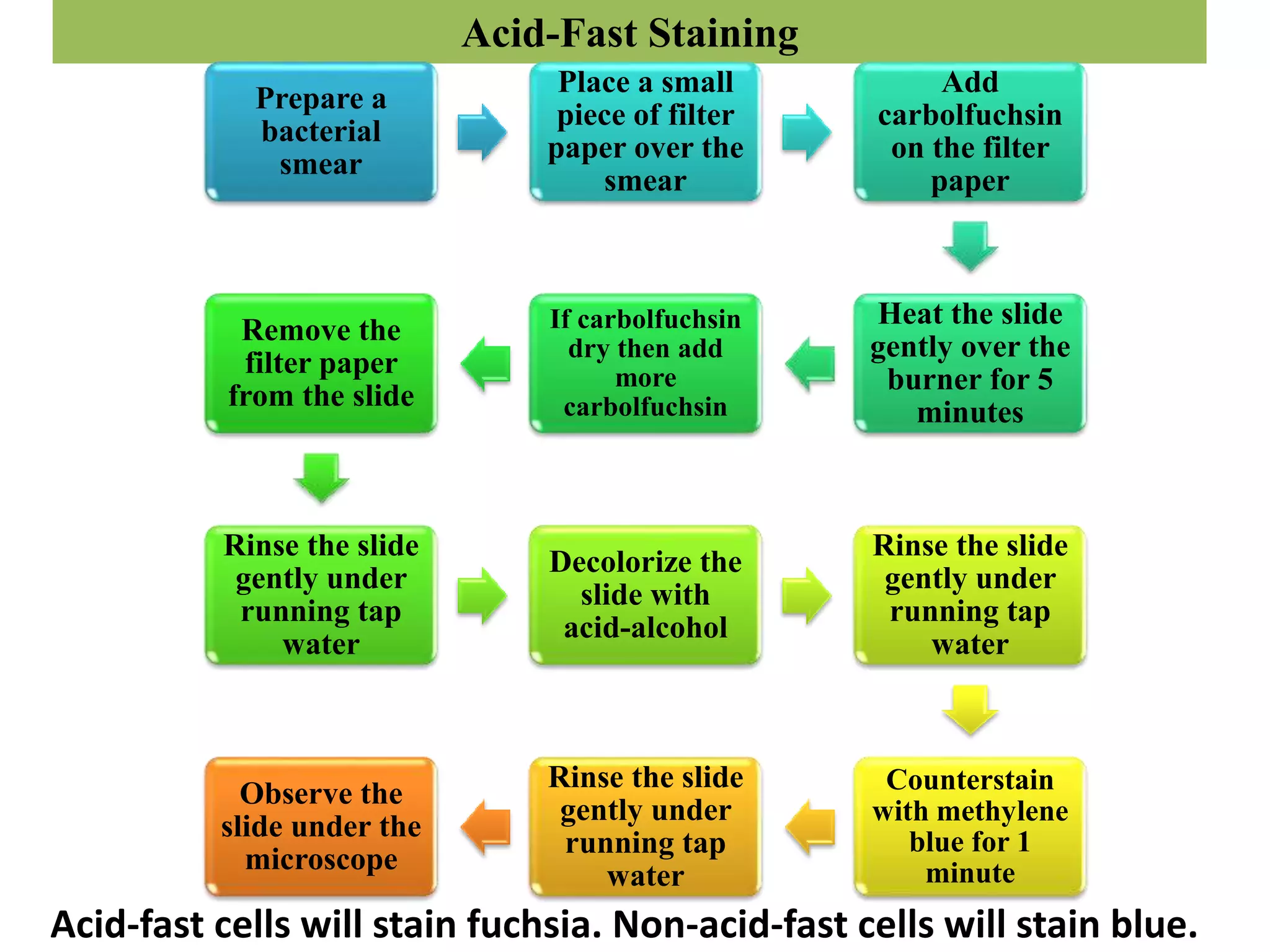
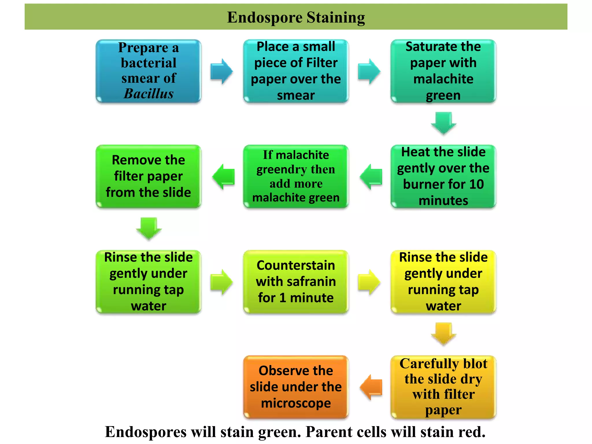
• Staining is a technique used to improve the contrast of samples
at microscopic level.
• Stains and dyes are commonly used to study the tissue or cells under
the microscope.
• In the medical fields of histopathology, hematology,
and cytopathology which will be focus on the study
and diagnose disease at a microscopic level.
• Biological staining is also used to mark cells in flow cytometry.
• Staining is not restricted to only biological materials, it can also be
used to study the structure of other materials like lamellar structures
of semi-crystalline polymers.
Types of stain/dye
• Acidic – Eosin, Nigrosin, India ink.
• Basic – Malachite green, crystal violet, basic fuschin,
safranin, methylene blue.
• Neutral – Giemsa stain.
Prepare a bacterial smear and heat fix it
Add basic dye on the smear for
approximately 1 minute
Rinse the slide gently under running tap
water
Carefully blot dry with Filter paper.
Observe the slide under the microscope, using
proper microscope technique.
Simple/Positive Staining
Simple staining provide a quick and easy way to determine
cell shape, size, and arrangement.
Negative Staining
Negative staining is an excellent way to determine an organism’s cellular
morphology. The nigrosine provides a dark background against which the shapes
of the unstained cells are clearly visible.
Place a single drop of Nigrosin on a clean glass slide
Take a loopful of culture suspension and mix it
into the drop of Nigrosin
Place the end of another clean slide at an angle to
the end of the slide containing the organism and
spread the drop out into a film.
Allow the film to air dry.
Observe the slide under the microscope
Gram Staining
Prepare a
bacterial
smear and
heat fix it
Flood the
smear with
crystal violet
for 1 minute.
Rinse the slide
gently under
running tap
water
Flood the
smear with
iodine for 1
minute.
Rinse the slide
gently under
running tap
water
Decolorize with
decolorizer
(acetone/alcohol)
for 30-45 seconds
Rinse the slide
gently under
running tap
water
Counterstain
with safranin
for 1 minute
Rinse the slide
gently with
water
Carefully blot
the slide dry
with filter
paper
Observe the
slide under
the
microscope
With proper staining technique…
Gram positive bacteria will stain
purple.
Gram negative bacteria will stain
red/pink.
Capsule Staining
Place a single
drop of India ink
on a clean
microscope slide
Take a loopful of
K. pneumoniae
culture suspension
and mix it into the
drop.
Place the end of another
clean slide at an angle to
the end of the slide
containing the organism
and spread the drop out
into a film
Allow the film
to air dry
Saturate the
slide with
crystal violet
for 1 minute
Rinse the slide
gently under
running tap
water
Allow the slide
to air dry
Observe the
slide under the
microscope
The
background
will be dark.
The bacterial
cells will be
stained purple.
The capsule will
appear clear
against the dark
background.
Acid-Fast Staining
Prepare a
bacterial
smear
Place a small
piece of filter
paper over the
smear
Add
carbolfuchsin
on the filter
paper
Heat the slide
gently over the
burner for 5
minutes
If carbolfuchsin
dry then add
more
carbolfuchsin
Remove the
filter paper
from the slide
Rinse the slide
gently under
running tap
water
Decolorize the
slide with
acid-alcohol
Rinse the slide
gently under
running tap
water
Counterstain
with methylene
blue for 1
minute
Rinse the slide
gently under
running tap
water
Observe the
slide under the
microscope
Acid-fast cells will stain fuchsia. Non-acid-fast cells will stain blue.
Endospore Staining
Prepare a
bacterial
smear of
Bacillus
Place a small
piece of Filter
paper over the
smear
Saturate the
paper with
malachite
green
Heat the slide
gently over the
burner for 10
minutes
If malachite
greendry then
add more
malachite green
Remove the
filter paper
from the slide
Rinse the slide
gently under
running tap
water
Counterstain
with safranin
for 1 minute
Rinse the slide
gently under
running tap
water
Carefully blot
the slide dry
with filter
paper
Observe the
slide under the
microscope
Endospores will stain green. Parent cells will stain red.
THANK
YOU

Staining techniques

  • 1.
    Basic Staining Techniquesin Microbiology Mr. Vikas Lalji Gupta Asst. Professor, VIVA College,MH-India
  • 2.
    Staining • Staining isa technique used to improve the contrast of samples at microscopic level. • Stains and dyes are commonly used to study the tissue or cells under the microscope. • In the medical fields of histopathology, hematology, and cytopathology which will be focus on the study and diagnose disease at a microscopic level. • Biological staining is also used to mark cells in flow cytometry. • Staining is not restricted to only biological materials, it can also be used to study the structure of other materials like lamellar structures of semi-crystalline polymers.
  • 3.
    Types of stain/dye •Acidic – Eosin, Nigrosin, India ink. • Basic – Malachite green, crystal violet, basic fuschin, safranin, methylene blue. • Neutral – Giemsa stain.
  • 4.
    Prepare a bacterialsmear and heat fix it Add basic dye on the smear for approximately 1 minute Rinse the slide gently under running tap water Carefully blot dry with Filter paper. Observe the slide under the microscope, using proper microscope technique. Simple/Positive Staining Simple staining provide a quick and easy way to determine cell shape, size, and arrangement.
  • 5.
    Negative Staining Negative stainingis an excellent way to determine an organism’s cellular morphology. The nigrosine provides a dark background against which the shapes of the unstained cells are clearly visible. Place a single drop of Nigrosin on a clean glass slide Take a loopful of culture suspension and mix it into the drop of Nigrosin Place the end of another clean slide at an angle to the end of the slide containing the organism and spread the drop out into a film. Allow the film to air dry. Observe the slide under the microscope
  • 6.
    Gram Staining Prepare a bacterial smearand heat fix it Flood the smear with crystal violet for 1 minute. Rinse the slide gently under running tap water Flood the smear with iodine for 1 minute. Rinse the slide gently under running tap water Decolorize with decolorizer (acetone/alcohol) for 30-45 seconds Rinse the slide gently under running tap water Counterstain with safranin for 1 minute Rinse the slide gently with water Carefully blot the slide dry with filter paper Observe the slide under the microscope With proper staining technique… Gram positive bacteria will stain purple. Gram negative bacteria will stain red/pink.
  • 7.
    Capsule Staining Place asingle drop of India ink on a clean microscope slide Take a loopful of K. pneumoniae culture suspension and mix it into the drop. Place the end of another clean slide at an angle to the end of the slide containing the organism and spread the drop out into a film Allow the film to air dry Saturate the slide with crystal violet for 1 minute Rinse the slide gently under running tap water Allow the slide to air dry Observe the slide under the microscope The background will be dark. The bacterial cells will be stained purple. The capsule will appear clear against the dark background.
  • 8.
    Acid-Fast Staining Prepare a bacterial smear Placea small piece of filter paper over the smear Add carbolfuchsin on the filter paper Heat the slide gently over the burner for 5 minutes If carbolfuchsin dry then add more carbolfuchsin Remove the filter paper from the slide Rinse the slide gently under running tap water Decolorize the slide with acid-alcohol Rinse the slide gently under running tap water Counterstain with methylene blue for 1 minute Rinse the slide gently under running tap water Observe the slide under the microscope Acid-fast cells will stain fuchsia. Non-acid-fast cells will stain blue.
  • 9.
    Endospore Staining Prepare a bacterial smearof Bacillus Place a small piece of Filter paper over the smear Saturate the paper with malachite green Heat the slide gently over the burner for 10 minutes If malachite greendry then add more malachite green Remove the filter paper from the slide Rinse the slide gently under running tap water Counterstain with safranin for 1 minute Rinse the slide gently under running tap water Carefully blot the slide dry with filter paper Observe the slide under the microscope Endospores will stain green. Parent cells will stain red.
  • 10.