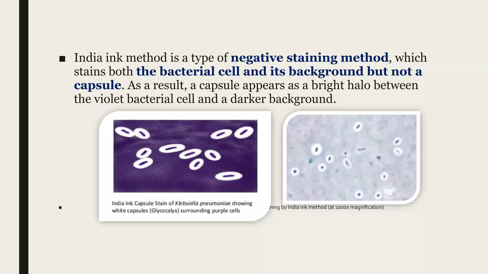
The document discusses capsule staining techniques used to identify the presence of capsules in bacteria under a microscope. There are two main methods - India ink negative staining and Anthony's positive staining. India ink stains the bacterial cell and background violet and black, leaving the non-stained capsule visible as a clear halo. Anthony's stains the cell and capsule blue and leaves the background light violet, again showing the capsule as a halo. Identifying capsules is important as their presence indicates more pathogenic bacteria.
Capsule is an layer around the bacteria cell which gives bacteria the protection and pathogenicity. Staining such an layer is difficult with the normal stains so it is necessary to stain the background and the cell itself which makes the capsule appear colourless.
Oxidase Test Microbiology - Principle, Procedure, Limitations, Results, QC - in lab #Oxidase Test
As the channel name suggests, our channel will be a perfect lounge for the malayali medicos..we wil be covering videos which will be like lecture classes related to the subjects biochemistry and microbiology in which we are specialised.. It will be a better learning experience for the students especially for those who are not able to understand and follow the normal classes in college..we assure the students that you will get a basic idea regarding the topic and extra reading can be done from the reference textbooks...
If you like my video
#like
#comment
#subscribe my channel
don't forget to subscribe my channel
Qualification
Maneesha M Joseph
MSc MLT (Microbiology)
Assistant Professor
Baby memorial college of allied Health science
Kozhikode
Our Partner Channel
Health & Voyage channel link - https://youtu.be/nzKqRVjlwc0
#Oxidase Test
#Medical
#Microbiology
# malayalam lecturer
#Mallu Medicos Lounge
#MalluMedicosLounge
#MLT
this presentation involves a comprehensive outlines regarding the most common different methods used in diagnostic microbiology to stain bacteria and their structures
COLLECTION AND TRANSPORTATION OF CLINICAL SAMPLESNCRIMS, Meerut
Principles of Sample Collection:
Aseptic precautions to minimize chances of
contamination.
Appropriate anatomic sites
Adequate volume
Adequate no. of samples
Appropriate time
Appropriate container with proper labelling
Before initiation of anti-microbials
Adequate information in request form
Acid fast staining is differential staining technique which differentiate bacteria into two group- acid fast bacteria and non acid bacteria. It used to identify acid-fast organisms such as members of the genus Mycobacterium .
This is a rundown of some staining techniques used in microbiology, including simple staining, negative staining, gram staining, acid-fast staining, endospore staining, and flagellar staining.
INTRODUCTION TO MICRO LAB, STAINING TECHNIQUES & MORPHOLOGY OF BACTERIADrBhavikapatel
This PPT is helpful to understand first practical to 2nd year MBBS student.
I have added 2 video in this PPT to understand staining techniques properly.
Reference: 1 Gram stain video: Dr.G Bhanu prakash animated medical videos
2. Zn stain video: sridhar Rao
Laboratory diagnosis of bacteria in 5 parts/ dental implant coursesIndian dental academy
The Indian Dental Academy is the Leader in continuing dental education , training dentists in all aspects of dentistry and
offering a wide range of dental certified courses in different formats.for more details please visit
www.indiandentalacademy.com
What is greenhouse gasses and how many gasses are there to affect the Earth.moosaasad1975
What are greenhouse gasses how they affect the earth and its environment what is the future of the environment and earth how the weather and the climate effects.
This pdf is about the Schizophrenia.
For more details visit on YouTube; @SELF-EXPLANATORY;
https://www.youtube.com/channel/UCAiarMZDNhe1A3Rnpr_WkzA/videos
Thanks...!
Slide 1: Title Slide
Extrachromosomal Inheritance
Slide 2: Introduction to Extrachromosomal Inheritance
Definition: Extrachromosomal inheritance refers to the transmission of genetic material that is not found within the nucleus.
Key Components: Involves genes located in mitochondria, chloroplasts, and plasmids.
Slide 3: Mitochondrial Inheritance
Mitochondria: Organelles responsible for energy production.
Mitochondrial DNA (mtDNA): Circular DNA molecule found in mitochondria.
Inheritance Pattern: Maternally inherited, meaning it is passed from mothers to all their offspring.
Diseases: Examples include Leber’s hereditary optic neuropathy (LHON) and mitochondrial myopathy.
Slide 4: Chloroplast Inheritance
Chloroplasts: Organelles responsible for photosynthesis in plants.
Chloroplast DNA (cpDNA): Circular DNA molecule found in chloroplasts.
Inheritance Pattern: Often maternally inherited in most plants, but can vary in some species.
Examples: Variegation in plants, where leaf color patterns are determined by chloroplast DNA.
Slide 5: Plasmid Inheritance
Plasmids: Small, circular DNA molecules found in bacteria and some eukaryotes.
Features: Can carry antibiotic resistance genes and can be transferred between cells through processes like conjugation.
Significance: Important in biotechnology for gene cloning and genetic engineering.
Slide 6: Mechanisms of Extrachromosomal Inheritance
Non-Mendelian Patterns: Do not follow Mendel’s laws of inheritance.
Cytoplasmic Segregation: During cell division, organelles like mitochondria and chloroplasts are randomly distributed to daughter cells.
Heteroplasmy: Presence of more than one type of organellar genome within a cell, leading to variation in expression.
Slide 7: Examples of Extrachromosomal Inheritance
Four O’clock Plant (Mirabilis jalapa): Shows variegated leaves due to different cpDNA in leaf cells.
Petite Mutants in Yeast: Result from mutations in mitochondrial DNA affecting respiration.
Slide 8: Importance of Extrachromosomal Inheritance
Evolution: Provides insight into the evolution of eukaryotic cells.
Medicine: Understanding mitochondrial inheritance helps in diagnosing and treating mitochondrial diseases.
Agriculture: Chloroplast inheritance can be used in plant breeding and genetic modification.
Slide 9: Recent Research and Advances
Gene Editing: Techniques like CRISPR-Cas9 are being used to edit mitochondrial and chloroplast DNA.
Therapies: Development of mitochondrial replacement therapy (MRT) for preventing mitochondrial diseases.
Slide 10: Conclusion
Summary: Extrachromosomal inheritance involves the transmission of genetic material outside the nucleus and plays a crucial role in genetics, medicine, and biotechnology.
Future Directions: Continued research and technological advancements hold promise for new treatments and applications.
Slide 11: Questions and Discussion
Invite Audience: Open the floor for any questions or further discussion on the topic.
Observation of Io’s Resurfacing via Plume Deposition Using Ground-based Adapt...Sérgio Sacani
Since volcanic activity was first discovered on Io from Voyager images in 1979, changes
on Io’s surface have been monitored from both spacecraft and ground-based telescopes.
Here, we present the highest spatial resolution images of Io ever obtained from a groundbased telescope. These images, acquired by the SHARK-VIS instrument on the Large
Binocular Telescope, show evidence of a major resurfacing event on Io’s trailing hemisphere. When compared to the most recent spacecraft images, the SHARK-VIS images
show that a plume deposit from a powerful eruption at Pillan Patera has covered part
of the long-lived Pele plume deposit. Although this type of resurfacing event may be common on Io, few have been detected due to the rarity of spacecraft visits and the previously low spatial resolution available from Earth-based telescopes. The SHARK-VIS instrument ushers in a new era of high resolution imaging of Io’s surface using adaptive
optics at visible wavelengths.
Multi-source connectivity as the driver of solar wind variability in the heli...Sérgio Sacani
The ambient solar wind that flls the heliosphere originates from multiple
sources in the solar corona and is highly structured. It is often described
as high-speed, relatively homogeneous, plasma streams from coronal
holes and slow-speed, highly variable, streams whose source regions are
under debate. A key goal of ESA/NASA’s Solar Orbiter mission is to identify
solar wind sources and understand what drives the complexity seen in the
heliosphere. By combining magnetic feld modelling and spectroscopic
techniques with high-resolution observations and measurements, we show
that the solar wind variability detected in situ by Solar Orbiter in March
2022 is driven by spatio-temporal changes in the magnetic connectivity to
multiple sources in the solar atmosphere. The magnetic feld footpoints
connected to the spacecraft moved from the boundaries of a coronal hole
to one active region (12961) and then across to another region (12957). This
is refected in the in situ measurements, which show the transition from fast
to highly Alfvénic then to slow solar wind that is disrupted by the arrival of
a coronal mass ejection. Our results describe solar wind variability at 0.5 au
but are applicable to near-Earth observatories.
This presentation explores a brief idea about the structural and functional attributes of nucleotides, the structure and function of genetic materials along with the impact of UV rays and pH upon them.
A brief information about the SCOP protein database used in bioinformatics.
The Structural Classification of Proteins (SCOP) database is a comprehensive and authoritative resource for the structural and evolutionary relationships of proteins. It provides a detailed and curated classification of protein structures, grouping them into families, superfamilies, and folds based on their structural and sequence similarities.
Cancer cell metabolism: special Reference to Lactate PathwayAADYARAJPANDEY1
Normal Cell Metabolism:
Cellular respiration describes the series of steps that cells use to break down sugar and other chemicals to get the energy we need to function.
Energy is stored in the bonds of glucose and when glucose is broken down, much of that energy is released.
Cell utilize energy in the form of ATP.
The first step of respiration is called glycolysis. In a series of steps, glycolysis breaks glucose into two smaller molecules - a chemical called pyruvate. A small amount of ATP is formed during this process.
Most healthy cells continue the breakdown in a second process, called the Kreb's cycle. The Kreb's cycle allows cells to “burn” the pyruvates made in glycolysis to get more ATP.
The last step in the breakdown of glucose is called oxidative phosphorylation (Ox-Phos).
It takes place in specialized cell structures called mitochondria. This process produces a large amount of ATP. Importantly, cells need oxygen to complete oxidative phosphorylation.
If a cell completes only glycolysis, only 2 molecules of ATP are made per glucose. However, if the cell completes the entire respiration process (glycolysis - Kreb's - oxidative phosphorylation), about 36 molecules of ATP are created, giving it much more energy to use.
IN CANCER CELL:
Unlike healthy cells that "burn" the entire molecule of sugar to capture a large amount of energy as ATP, cancer cells are wasteful.
Cancer cells only partially break down sugar molecules. They overuse the first step of respiration, glycolysis. They frequently do not complete the second step, oxidative phosphorylation.
This results in only 2 molecules of ATP per each glucose molecule instead of the 36 or so ATPs healthy cells gain. As a result, cancer cells need to use a lot more sugar molecules to get enough energy to survive.
introduction to WARBERG PHENOMENA:
WARBURG EFFECT Usually, cancer cells are highly glycolytic (glucose addiction) and take up more glucose than do normal cells from outside.
Otto Heinrich Warburg (; 8 October 1883 – 1 August 1970) In 1931 was awarded the Nobel Prize in Physiology for his "discovery of the nature and mode of action of the respiratory enzyme.
WARNBURG EFFECT : cancer cells under aerobic (well-oxygenated) conditions to metabolize glucose to lactate (aerobic glycolysis) is known as the Warburg effect. Warburg made the observation that tumor slices consume glucose and secrete lactate at a higher rate than normal tissues.
Deep Behavioral Phenotyping in Systems Neuroscience for Functional Atlasing a...Ana Luísa Pinho
Functional Magnetic Resonance Imaging (fMRI) provides means to characterize brain activations in response to behavior. However, cognitive neuroscience has been limited to group-level effects referring to the performance of specific tasks. To obtain the functional profile of elementary cognitive mechanisms, the combination of brain responses to many tasks is required. Yet, to date, both structural atlases and parcellation-based activations do not fully account for cognitive function and still present several limitations. Further, they do not adapt overall to individual characteristics. In this talk, I will give an account of deep-behavioral phenotyping strategies, namely data-driven methods in large task-fMRI datasets, to optimize functional brain-data collection and improve inference of effects-of-interest related to mental processes. Key to this approach is the employment of fast multi-functional paradigms rich on features that can be well parametrized and, consequently, facilitate the creation of psycho-physiological constructs to be modelled with imaging data. Particular emphasis will be given to music stimuli when studying high-order cognitive mechanisms, due to their ecological nature and quality to enable complex behavior compounded by discrete entities. I will also discuss how deep-behavioral phenotyping and individualized models applied to neuroimaging data can better account for the subject-specific organization of domain-general cognitive systems in the human brain. Finally, the accumulation of functional brain signatures brings the possibility to clarify relationships among tasks and create a univocal link between brain systems and mental functions through: (1) the development of ontologies proposing an organization of cognitive processes; and (2) brain-network taxonomies describing functional specialization. To this end, tools to improve commensurability in cognitive science are necessary, such as public repositories, ontology-based platforms and automated meta-analysis tools. I will thus discuss some brain-atlasing resources currently under development, and their applicability in cognitive as well as clinical neuroscience.
2. WHAT DOYOU MEAN BY STAINING?
■ Artificial coloration of a substance to facilitate examination of
tissues, microorganisms, or other cells under the microscope. For
various techniques, see under stain.
■ Staining is a technique used to enhance contrast in samples,
generally at the microscopic level.
■ Staining A technique in which cells or thin sections of biological
tissue that are normally transparent are immersed in one or more
coloured dyes (stains) to make them more clearly visible
through a microscope. Staining heightens the contrast between
the various cell or tissue components.
■ Why Stain Cells? The most basic reason that cells are stained is
to enhance visualization of the cell or certain cellular components
under a microscope. Cells may also be stained to highlight
metabolic processes or to differentiate between live and dead cells
in a sample.
3. CAPSULE STAINING
■ Capsule staining is a type of differential staining method which
creates contrast in the microscopic image either by staining
bacterial cell and background or by staining the capsule only.
Therefore, capsule staining can be done by two methods, namely
positive and negative staining methods.
■ The capsule staining is employed to know the presence or absence
of capsule, based on which we could classify the type of bacteria,
i.e. whether a bacteria is capsulated or non-capsulated.
■ It is necessary to perform capsule staining because the presence of
capsule indicates a virulent strain of bacteria that can cause
disease. Thus, by knowing the presence of the capsule, we can
determine the degree of pathogenicity.
4. DEFINATION OF CAPSULE STAINING
■ A capsule staining can define as the special staining method that
makes the use of differential capsule stain which either highlights
or stains the capsule or stains both the bacterial cell and its
background.
■ It is an important staining method, because some bacteria like
Bacillus anthracis, Streptococcus pneumoniae etc. have a capsule,
which can cause pathogenicity in many humans and animals. Thus,
it becomes necessary to identify the presence of an extracellular
capsule. Capsule staining can be performed by India ink,
Anthony’s, Maneval’s and Hiss method.
5. Principle
■ The principle of capsule staining is based on staining of
background with an acidic stain and staining of bacterial
cell with a basic stain. As a capsule is non-ionic, it will
not stain by either of the two dyes. Thus a capsule
staining creates a contrast by staining a bacterial cell and
its background in between which, capsule appears as a
colourless halo.
6. Methods of Capsule Staining
■ There are different methods for capsule staining, among which the most common
methods are:
■ India ink method negative staining method
■ Anthony’s method positive staining method
■ Maneval’s method negative staining method
■ Hiss method positive staining method
■ We study above 2 method.
7. Materials required
■ Microscopic slides
■ Inoculating loop
■ Microscope with 100x objective lens (oil immersion)
■ Immersion oil
■ Gas burner
■ Test bacteria culture
8. India ink Method
■ India ink method uses two types of stain, i.e. a basic stain (Crystal violet) and an acidic
stain (India ink).
■ Crystal violet being positive stain will stain the negatively charged bacterial cell. India
ink being negative stain will stain the positively charged background.After staining.
■ The Background appears darker or black.
■ A Bacterial cell appears violet.
■ The capsule appears as a clear halo.
10. ■ India ink method is a type of negative staining method, which
stains both the bacterial cell and its background but not a
capsule. As a result, a capsule appears as a bright halo between
the violet bacterial cell and a darker background.
■ Capsule staining by India ink method (at 1000x magnification)
11. Anthony’s Method
■ This method makes the use of two reagents, namely crystal violet
as primary stain and 20% of CuSO4 solution as a decolouring agent
and counterstain.
■ Crystal violet will stain the bacterial cell and background. The
CuSO4 solution will stain the non-ionic capsule. After staining.
■ Bacterial cell appears violet.
■ The background appears light violet.
■ The capsule appears as a faint blue halo.
12. Procedure
■ 1.Take a clean, sterilized or grease free slide.
■ 2.Add a drop of crystal violet to the centre of the glass slide.
■ 3.Prepare a smear, by taking an inoculum from the bacterial culture and mix it with a
drop of crystal violet.
■ 4.Then, allow the smear to air dry (Do not heat fix, as it can cause cell shrinkage and
distortion of the bacteria).
■ 5.Flood a smear with 20% of CuSO4 solution for at least 30 seconds and remove the
extra stain by tilting a glass slide.
■ 6.Add oil immersion to the stained area and observe it under the microscope having a
100X objective.
13. ■ Anthony’s method is a type of positive staining method that
stains the capsule along with the bacterial cell but not stains the
background. As a result, a capsule appears as faint blue halo
between the violet bacterial cell and purple background.