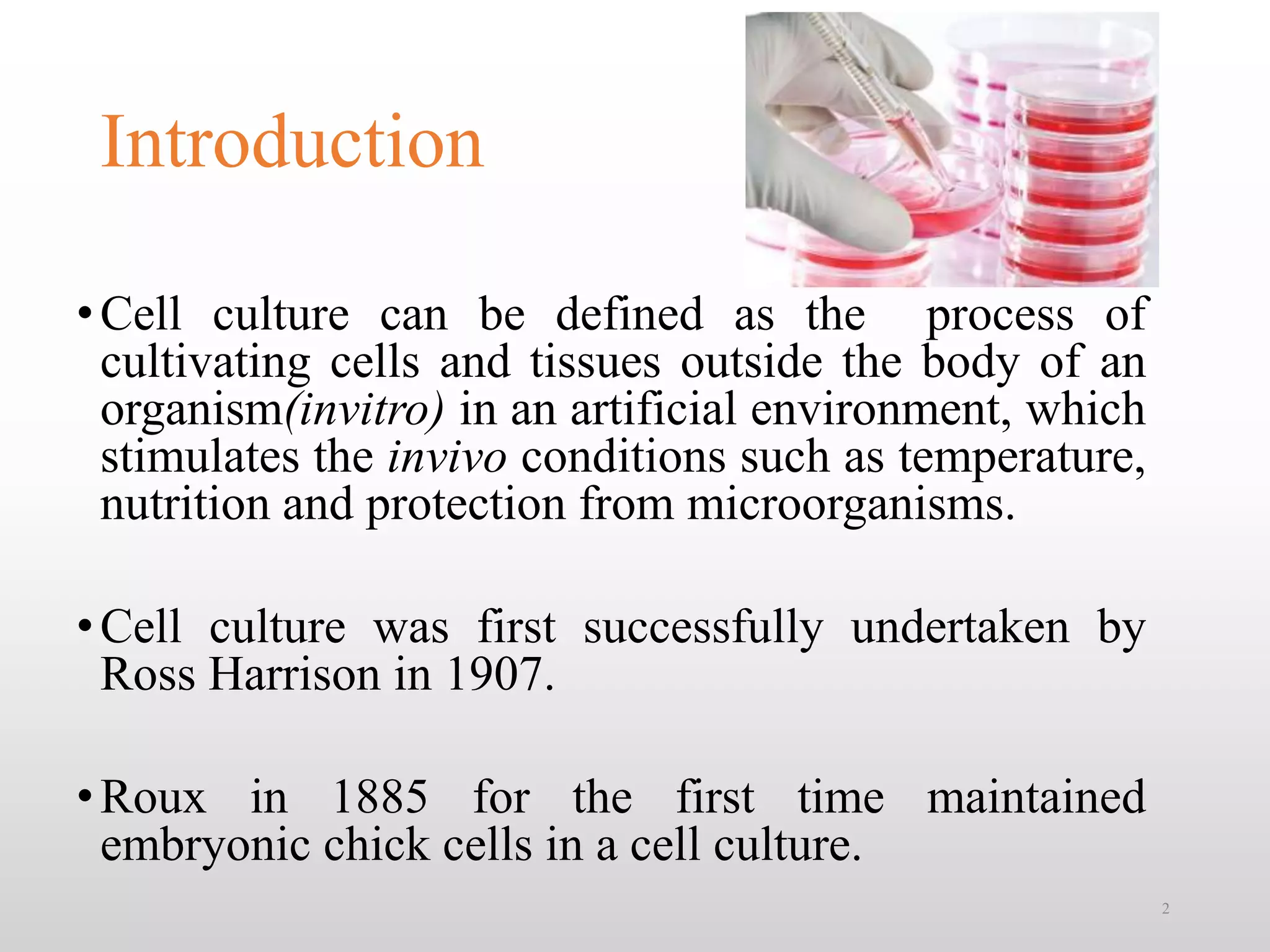
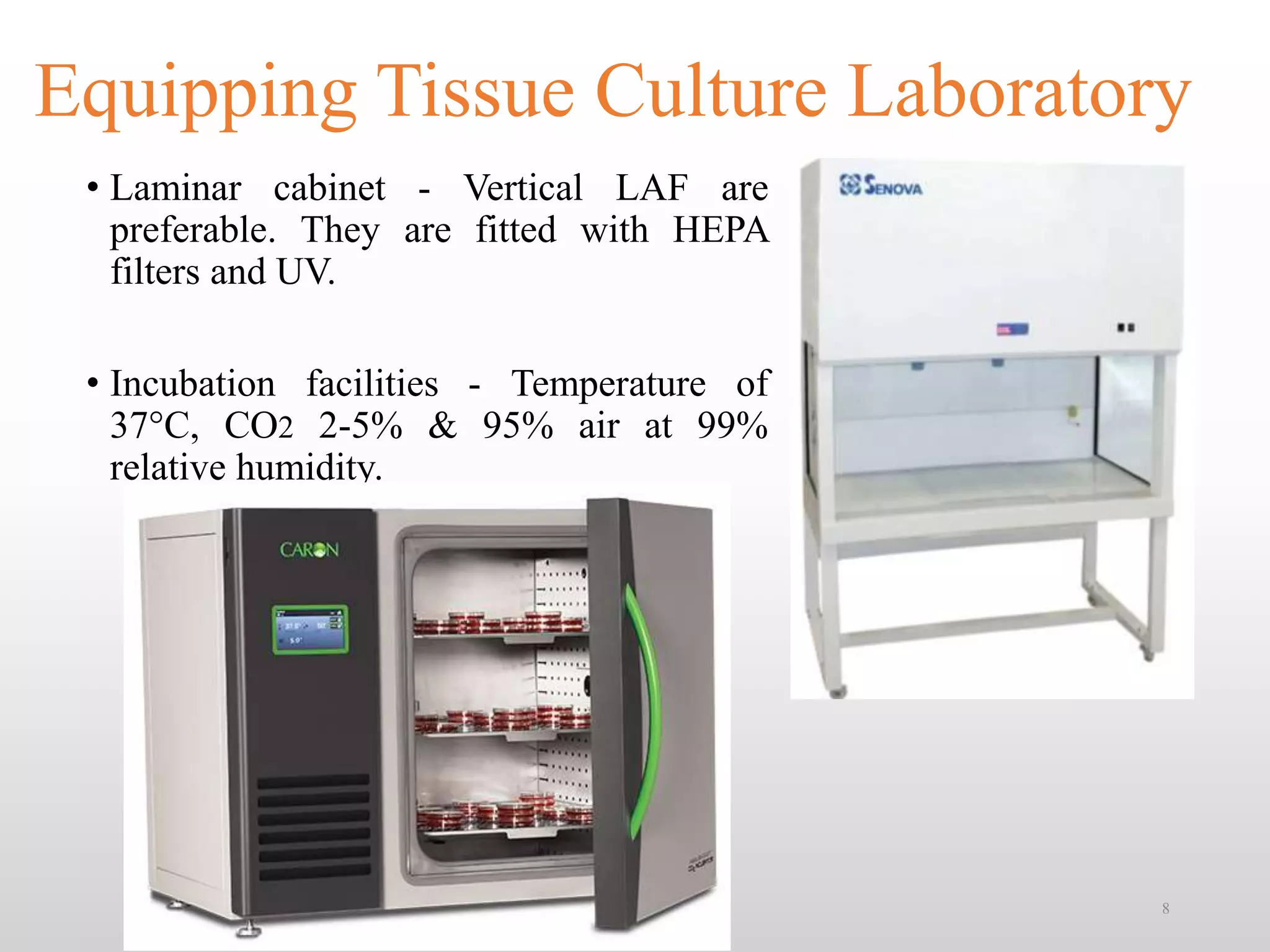
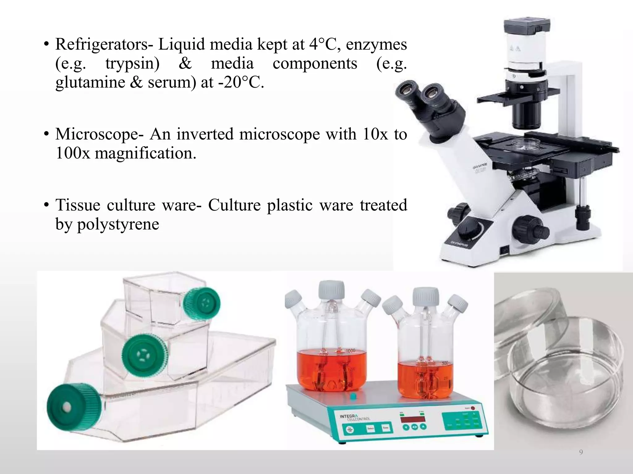
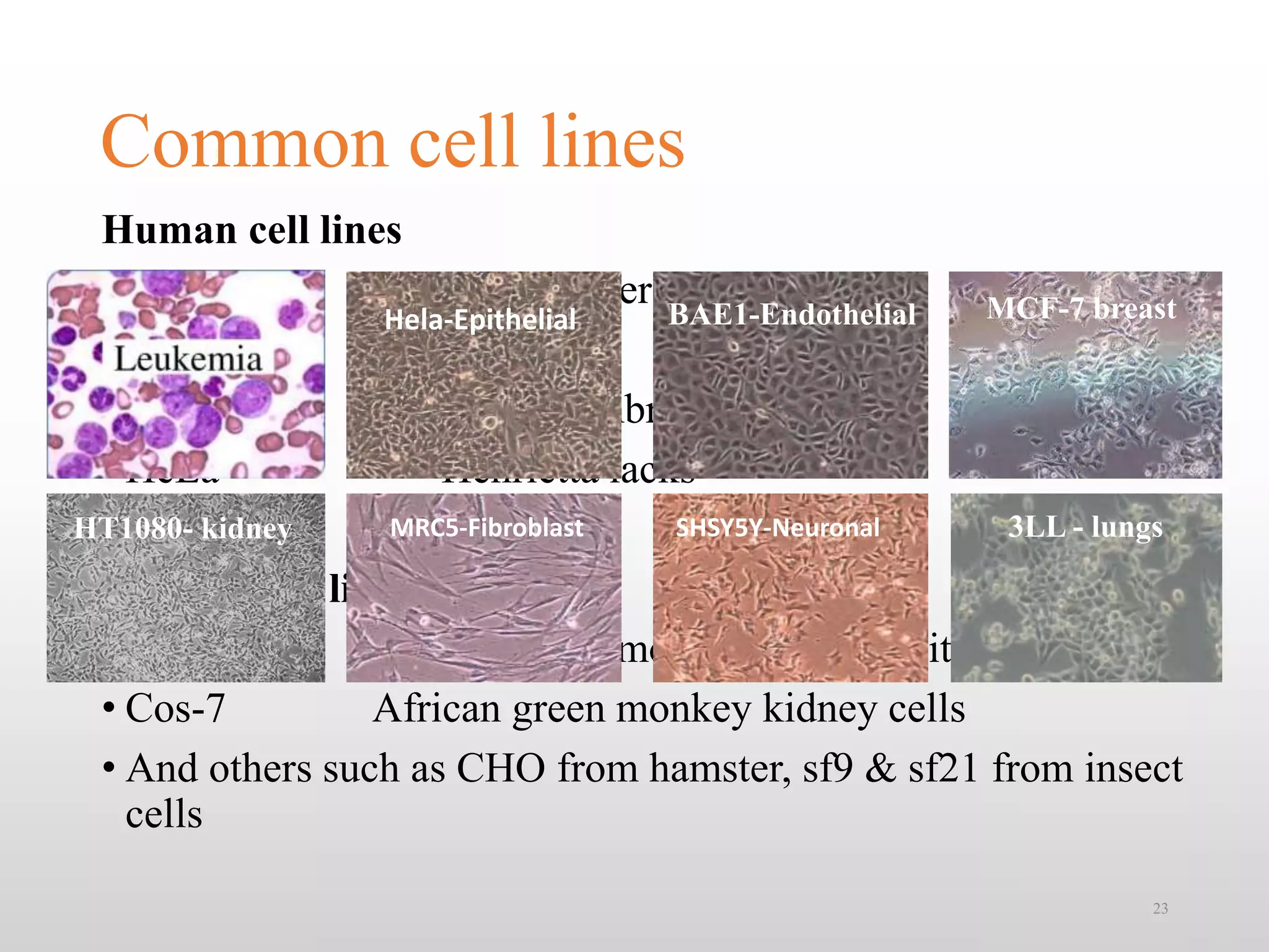
Artificial Intelligence In Microbiology by Dr. Prince C P
Ā
Animal cell culture techniques
1. Animal Cell Culture Techniques &
Its Applications
Presented By:
Nagendra P
16PBT204
M.Tech Pharmaceutical Biotechnology
1
2. Introduction
ā¢Cell culture can be defined as the process of
cultivating cells and tissues outside the body of an
organism(invitro) in an artificial environment, which
stimulates the invivo conditions such as temperature,
nutrition and protection from microorganisms.
ā¢Cell culture was first successfully undertaken by
Ross Harrison in 1907.
ā¢Roux in 1885 for the first time maintained
embryonic chick cells in a cell culture.
2
3. Historical events in the
development of cell culture
ā¢ 1878: Claude Bernard proposed that physiological systems
of an organism can be maintained in a living system after
the death of an organism.
ā¢ 1897: Loeb demonstrated the survival of cells isolated from
blood and connective tissue in serum and plasma.
ā¢ 1907: Harrison cultivated frog nerve cells in a lymph clot
held by the 'hanging drop' method and observed the growth
of nerve fibers in vitro for several weeks. He was
considered by some as the father of cell culture.
3
4. ā¢ 1916: Rous and Jones introduced proteolytic enzyme trypsin
for the subculture of adherent cells.
ā¢ 1940s: The use of the antibiotics penicillin and streptomycin in
culture medium decreased the problem of contamination in cell
culture.
ā¢ 1952: George Gey established a continuous cell line from a
human cervical carcinoma known as HeLa (Helen Lane) cells.
ā¢ Dulbecco developed plaque assay for animal viruses using
confluent monolayers of cultured cells.
ā¢ 1955: Eagle studied the nutrient requirements of selected cells
in culture and established the first widely used chemically
defined medium.
ā¢ 1961: Hayflick and Moorhead isolated human fibroblasts (WI-
38) and showed that they have a finite lifespan in culture. 4
5. Major developmentās in cell culture
technology
ā¢ First development was the use of antibiotics which inhibits
the growth of contaminants.
ā¢ Second was the use of trypsin to remove adherent cells to
subculture further from the culture vessel.
ā¢ Third was the use of chemically defined culture medium.
5
6. Terminologies
ā¢ Primary Cell Culture: When cells are surgically removed from an
organism and placed into a suitable culture environment they will
attach, divide and grow.
ā¢ Cell Line: When the primary culture is subcultured and they show an
ability to continuously propagate.
ā¢ Anchorage dependency: Cells grow as monolayers adhering to the
substrate (glass/ plastic)
ā¢ Passaging/ subculturing: The process of splitting the cells.
ā¢ Finite cells: When the cells has finite life span.
ā¢ Continuous cell lines: When the cells can grow upto infinite
lifespan. 6
8. Equipping Tissue Culture Laboratory
ā¢ Laminar cabinet - Vertical LAF are
preferable. They are fitted with HEPA
filters and UV.
ā¢ Incubation facilities - Temperature of
37Ā°C, CO2 2-5% & 95% air at 99%
relative humidity.
8
9. ā¢ Refrigerators- Liquid media kept at 4Ā°C, enzymes
(e.g. trypsin) & media components (e.g.
glutamine & serum) at -20Ā°C.
ā¢ Microscope- An inverted microscope with 10x to
100x magnification.
ā¢ Tissue culture ware- Culture plastic ware treated
by polystyrene
9
11. Tissue Culture Media
ā¢ Cells have complex nutritional requirements that must be met to
permit their propagation in vitro.
ā¢ Previously, scientists employed chick embryo extract, plasma,
sera, lymph etc.,
ā¢ However, they varied in their growth promoting characteristics
and thus hampering the reproducibility of the experiments.
ā¢ Today, a number of chemically-defined formulations have been
developed that support the growth of a variety of established
cell lines
11
13. ā¢ Eagleās basal media
ā¢ Eaglesās Minimum Essential Media (MEM)
ā¢ Dulbeccoās Modified Essential Media (DMEM)
ā¢ Iscoveās Modified Dulbeccoās Medium (IMDM)
ā¢ Roosevelt Park Memorial Institute (RPMI 1640)
ā¢ HAMās F12
ā¢ The various nutrients required are:
ā¢ glucose,
ā¢ fats and fatty acids,
ā¢ lipids, phospholipids and sulpholipids,
ā¢ ATP and amino acids
ā¢ Vitamins
ā¢ Minerals
ā¢ Serum:
Serum can provide various growth factors, hormones, cell
adhesion factor and other factors needed by the most
mammalian cells for their long term growth and metabolism.
ā¢ FCS, FBS, CS, HS, HoS.
13
14. Properties and Special Requirements of
Media
ā¢ pH: Optimum pH between 7.2 to 7.4 is generally needed for
mammalian cells. Phenol red is used as an internal indicator.
ā¢ CO2 and Bicarbonate: NaHCO3 and 20 mM HEPES.
Addition of pyruvate increases the endogenous production of
CO2.
14
pH Colour of the medium
7.8 Pink / purple
7.6 Pink / purple
7.4 Red
7.0 Orange
5.5 Yellow
< 6.5 Lemon Yellow
15. ā¢ Oxygen
ā¢ Cells depend upon glycolysis for the supply of O2
ā¢ Selenium controls O2 diffusion
ā¢ Glutathione acts as free radical scavenger
ā¢ Temperature
ā¢ The optimum temperature of mammal is 37oC.
ā¢ Change of Ā± 5Ā°C is acceptable.
ā¢ Humidity
ā¢ For cell growth 100% humidity is essential to reduce evaporation of the
media.
ā¢ Antibiotics
ā¢ penicillin (100 U/ml) for bacteria,
ā¢ streptomycin (100 mg/ml) for bacteria,
ā¢ or gentamycin (50mg/ ml) for bacteria,
ā¢ and nystatin (50mg/ml) for fungi and yeast
15
16. Limitations of Serum
ā¢ It is undefined with respect to its chemical composition.
ā¢ It can be a source of adventitious agents and their by-products.
ā¢ Serum also presents a variable performance of cell growth and
adds a substantial cost.
ā¢ Problem during downstream processing.
ā¢ Availability.
Serum Free Media
ā¢ Important amino acids, some trace elements, growth factor,
hormone, transport protein and adhesion factor are added.
ā¢ Adhesion factor added are main components of intercellular
substance and serum, such as fibronectin, collagen, and laminin.
ā¢ Primary purpose of introducing SFM is to promote the specific
growth of a particular type of cell.
16
17. Types of tissue culture
ā¢ CELL CULTURE
ā¢ Tissue from an explant is dispersed,
mostly enzymatically, into a cell
suspension which may then be
cultured as a monolayer or
suspension culture
Advantages
ā¢ Development of a cell line over
several generations
ā¢ Scale-up is possible
ā¢ Absolute control of physical
environment
ā¢ Homogeneity of sample
ā¢ Less compound needed than in
animal models
Disadvantages
ā¢ Cells may lose some differentiated
characteristics.
ā¢ Hard to maintain
ā¢ Only grow small amount of tissue at
high cost
ā¢ Dedifferentiation
ā¢ Instability, aneuploidy
ā¢ EXPLANT CULTURE
ā¢ Is the growth
of tissues or cells separate from the
organism.
ā¢ This is typically facilitated via use of
a liquid, semi-solid, or solid growth
medium, such as broth or agar.
Advantages
ā¢ Some normal functions may be
maintained.
ā¢ Better than organ culture for scale-up
but not ideal.
Disadvantages
ā¢ Original organization of tissue is lost.
17
ORGAN CULTURE
The entire embryos or organs are
excised from the body and culture
Advantages
Normal physiological functions
are maintained.
Cells remain fully differentiated.
Disadvantages
Scale-up is not recommended.
Growth is slow.
Fresh explantation is required for
every experiment.
19. Primary Cultures
ā¢ When cells are surgically removed from an organism and
placed into a suitable culture environment they will attach,
divide and grow.
ā¢ Most of the primary culture cells have a finite lifespan of 50-
60 divisions invitro.
ā¢ Primary cells are considered by many researchers to be more
physiologically similar to in vivo cells
ā¢ Due to their limited lifespan, one cannot do long-term
experiments with these cells
19
20. Tissue explants are excised using sharp scalpel.
Mechanical disruption by pestle and mortar. Then filtered using a 0.22Āµ Filter fitted
to a syringe.
Enzymatic digestion by Trysin or collagenase
Cells are counted on a Haemocytometer. 1-2 Ć 10^5 cells / mL is seeded in to the
media.
5mL of cells is suspended into 25cm^2 flask.
The flasks are incubated in a CO2 incubator.
The flasks are observed daily for their normal growth characteristics.
Media is changed every 2-3 days until the cells attain 80% confluent. 20
22. Continuous cell lines
ā¢ Most cell lines grow for a limited number of generations after which
they ceases.
ā¢ Cell lines which either occur spontaneously or induced virally or
chemically gets transformed into Continuous cell lines.
ā¢ Characteristics of continuous cell lines
-smaller, more rounded, less adherent with a higher nucleus
/cytoplasm ratio
-Fast growth and have aneuploid chromosome number
-reduced serum and anchorage dependence and grow more in
suspension conditions
-ability to grow upto higher cell density
-different in phenotypes from donar tissue
-stop expressing tissue specific markers
- Loss of Contact inhibition 22
23. Common cell lines
Human cell lines
ā¢ MCF-7 breast cancer
ā¢ HL 60 Leukemia
ā¢ HEK-293 Human embryonic kidney
ā¢ HeLa Henrietta lacks
Primate cell lines
ā¢ Vero African green monkey kidney epithelial cells
ā¢ Cos-7 African green monkey kidney cells
ā¢ And others such as CHO from hamster, sf9 & sf21 from insect
cells
23
Hela-Epithelial MCF-7 breast
MRC5-FibroblastHT1080- kidney SHSY5Y-Neuronal 3LL - lungs
BAE1-Endothelial
24. Subculturing
Incubate the cells.
Then split the cells into 2 or 3 flasks containing complete media.
Add equivalent of 2 volumes of pre-warmed complete growth
medium. Disperse the medium by pipetting over the cell layer surface
several times.
Incubate the culture vessel at room temperature for approximately 2
minutes.
Add the pre-warmed dissociation reagent such as trypsin. Gently rock the
container to get complete coverage of the cell layer.
Remove spent media from the culture vessel.
24
25. Cryopreservation
Next day transfer the cryovials to Liquid nitrogen.
Transfer the cells to cryovials, incubate the cryovials at -80Ā°C overnight
Resuspend the cells in 1-2ml of freezing medium containing DMSO.
Centrifuge at 200g for 5 min at RT and remove the growth medium by aspiration
Dilute the cells with growth medium.
Dissociate the cells by trypsin
Remove the growth medium, wash the cells by PBS and remove the PBS by
aspiration.
25
26. Detection of contaminants
ā¢ In general indicators of contamination are turbid culture media,
change in growth rates, abnormally high pH, poor attachment, multi-
nucleated cells, graining cellular appearance, vacuolization,
inclusion bodies and cell lysis
ā¢ Yeast, bacteria & fungi usually shows visible effect on the culture
(changes in medium turbidity or pH)
ā¢ Mycoplasma detected by direct DNA staining with intercalating
fluorescent substances e.g. Hoechst 33258
ā¢ Mycoplasma also detected by enzyme immunoassay by specific
antisera or monoclonal abs or by PCR amplification of
mycoplasmal RNA
ā¢ The best and the oldest way to eliminate contamination is to discard
the infected cell lines directly
26
27. Invitro Transformation of Cells
ā¢ Transformed, Infinite or Established Cells
ā¢ Changed from normal cells to cells with many of the properties of cancer
cells
ā¢ Some of these cell lines have actually been derived from tumors or are
transformed spontaneously in culture by mutations
ā¢ Chemical or gamma ray treated cells can become infinite with loss of growth
factors
ā¢ Viral infection with SV40 T antigen can insert oncogenes and lead to gene
alteration
ā¢ No matter how transformation occurred, the result is a cell with altered
functional, morphological, and growth characteristics
28. Advantages of Tissue Culture
ļ¼Control of the environment.
ļ¼Characterization and Homogenity of sample.
ļ¼Economy, Scale and Mechanization.
ļ¼Invitro modelling of Invivo Conditions.
28
29. Limitations
ā¢ To grow cells outside their normal environment, three major controls are
involved.
ā¢ Observing scrict asepsis
ā¢ Providing the right kind of physic-chemical environment
ā¢ Nutrients in its simplest absorbable form
ā¢ Culturing technique needs a great deal of expertise.
ā¢ Tissue samples consists of a mixture of heterogenous cell populations
ā¢ Continuously growing cells often show genetic instability.
ā¢ Differences in the behavior or cells in cultured and in its natural form.
ā¢ Should include proper balance of the hormones.
29
30. Applications of Cell Culture
ā¢ Excellent model systems for studying:
ļ¼ The normal physiology, cell biology and biochemistry of cells
ļ¼ The effects of drugs, radiation and toxic compounds on the cells
ļ¼ Study mutagenesis and carcinogenesis
ā¢ Used for gene transfer studies.
ā¢ Large scale manufacturing of biological compounds
ā¢ (vaccines, insulin, interferon, other therapeutic protein)
30
31. Products
Monoclonal
Antibodies
(Mabās)
Immuno-
biological
Regulators
Virus
vaccines
Hormones
ļ±Produced by hybridoma cell
ļ±Used for diagnostic assay systems
(determine drugs, toxins & vitamin);
therapeutic purposes & biological
separations ā chromatographic
separations to purify protein
molecules
Interferon ā anticancer
glycoprotein (secreted
animal cell or recombinant
bacteria)
Lymphokines
Interleukines (anticancer
agent)
Prophylactics
Virus is collected, inactivated
and used as vaccine
A weakened form will induce a
protective response but no
disease
Large molecules: 50-200 amino
acids
Produce by hormone-
synthesizing organ
May also produce by chemical
synthesis
Example: Erythropoietin
33. References
1) Baksh, D., Song, L., & Tuan, R. S. (2004). Adult mesenchymal stem cells: characterization,
differentiation, and application in cell and gene therapy. Journal of cellular and molecular
medicine, 8(3), 301-316
2) Butler, M. (2005). Animal cell cultures: recent achievements and perspectives in the
production of biopharmaceuticals. Applied microbiology and biotechnology, 68(3), 283-291.
3) Freshney, R. I. (2010). Culture of animal cells. Hoboken.
4) Gangal, S. (2007). Principles and Practice of Animal Tissue Culture. Universities Press.
5) http://www.biotechnology4u.com/animal_biotechnology_applications_animal_cell_culture.ht
ml
6) Montagnon, B. J. (1988). Polio and rabies vaccines produced in continuous cell lines: a reality
for Vero cell line. Developments in biological standardization, 70, 27-47.
7) Park, T. H., & Shuler, M. L. (2003). Integration of cell culture and microfabrication
technology. Biotechnology progress, 19(2), 243-253.
8) Pau, M. G., Ophorst, C., Koldijk, M. H., Schouten, G., Mehtali, M., & Uytdehaag, F. (2001).
The human cell line PER. C6 provides a new manufacturing system for the production of
influenza vaccines. Vaccine, 19(17), 2716-2721.
9) Seliktar, D. (2012). Designing cell-compatible hydrogels for biomedical
applications. Science, 336(6085), 1124-1128.
10) Wurm, F. M. (2004). Production of recombinant protein therapeutics in cultivated mammalian
cells. Nature biotechnology, 22(11), 1393-1398.
33