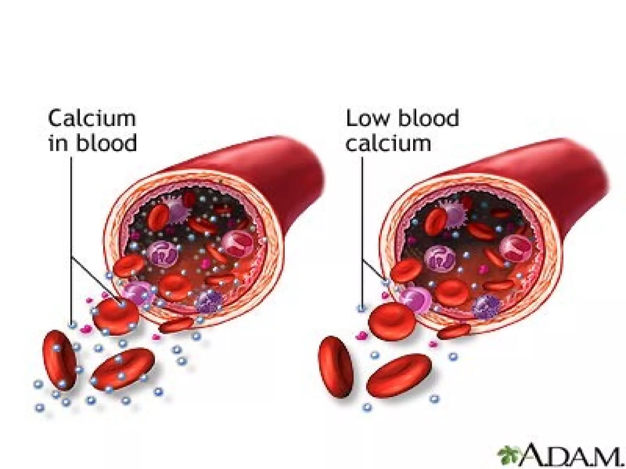
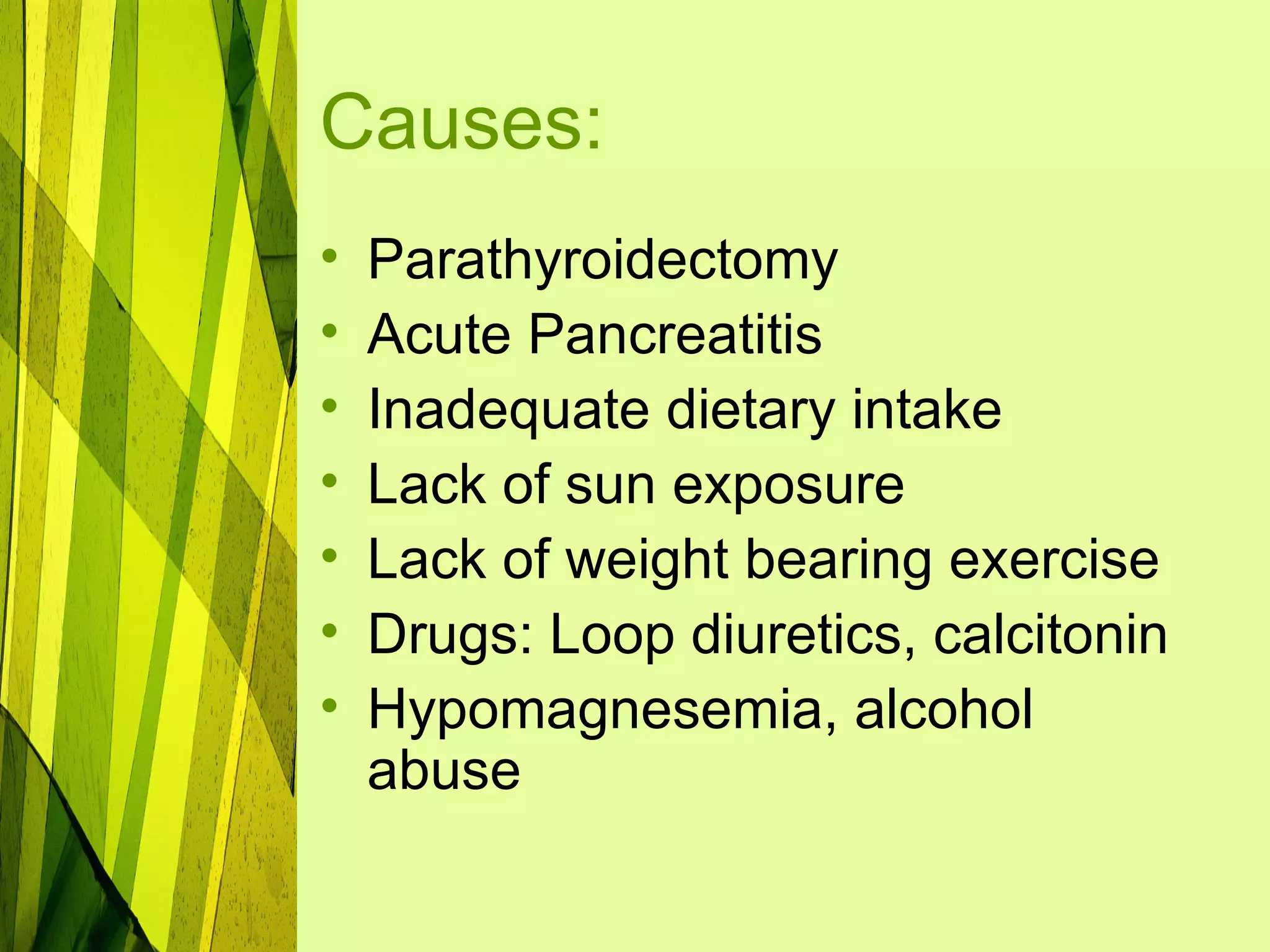
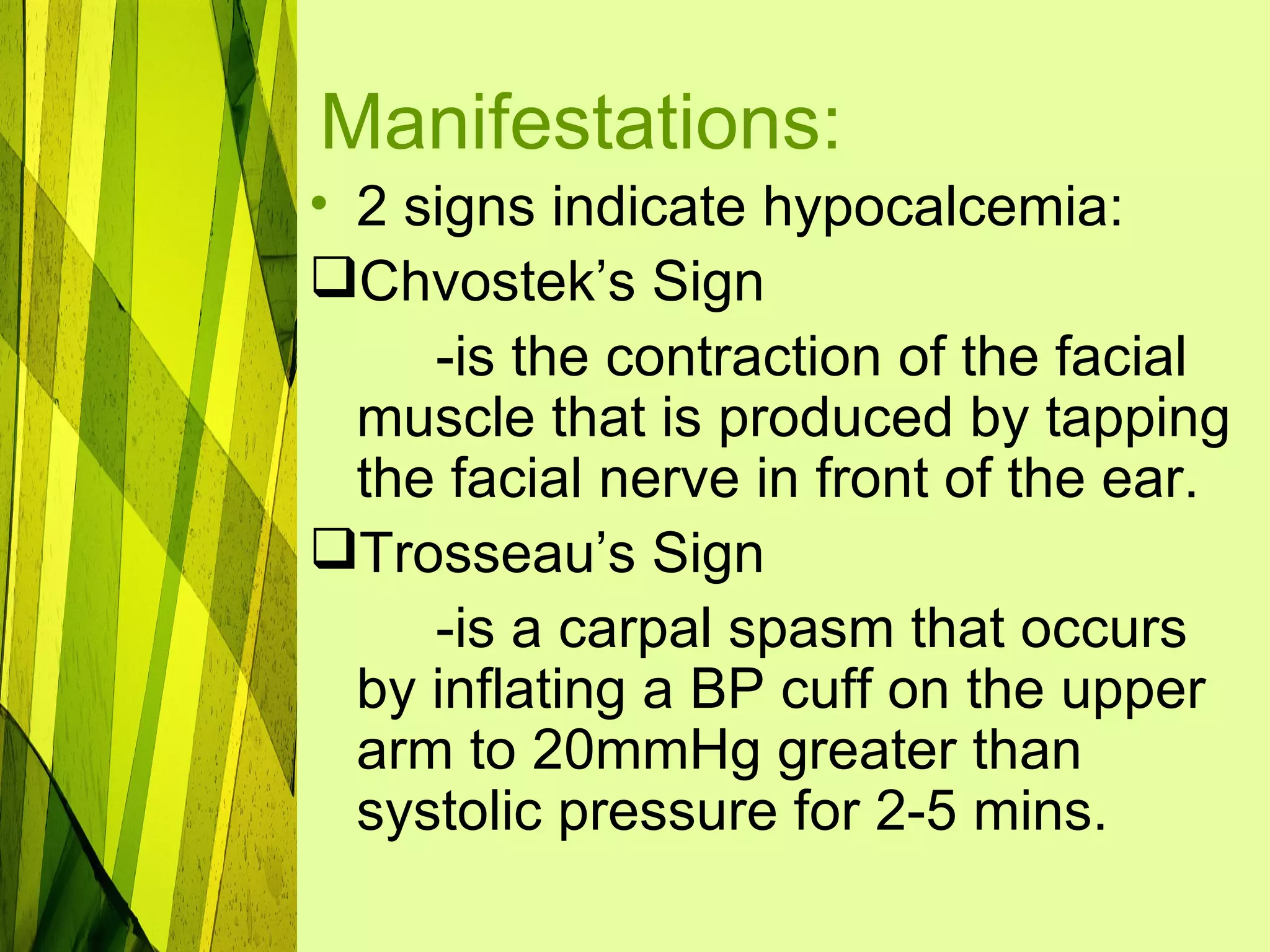
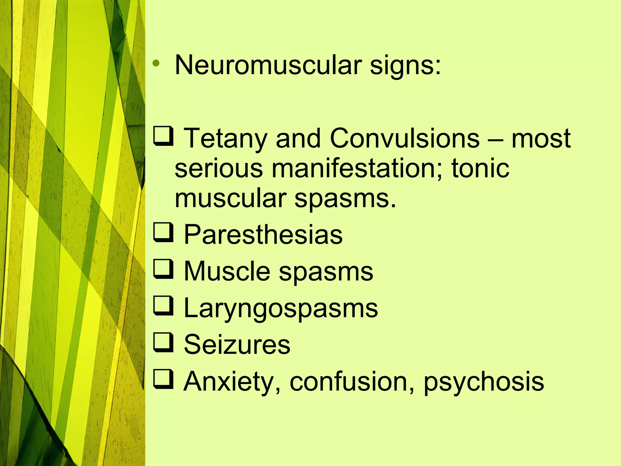
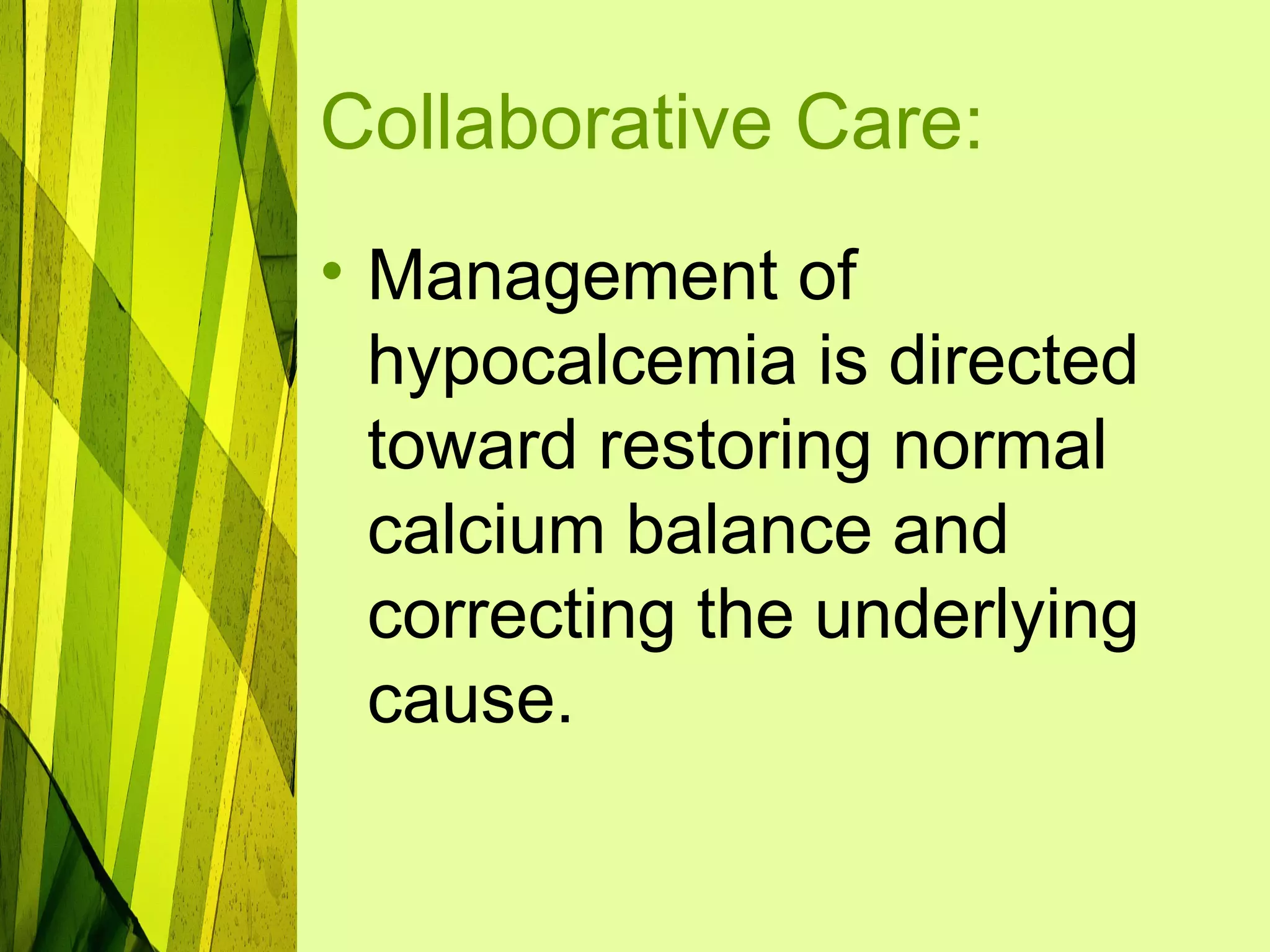
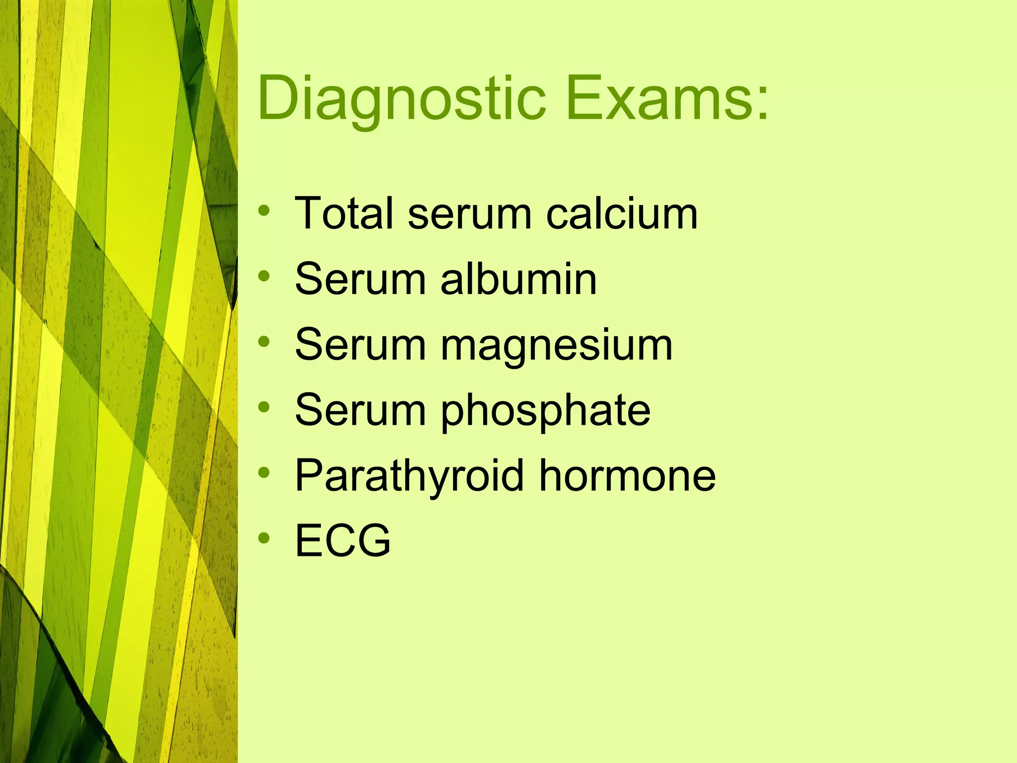
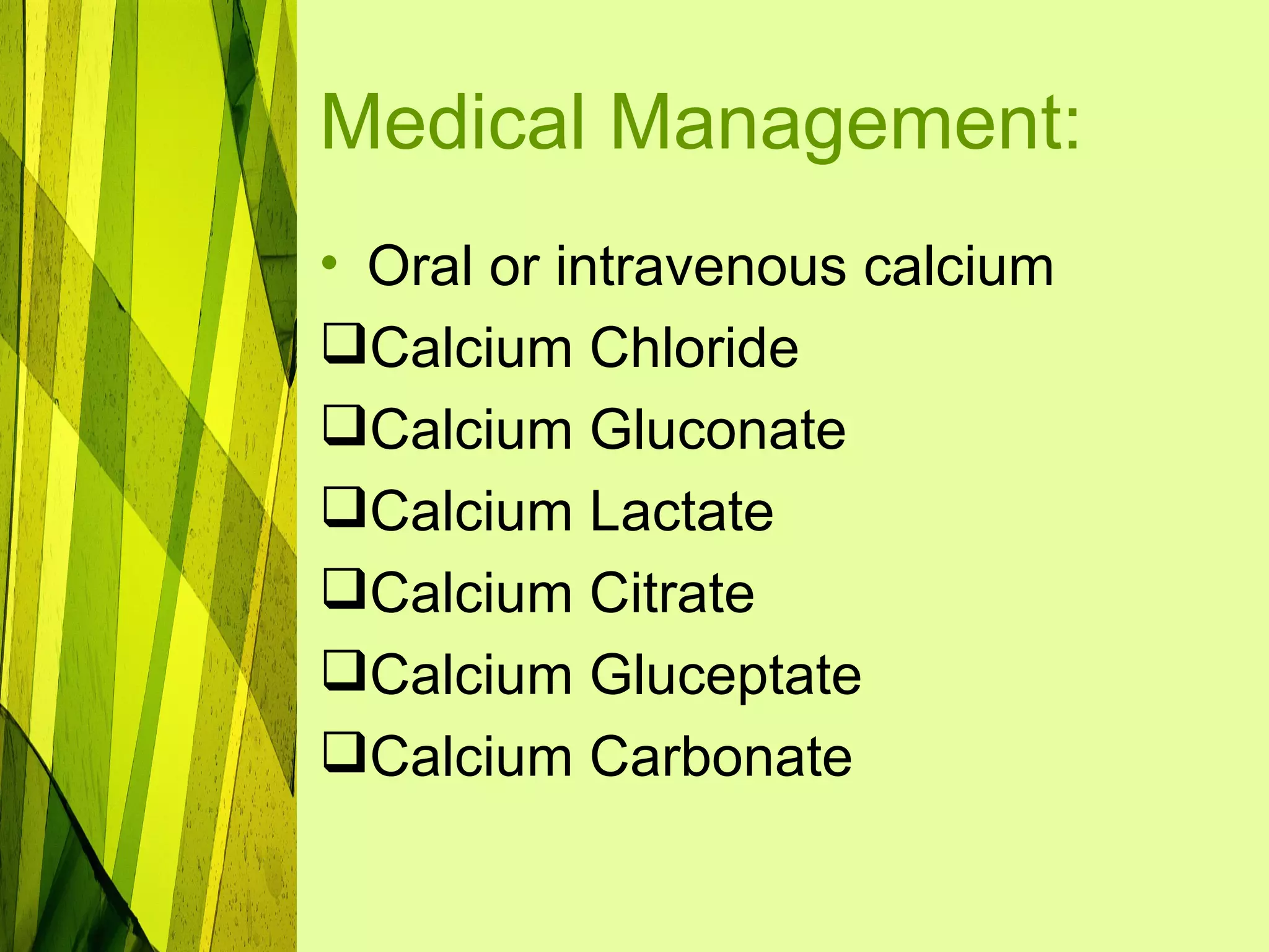
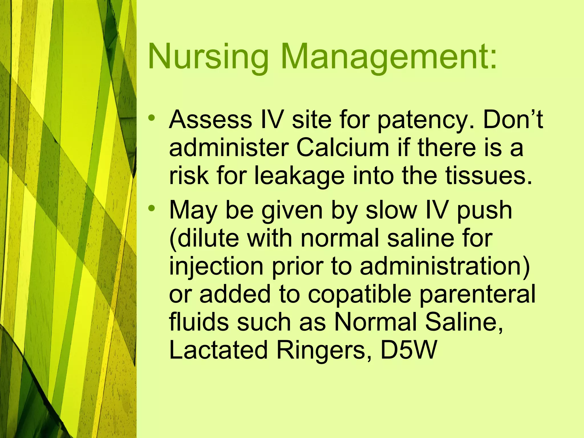
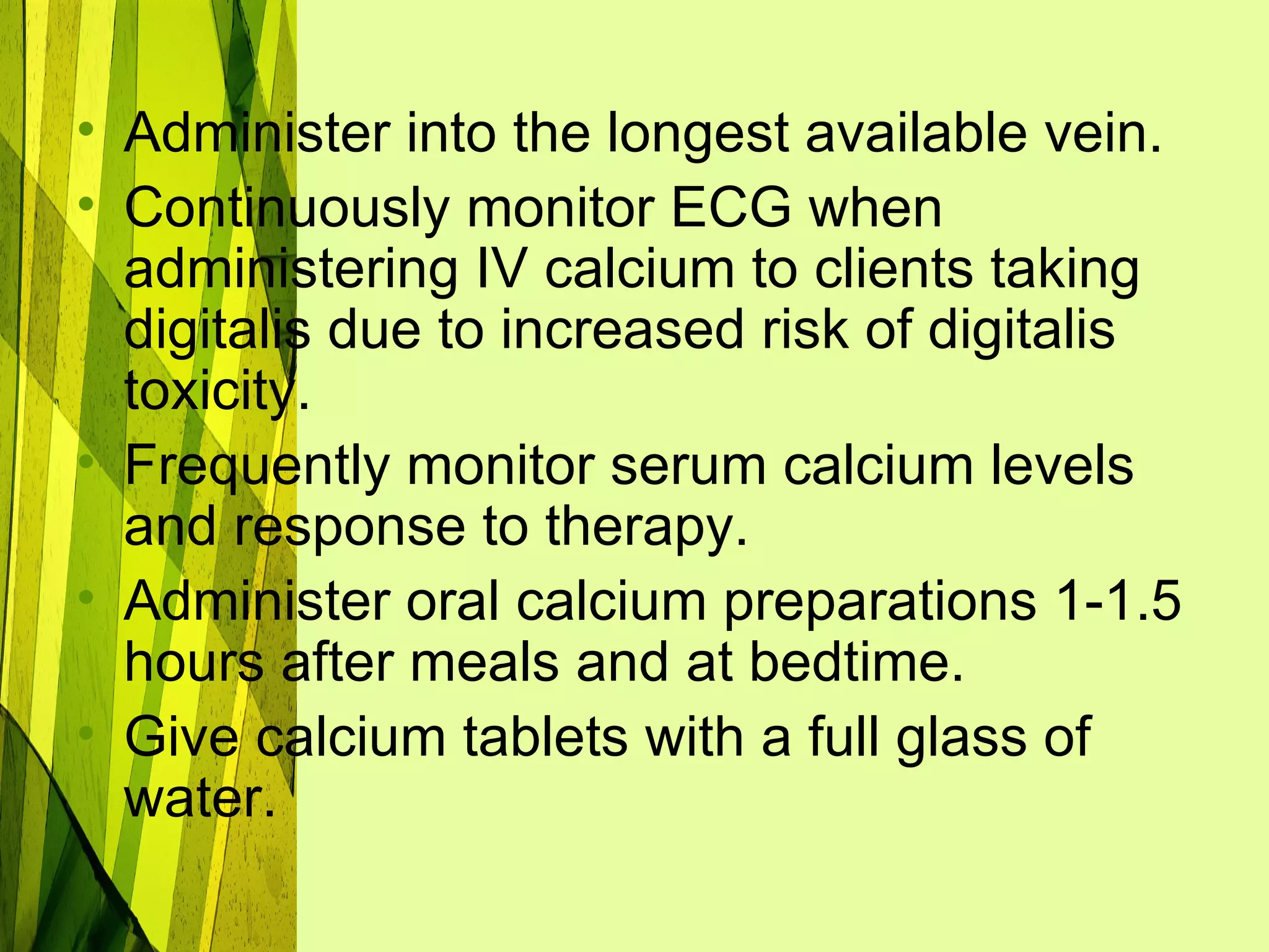
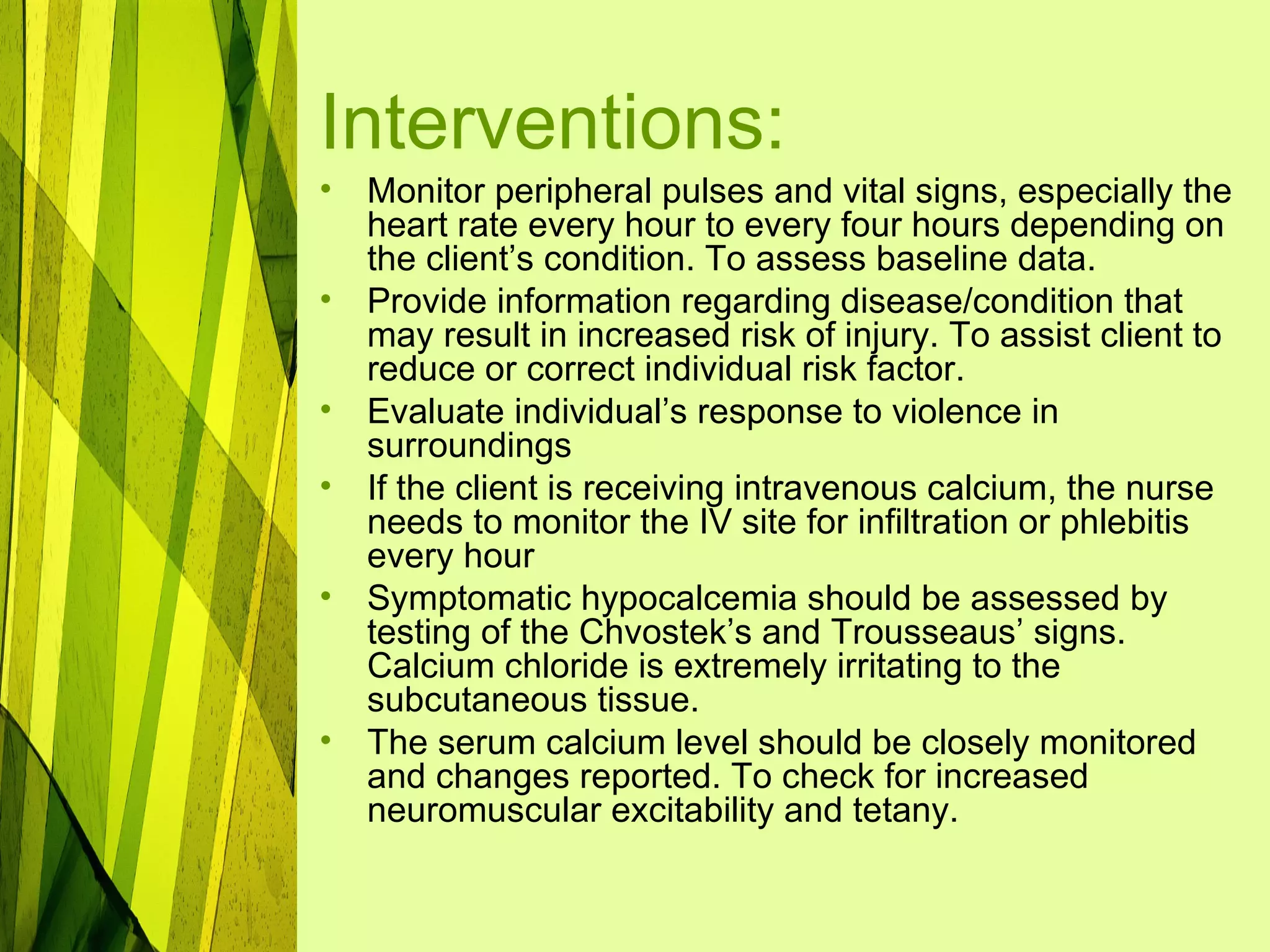
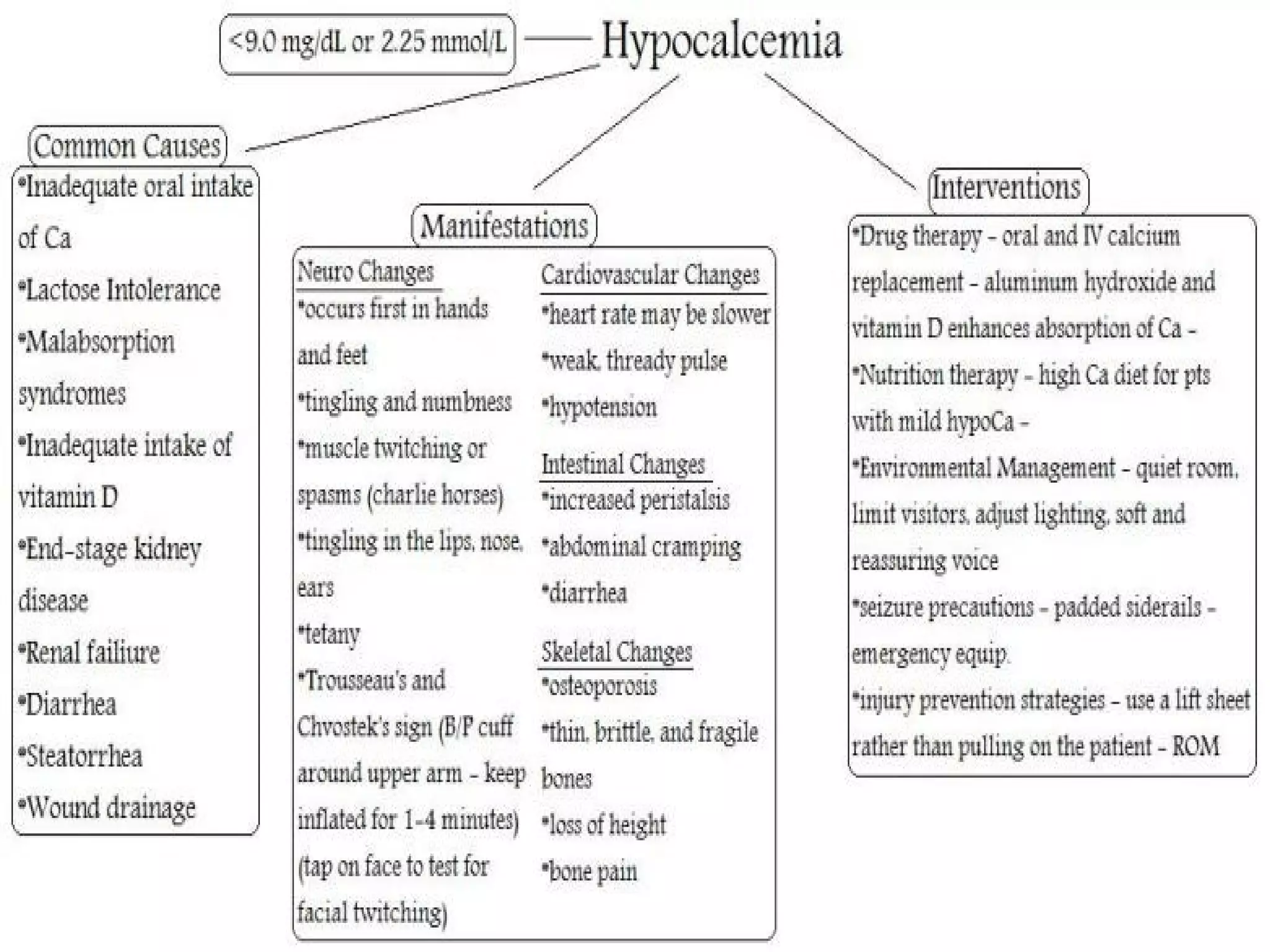
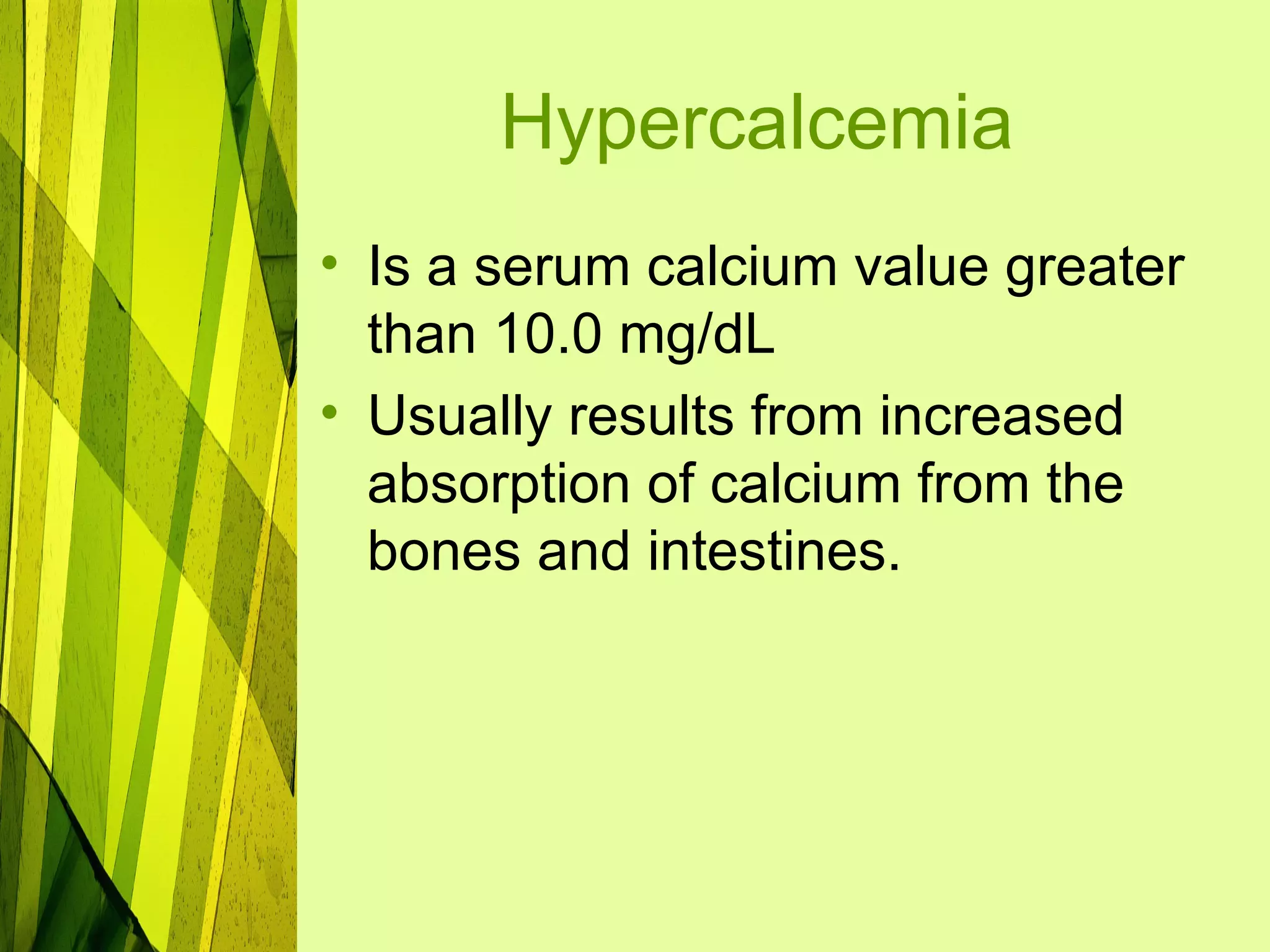
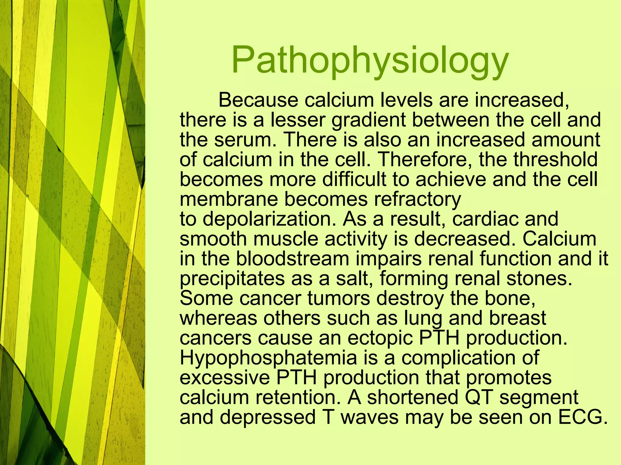
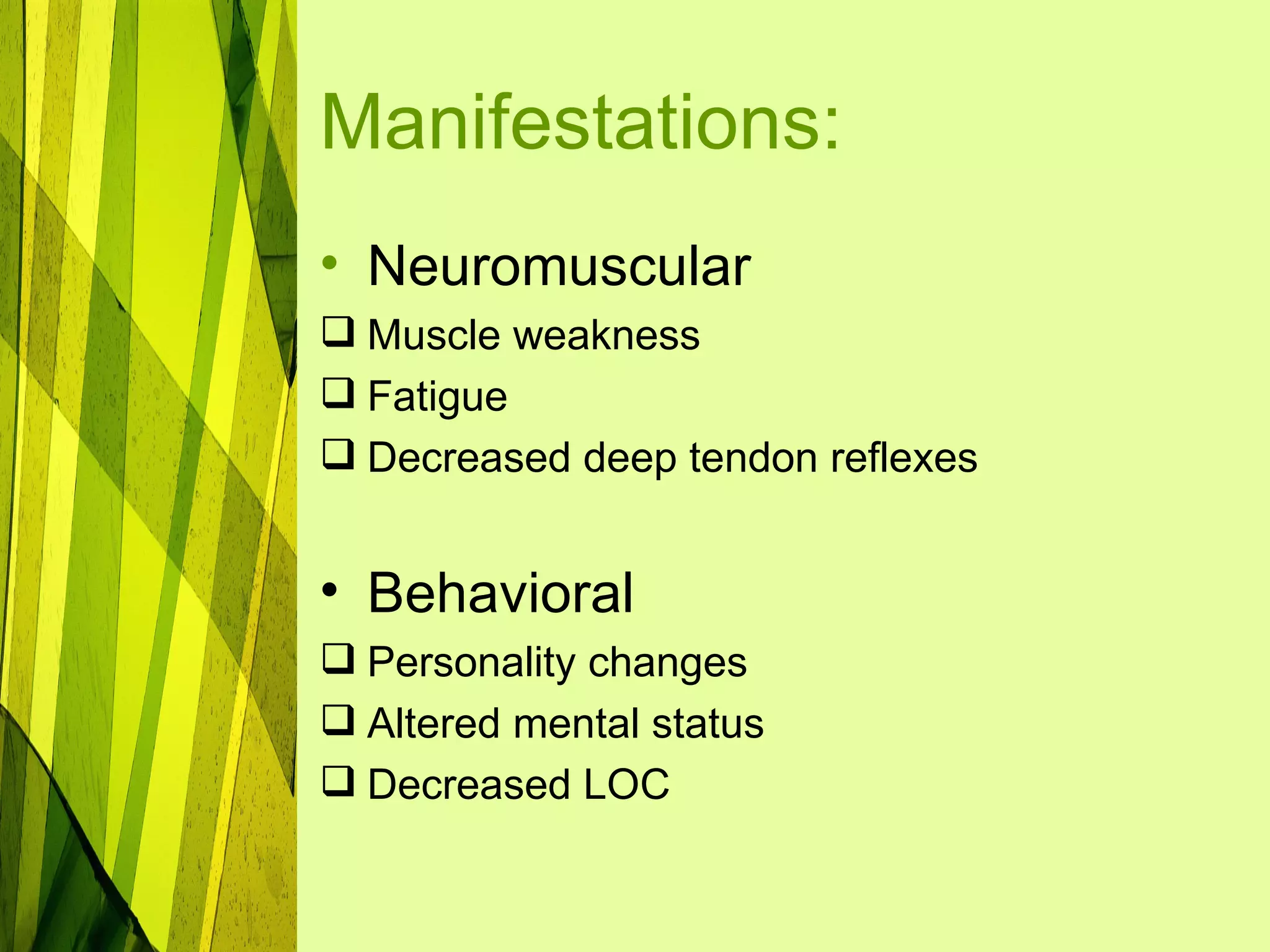
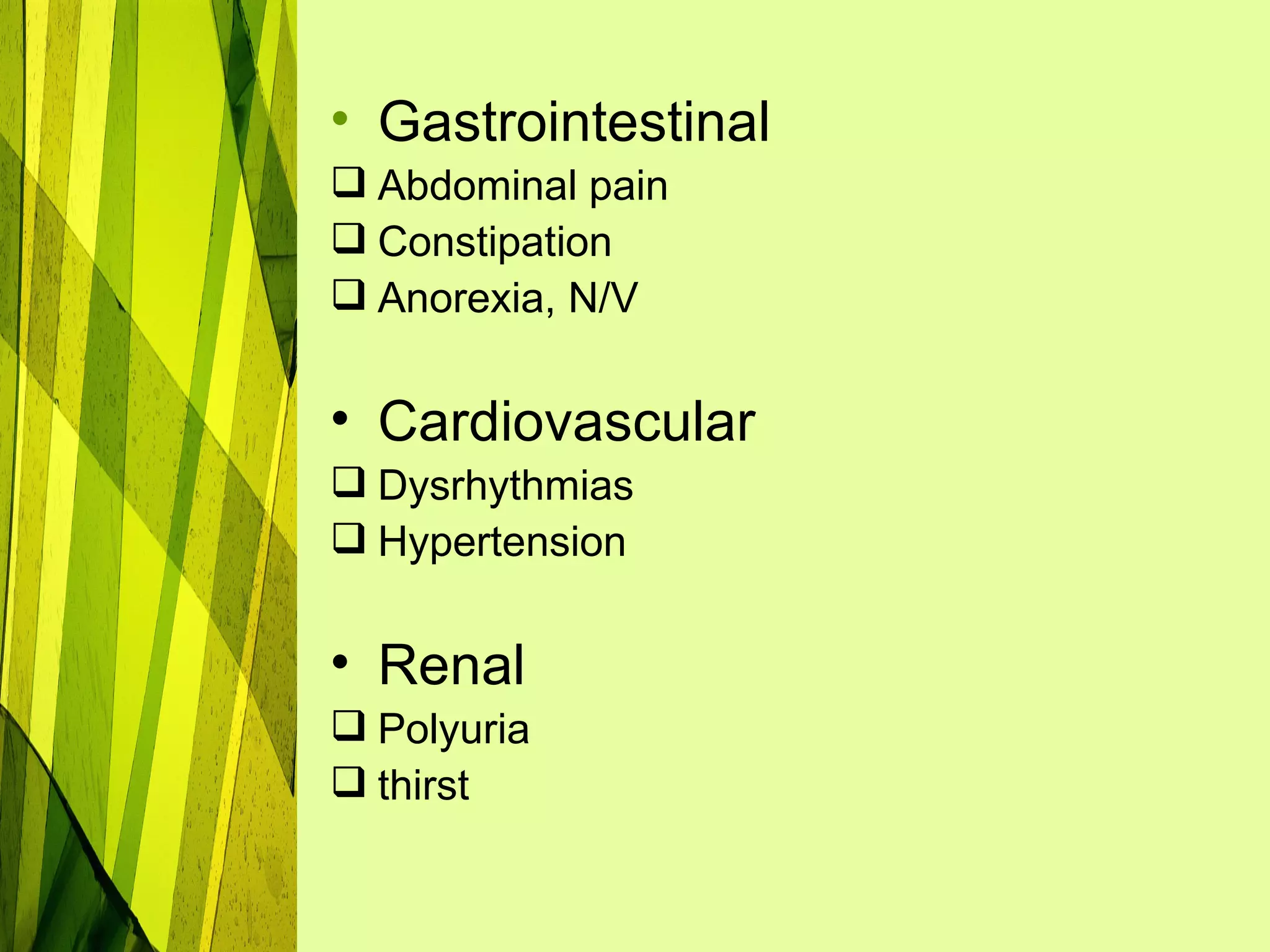
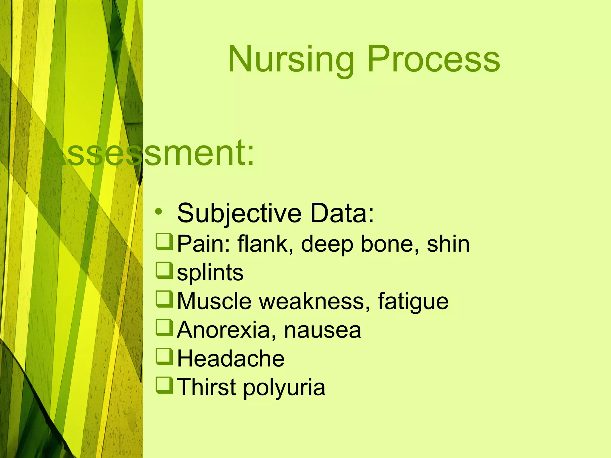
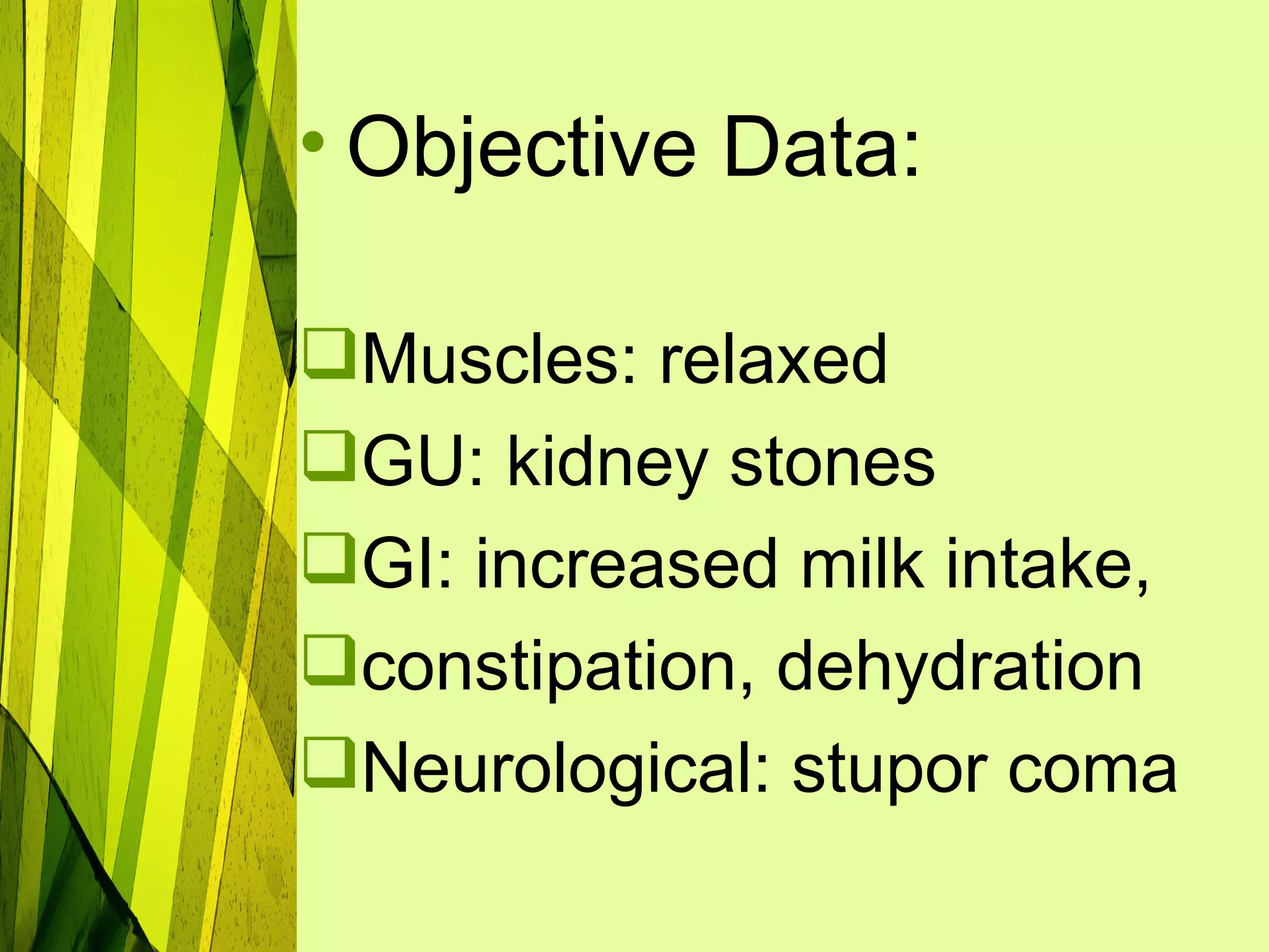
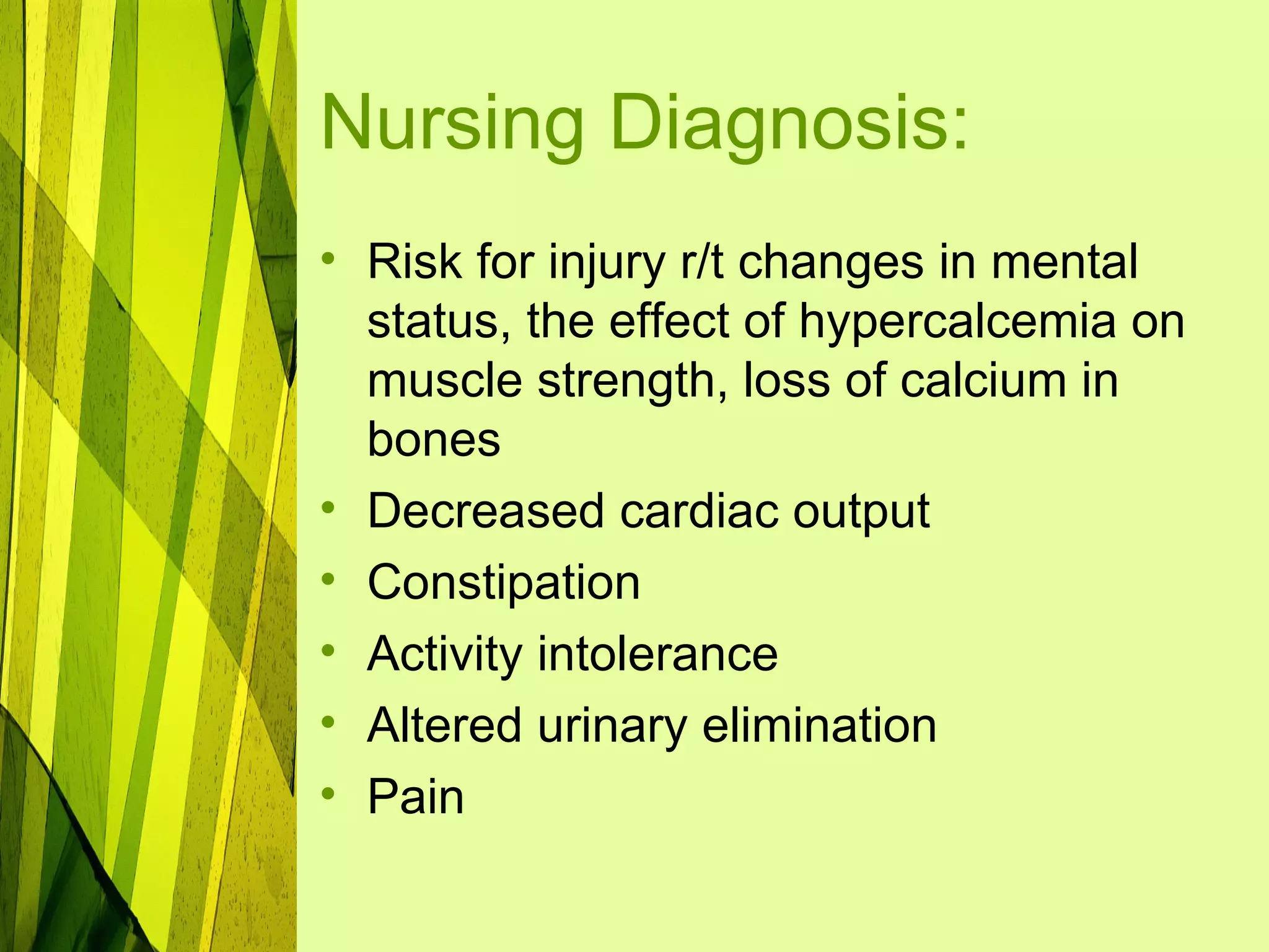
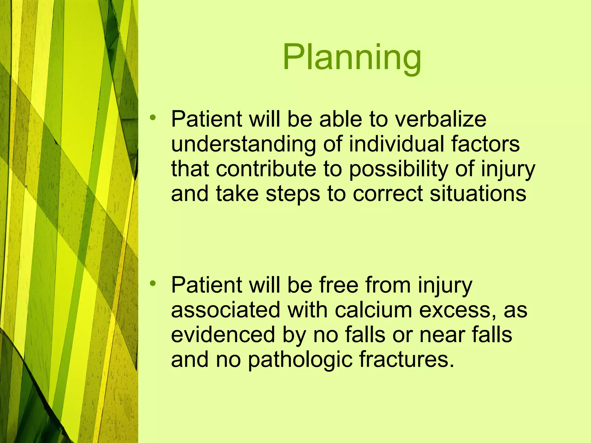
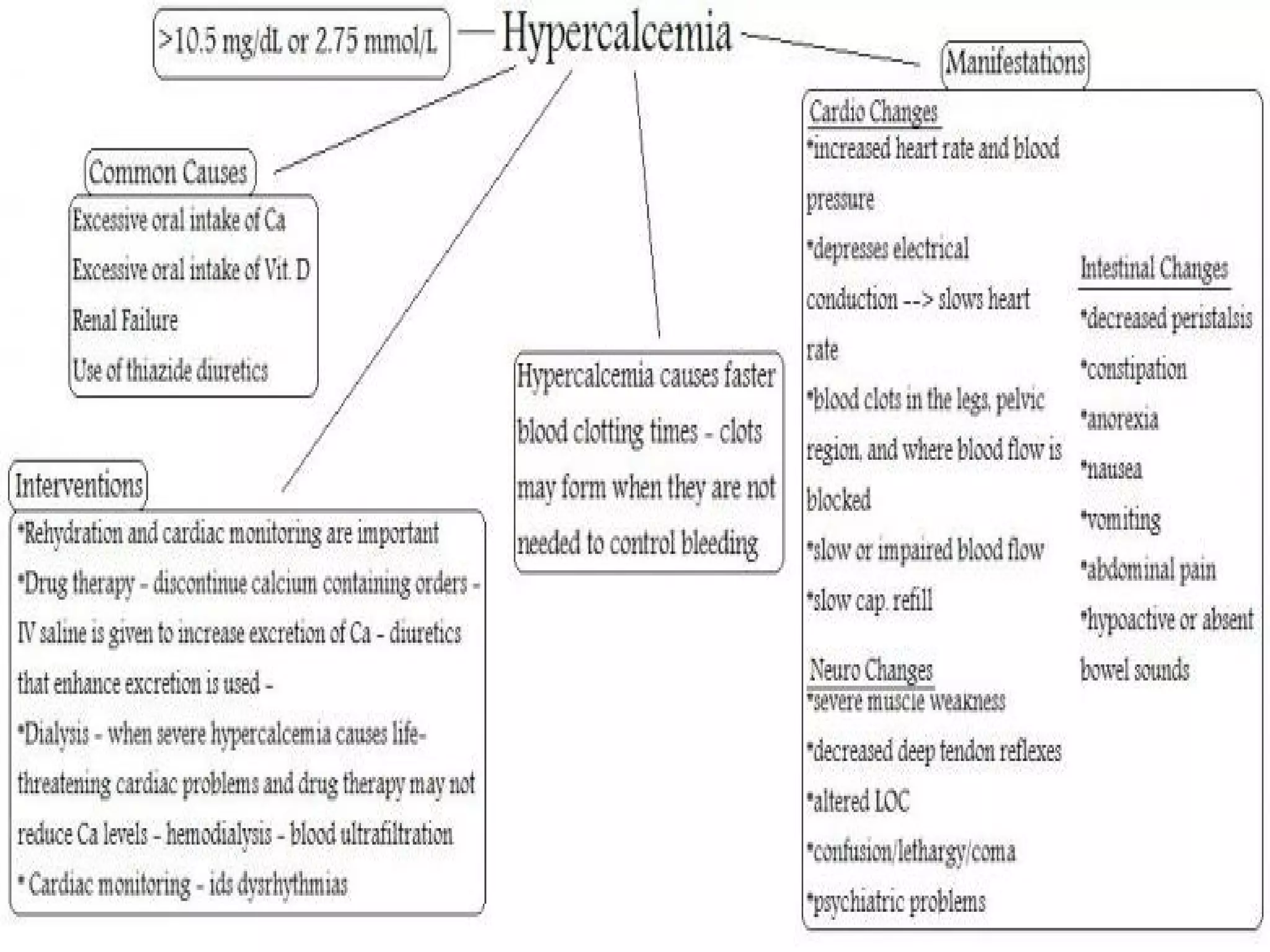
Calcium
Imbalances
Hypocalcemia
• Is a total serum level of less than
  8.5 mg/dl
• It can result for decreased total
  body calcium stores or low
  levels of extracellular calcium
  with normal amounts of Calcium
  stored in bones.
Pathophysiology
A lack of PTH results in inactivity of osteoclasts and a
consequent fall in serum calcium levels. Nerve fibers
become more and more excitable and may discharge
spontaneously, causing muscles to twitch and to go
into spasms or even tetany. Spasms of the muscles
of the larynx interfere with respiration and may lead
to death. During hypocalcemia, the bone is stimulated
to release calcium, which makes the bone
osteoporotic and subject to fracture. Hypocalcemia
increases capillary permeability; causes
neuromuscular excitability of skeletal, smooth, and
cardiac muscles; and decreases blood coagulation,
which results in bleeding. Severe hypocalcemia
causes neuromuscular excitability that result in
tetany. If it is untreated, convulsions and death can
occur. Acute hypocalcemia may cause cardiac
insufficiency and cardiac dysrhythmias.
Risk Factors:
• People who have had
  parathyroidectomy
• Older adults (especially women)
• People with lactose intolerance
• Alcoholic people
Causes:
•   Parathyroidectomy
•   Acute Pancreatitis
•   Inadequate dietary intake
•   Lack of sun exposure
•   Lack of weight bearing exercise
•   Drugs: Loop diuretics, calcitonin
•   Hypomagnesemia, alcohol
    abuse
Manifestations:
• 2 signs indicate hypocalcemia:
Chvostek’s Sign
     -is the contraction of the facial
  muscle that is produced by tapping
  the facial nerve in front of the ear.
Trosseau’s Sign
     -is a carpal spasm that occurs
  by inflating a BP cuff on the upper
  arm to 20mmHg greater than
  systolic pressure for 2-5 mins.
• Neuromuscular signs:

 Tetany and Convulsions – most
 serious manifestation; tonic
 muscular spasms.
 Paresthesias
 Muscle spasms
 Laryngospasms
 Seizures
 Anxiety, confusion, psychosis
Collaborative Care:
• Management of
  hypocalcemia is directed
  toward restoring normal
  calcium balance and
  correcting the underlying
  cause.
Diagnostic Exams:
•   Total serum calcium
•   Serum albumin
•   Serum magnesium
•   Serum phosphate
•   Parathyroid hormone
•   ECG
Medical Management:
• Oral or intravenous calcium
Calcium Chloride
Calcium Gluconate
Calcium Lactate
Calcium Citrate
Calcium Gluceptate
Calcium Carbonate
Nursing Management:
• Assess IV site for patency. Don’t
  administer Calcium if there is a
  risk for leakage into the tissues.
• May be given by slow IV push
  (dilute with normal saline for
  injection prior to administration)
  or added to copatible parenteral
  fluids such as Normal Saline,
  Lactated Ringers, D5W
• Administer into the longest available vein.
• Continuously monitor ECG when
  administering IV calcium to clients taking
  digitalis due to increased risk of digitalis
  toxicity.
• Frequently monitor serum calcium levels
  and response to therapy.
• Administer oral calcium preparations 1-1.5
  hours after meals and at bedtime.
• Give calcium tablets with a full glass of
  water.
• Diet high in calcium:
cottage cheese
Cheese
Milk
Cream
Yogurt
ice cream
Spinach
Tofu
broccoli
Nursing Process

Assessment:
•   Subjective Data:
•   Fatigue
•   Tingling/numbness; fingers and
•   circumoral
•   Abdominal cramps
•   Palpitations
•   •Dyspnea
• Objective Data:

 Muscle spasms: tonic muscles,
 carpopedal, laryngeal
 Neuromuscular: grimacing,
 hyperirritable facial nerves
 Tetany convulsions
 Orthopedic: osteoporosis fractures
 Cardiac: arrhythmias arrest
 GI: diarrhea
Nursing Diagnosis:
• Risk for injury r/t laryngospasm,
  cardiac dysrhythmias,
  convulsions, rapid administration
  of IV calcium, extravation of the
  medication into the
  subcutaneous tissue, increased
  neuromuscular irritability
  secondary to hypocalcemia
• Pain
• Diarrhea
• Hypocalcemia
• Altered nutrition, less than body
  requirements
• requirements
• Sensory-perceptual alteration
• (gustatory)
Planning:
• Patient will be able to verbalize
  understanding of individual factors
  that contribute to possibility of injury
  and take steps to correct situations


• Patient will be free from injury
  associated with calcium deficit, as
  evidenced by no falls or near falls
  and no pathologic fractures.
Interventions:
•   Monitor peripheral pulses and vital signs, especially the
    heart rate every hour to every four hours depending on
    the client’s condition. To assess baseline data.
•   Provide information regarding disease/condition that
    may result in increased risk of injury. To assist client to
    reduce or correct individual risk factor.
•   Evaluate individual’s response to violence in
    surroundings
•   If the client is receiving intravenous calcium, the nurse
    needs to monitor the IV site for infiltration or phlebitis
    every hour
•   Symptomatic hypocalcemia should be assessed by
    testing of the Chvostek’s and Trousseaus’ signs.
    Calcium chloride is extremely irritating to the
    subcutaneous tissue.
•   The serum calcium level should be closely monitored
    and changes reported. To check for increased
    neuromuscular excitability and tetany.
Hypercalcemia
• Is a serum calcium value greater
  than 10.0 mg/dL
• Usually results from increased
  absorption of calcium from the
  bones and intestines.
Pathophysiology
     Because calcium levels are increased,
there is a lesser gradient between the cell and
the serum. There is also an increased amount
of calcium in the cell. Therefore, the threshold
becomes more difficult to achieve and the cell
membrane becomes refractory
to depolarization. As a result, cardiac and
smooth muscle activity is decreased. Calcium
in the bloodstream impairs renal function and it
precipitates as a salt, forming renal stones.
Some cancer tumors destroy the bone,
whereas others such as lung and breast
cancers cause an ectopic PTH production.
Hypophosphatemia is a complication of
excessive PTH production that promotes
calcium retention. A shortened QT segment
and depressed T waves may be seen on ECG.
Causes:
•   Hyperparathyroidism
•   Cancer
•   Prolonged immobilization
•   Paget’s disease
•   Excess milk or antacid intake
•   Renal failure
Manifestations:
• Neuromuscular
 Muscle weakness
 Fatigue
 Decreased deep tendon reflexes


• Behavioral
 Personality changes
 Altered mental status
 Decreased LOC
• Gastrointestinal
 Abdominal pain
 Constipation
 Anorexia, N/V


• Cardiovascular
 Dysrhythmias
 Hypertension


• Renal
 Polyuria
 thirst
Complications:
•   Peptic ulcer
•   Pancreatitis
•   Kidney stones
•   Hypercalcemic crisis
Diagnostic Exams:
•   Serum electrolytes
•   Serum PTH levels
•   ECG
•   Sulkowitch’s urine test
Collaborative Care:
• The management of
  hypercalcemia focuses on
  correcting the underlying cause
  and reducing serum calcium
  levels.
Medical Management:
•   Loop Diuretics (Furosemide)
•   Calcitonin
•   Biphosphonates
•   Glucocorticoid drugs
Nursing Process

Assessment:
     • Subjective Data:
      Pain: flank, deep bone, shin
      splints
      Muscle weakness, fatigue
      Anorexia, nausea
      Headache
      Thirst polyuria
• Objective Data:

Muscles: relaxed
GU: kidney stones
GI: increased milk intake,
constipation, dehydration
Neurological: stupor coma
Nursing Diagnosis:
• Risk for injury r/t changes in mental
  status, the effect of hypercalcemia on
  muscle strength, loss of calcium in
  bones
• Decreased cardiac output
• Constipation
• Activity intolerance
• Altered urinary elimination
• Pain
Planning
• Patient will be able to verbalize
  understanding of individual factors
  that contribute to possibility of injury
  and take steps to correct situations


• Patient will be free from injury
  associated with calcium excess, as
  evidenced by no falls or near falls
  and no pathologic fractures.
Interventions
• decrease foods high in calcium;
• identify cause of imbalance
• give steroids as indicated
• diuretics as ordered
• isotonic
• saline IV
• Prevent injury: prevent pathological
  fractures (e.g. advanced cancer)
• prevent renal calculi by increasing
  fluid intake
Calcium imbalances

Calcium imbalances

  • 1.
  • 2.
    Hypocalcemia • Is atotal serum level of less than 8.5 mg/dl • It can result for decreased total body calcium stores or low levels of extracellular calcium with normal amounts of Calcium stored in bones.
  • 3.
    Pathophysiology A lack ofPTH results in inactivity of osteoclasts and a consequent fall in serum calcium levels. Nerve fibers become more and more excitable and may discharge spontaneously, causing muscles to twitch and to go into spasms or even tetany. Spasms of the muscles of the larynx interfere with respiration and may lead to death. During hypocalcemia, the bone is stimulated to release calcium, which makes the bone osteoporotic and subject to fracture. Hypocalcemia increases capillary permeability; causes neuromuscular excitability of skeletal, smooth, and cardiac muscles; and decreases blood coagulation, which results in bleeding. Severe hypocalcemia causes neuromuscular excitability that result in tetany. If it is untreated, convulsions and death can occur. Acute hypocalcemia may cause cardiac insufficiency and cardiac dysrhythmias.
  • 5.
    Risk Factors: • Peoplewho have had parathyroidectomy • Older adults (especially women) • People with lactose intolerance • Alcoholic people
  • 6.
    Causes: • Parathyroidectomy • Acute Pancreatitis • Inadequate dietary intake • Lack of sun exposure • Lack of weight bearing exercise • Drugs: Loop diuretics, calcitonin • Hypomagnesemia, alcohol abuse
  • 7.
    Manifestations: • 2 signsindicate hypocalcemia: Chvostek’s Sign -is the contraction of the facial muscle that is produced by tapping the facial nerve in front of the ear. Trosseau’s Sign -is a carpal spasm that occurs by inflating a BP cuff on the upper arm to 20mmHg greater than systolic pressure for 2-5 mins.
  • 11.
    • Neuromuscular signs: Tetany and Convulsions – most serious manifestation; tonic muscular spasms.  Paresthesias  Muscle spasms  Laryngospasms  Seizures  Anxiety, confusion, psychosis
  • 12.
    Collaborative Care: • Managementof hypocalcemia is directed toward restoring normal calcium balance and correcting the underlying cause.
  • 13.
    Diagnostic Exams: • Total serum calcium • Serum albumin • Serum magnesium • Serum phosphate • Parathyroid hormone • ECG
  • 14.
    Medical Management: • Oralor intravenous calcium Calcium Chloride Calcium Gluconate Calcium Lactate Calcium Citrate Calcium Gluceptate Calcium Carbonate
  • 15.
    Nursing Management: • AssessIV site for patency. Don’t administer Calcium if there is a risk for leakage into the tissues. • May be given by slow IV push (dilute with normal saline for injection prior to administration) or added to copatible parenteral fluids such as Normal Saline, Lactated Ringers, D5W
  • 16.
    • Administer intothe longest available vein. • Continuously monitor ECG when administering IV calcium to clients taking digitalis due to increased risk of digitalis toxicity. • Frequently monitor serum calcium levels and response to therapy. • Administer oral calcium preparations 1-1.5 hours after meals and at bedtime. • Give calcium tablets with a full glass of water.
  • 17.
    • Diet highin calcium: cottage cheese Cheese Milk Cream Yogurt ice cream Spinach Tofu broccoli
  • 18.
    Nursing Process Assessment: • Subjective Data: • Fatigue • Tingling/numbness; fingers and • circumoral • Abdominal cramps • Palpitations • •Dyspnea
  • 19.
    • Objective Data: Muscle spasms: tonic muscles,  carpopedal, laryngeal  Neuromuscular: grimacing,  hyperirritable facial nerves  Tetany convulsions  Orthopedic: osteoporosis fractures  Cardiac: arrhythmias arrest  GI: diarrhea
  • 20.
    Nursing Diagnosis: • Riskfor injury r/t laryngospasm, cardiac dysrhythmias, convulsions, rapid administration of IV calcium, extravation of the medication into the subcutaneous tissue, increased neuromuscular irritability secondary to hypocalcemia
  • 21.
    • Pain • Diarrhea •Hypocalcemia • Altered nutrition, less than body requirements • requirements • Sensory-perceptual alteration • (gustatory)
  • 22.
    Planning: • Patient willbe able to verbalize understanding of individual factors that contribute to possibility of injury and take steps to correct situations • Patient will be free from injury associated with calcium deficit, as evidenced by no falls or near falls and no pathologic fractures.
  • 23.
    Interventions: • Monitor peripheral pulses and vital signs, especially the heart rate every hour to every four hours depending on the client’s condition. To assess baseline data. • Provide information regarding disease/condition that may result in increased risk of injury. To assist client to reduce or correct individual risk factor. • Evaluate individual’s response to violence in surroundings • If the client is receiving intravenous calcium, the nurse needs to monitor the IV site for infiltration or phlebitis every hour • Symptomatic hypocalcemia should be assessed by testing of the Chvostek’s and Trousseaus’ signs. Calcium chloride is extremely irritating to the subcutaneous tissue. • The serum calcium level should be closely monitored and changes reported. To check for increased neuromuscular excitability and tetany.
  • 25.
    Hypercalcemia • Is aserum calcium value greater than 10.0 mg/dL • Usually results from increased absorption of calcium from the bones and intestines.
  • 26.
    Pathophysiology Because calcium levels are increased, there is a lesser gradient between the cell and the serum. There is also an increased amount of calcium in the cell. Therefore, the threshold becomes more difficult to achieve and the cell membrane becomes refractory to depolarization. As a result, cardiac and smooth muscle activity is decreased. Calcium in the bloodstream impairs renal function and it precipitates as a salt, forming renal stones. Some cancer tumors destroy the bone, whereas others such as lung and breast cancers cause an ectopic PTH production. Hypophosphatemia is a complication of excessive PTH production that promotes calcium retention. A shortened QT segment and depressed T waves may be seen on ECG.
  • 27.
    Causes: • Hyperparathyroidism • Cancer • Prolonged immobilization • Paget’s disease • Excess milk or antacid intake • Renal failure
  • 28.
    Manifestations: • Neuromuscular  Muscleweakness  Fatigue  Decreased deep tendon reflexes • Behavioral  Personality changes  Altered mental status  Decreased LOC
  • 29.
    • Gastrointestinal  Abdominalpain  Constipation  Anorexia, N/V • Cardiovascular  Dysrhythmias  Hypertension • Renal  Polyuria  thirst
  • 30.
    Complications: • Peptic ulcer • Pancreatitis • Kidney stones • Hypercalcemic crisis
  • 31.
    Diagnostic Exams: • Serum electrolytes • Serum PTH levels • ECG • Sulkowitch’s urine test
  • 32.
    Collaborative Care: • Themanagement of hypercalcemia focuses on correcting the underlying cause and reducing serum calcium levels.
  • 33.
    Medical Management: • Loop Diuretics (Furosemide) • Calcitonin • Biphosphonates • Glucocorticoid drugs
  • 34.
    Nursing Process Assessment: • Subjective Data:  Pain: flank, deep bone, shin  splints  Muscle weakness, fatigue  Anorexia, nausea  Headache  Thirst polyuria
  • 35.
    • Objective Data: Muscles:relaxed GU: kidney stones GI: increased milk intake, constipation, dehydration Neurological: stupor coma
  • 36.
    Nursing Diagnosis: • Riskfor injury r/t changes in mental status, the effect of hypercalcemia on muscle strength, loss of calcium in bones • Decreased cardiac output • Constipation • Activity intolerance • Altered urinary elimination • Pain
  • 37.
    Planning • Patient willbe able to verbalize understanding of individual factors that contribute to possibility of injury and take steps to correct situations • Patient will be free from injury associated with calcium excess, as evidenced by no falls or near falls and no pathologic fractures.
  • 38.
    Interventions • decrease foodshigh in calcium; • identify cause of imbalance • give steroids as indicated • diuretics as ordered • isotonic • saline IV • Prevent injury: prevent pathological fractures (e.g. advanced cancer) • prevent renal calculi by increasing fluid intake