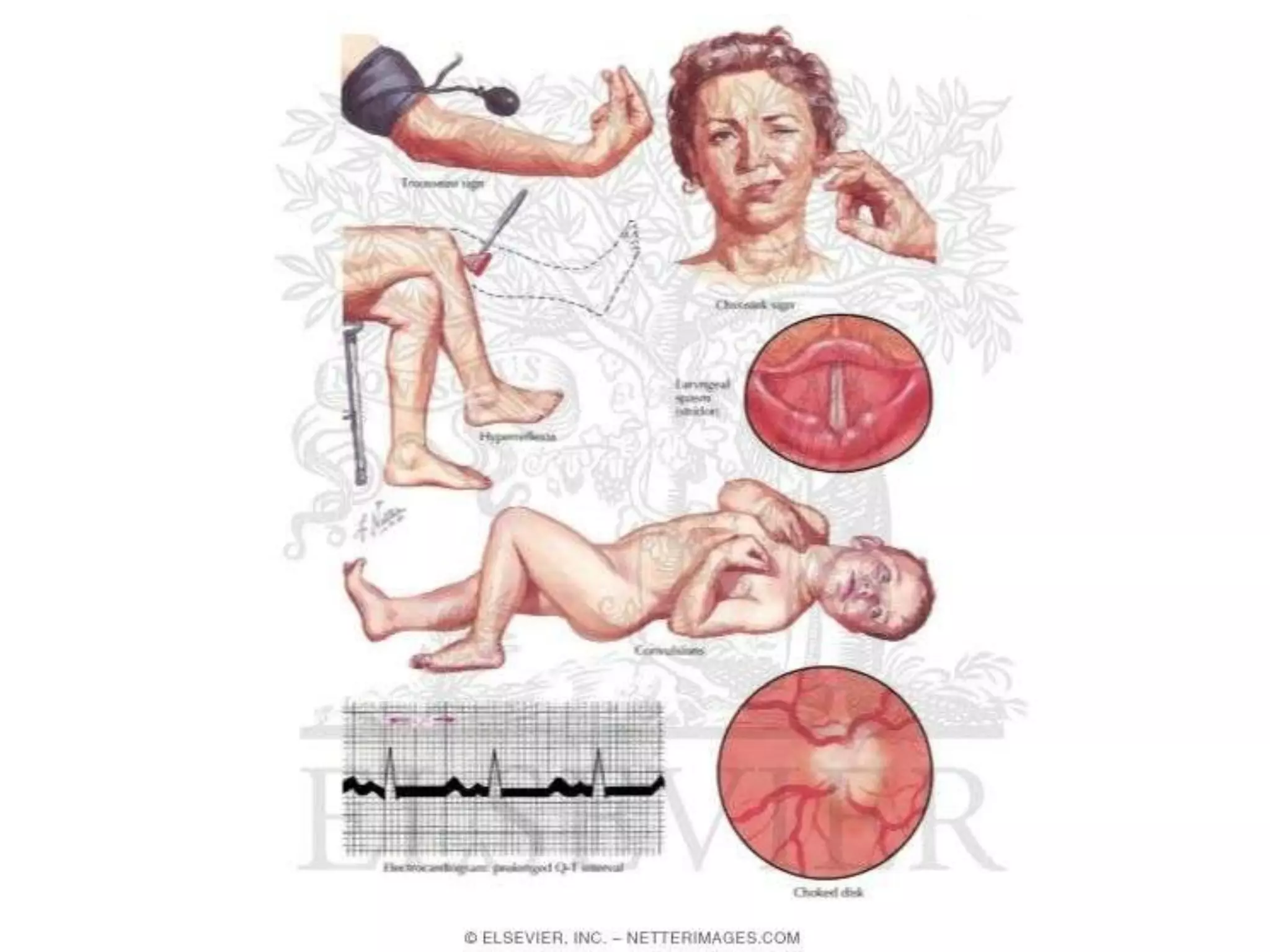
This document discusses hypocalcemia, defined as a serum calcium level below 8.5 mg/dl. It may be caused by low or high parathyroid hormone levels. Causes of low PTH include parathyroid gland agenesis, destruction, or dysfunction. Causes of high PTH include vitamin D deficiency, kidney disease impairing vitamin D activation, or drugs. Symptoms include increased neuromuscular excitability. Investigations include calcium, albumin, phosphorus, magnesium and PTH levels. Treatment involves vitamin D, calcium supplements, and magnesium as needed.
Hypercalcaemia is a common disorder we doctors from all faculties face in day to day clinical practice. This was a presentation done by me to give you an update regarding hypercalcaemia and it's management.
Explanation of what splenomegaly is in relation to its dimension deviation from normal spleen.Classification of splenomegaly according to it's size in adult and pediatric. The causes of splenomegaly along with the symptom that would manifest as a result of this anomaly. Lastly, diagnosis of splenomegaly
The Indian Dental Academy is the Leader in continuing dental education , training dentists in all aspects of dentistry and
offering a wide range of dental certified courses in different formats.
Indian Dental Academy: will be one of the most relevant and exciting training center with best faculty and flexible training programs for dental professionals who wish to advance in their dental practice,Offers certified courses in Dental implants,Orthodontics,Endodontics,Cosmetic Dentistry, Prosthetic Dentistry, Periodontics and General Dentistry.
4. LOW PARATHYROID HORMONE LEVELS
1. Parathyroid agenesis
a) Isolated
b) DiGeorge Syndrome
2. Parathyroid destruction
a) Surgical
b) Radiation
c) Infiltration by metastases or systemic diseases
d) Autoimmune
3. Reduced Parathyroid function
a) Hypomagnesemia
b) Activating CaSR mutations
5. HIGH PARATHYROID HORMONE LEVELS ( Secondary Hyperparathyroidism)
1. Vitamin D deficiency or impaired 1,25(OH)2*D production/action
a) Nutritional vitamin D deficiency
b) Renal insufficiency with impaired 1,25(OH)2*D
production
c) Vitamin D resistance
2. Parathyroid hormone resistance syndromes
a) PTH receptor mutations
b) Pseudohypoparathyroidism
3. Drugs
a) Calcium chelators
b) Inhibitors of bone resorption
c) Altered vitamin D metabolism (Phenytoin, Ketoconazole)
4. Miscellaneous
a) Acute Pancreatitis
b) Acute Rhabdomyolysis
c) Hungry bone syndrome
d) Osteoblastic metastases
6. FUNCTIONAL CLASSIFICATION
PTH Absent
1. Hereditary hypoparathyroidism
2. Acquired hypoparathyroidism
3. Hypomagnesaemia
PTH Ineffective
ACTIVE VITAMIN D LACKING
a) Dietary intake or sunlight
b) Defective metabolism:
c) Anticonvulsant therapy Chronic renal failure
d) Vitamin D–dependent rickets type I
ACTIVE VITAMIN D INEFFECTIVE
a. Intestinal malabsorption Pseudohypoparathyroidism
b. Vitamin D–dependent rickets type II
PTH Overwhelmed
1. Severe, acute hyperphosphatemia
2. Osteitis fibrosa after parathyroidectomy
3. Tumour lysis
4. Acute renal failure
5. Rhabdomyolysis
7. PATHOPHYSIOLOGY
Decrease in extracellular Ca2*+
The membrane potential on the outside becomes less negative
Less amount of depolarisation is required to initiate action potential
Increased excitability of muscle and nerve tissue
8. CLINICAL FEATURES
Onset
1. Acute hypocalcemia
i. Critically ill patients
ii. Drugs: Citrates, ACEI’s
2. Transient hypocalcemia
i. Sepsis, Burns, Acute renal failure, transfusions
ii. Drugs: Protamine, Heparin, Glucagon
3. Chronic hypocalcemia
12. HERIDITARY
Isolated With associated features
Autosomal Dominant Hypocalcemic Hypercalciuria
Barrter Syndrome type V
13. With associated features
Autosomal dominant Autosomal recessive Mitochondrial Autoimmune
Kenney-Caffey MELAS Polyglandular
DiGeorge Syndrome syndrome
Autoimmune
Kearns-Sayre Type I
HDR Syndrome Sanjad-Sakati syndrome deficiency
syndrome
14. ACQUIRED HYPOPARATHYROIDISM
• Inadvertent surgical removal
– Even if parathyroids retained, Hypoparathyroidism
sometimes resulted?
– Surgery for hyperparathyroidism – How much to
remove?
• Radiation induced
• Haemochromatosis
16. TREATMENT (ACQUIRED AND
HEREDITARY HYPOPARATHYROIDISM)
1. Vitamin D [40,000-120,000 U/d] or
1,25(OH)2*D3*(calcitriol) [0.5-
1microgm/day] ?
2. High oral calcium intake.
3. Thiazide diuretics?
(Hydrochlorothizide 12.5-50mg)
17. HYPOMAGNAESEMIA
• Effects of magnesium on PTH secretion?
• Severe hypomagnaesemia causes
hypocalcemia? (paradox?)
18. Chronic hypomagnesaemia
Intracellular magnesium deficiency
Interferes with secretion and peripheral response to PTH
Mechanism: Effects on adenylate cyclase proposed
19. TREATMENT
• Severe hypomagnaesemia (Parenteral
treatment)
IV MgCl2*, continuous infusion, 50 mmol/d
(GFR↓, 50-75% reduction in dose)
• During therapy monitor S. Mg every 12-24hr
23. CHRONIC RENAL FAILURE
Impaired production of 1,25(OH)2*D
Hypocalcemia
Secondary Hyperparathyroidism
Hyperphosphtemia (later stages)
FGF-23 increases
24. • Hyperphosphatemia lowers the blood calcium
1. Extraosseus deposition of calcium and
phosphate
2. Impairment in bone resorbing action of PTH
3. Reduction in the production of 1,25(OH)2*D
26. VITAMIN D DEFICIENCY
• Inadequte diet and/or exposure to sunlight
• Investigations my show: ↓ vitamin D
metabolites, ↓ calcium, ↑ PTH, ↑phosphate
• Hypocalcaemia itself causes steatorrhoea
• Treatment: Various metabolites can be given
depending on the disorder
27. DEFECTIVE VITAMIN D METABOLISM
1. Anticonvulsant therapy: Enzyme induction
2. Vitamin D-dependant rickets type 1:
a) Autosomal recessive
b) Mutations in genes coding 25-(OH)D-1α-
hydroxylase
c) Hypocalcemia, hyperphosphatemia, Hyperparath
yroidism, osteomalacia, ↑ ALP
d) Reversible on calcitriol supplementation
28. 3. Vitamin D-dependant rickets type 1:
a) End organ resistance to active metabolite
b) Mutations in Vitamin D receptor
c) More severe, associated partial or total alopecia.
d) Plasma 1,25(OH)2*D are elevated
Treatment: Regular calcium infusions