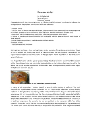
Vip Call Girls Anna Salai Chennai 👉 8250192130 ❣️💯 Top Class Girls Available
Â
C section
1. Caesarean section
Dr. Rekha Pathak
Senior scientist
Division of surgery
Caesarean section is also commonly termed as C-Section in which uterus is exteriorized to take out the
young one from the pregnant dam. Its indications are as follows:-
1. Uterine inertia
2. Various types of obstructive dystocia (for eg: Emphysematous fetus, Oversized fetus, small pelvic anal
of the dam, difficulty in parturition due to pelvic fractures, position and posture dystocia etc.)
3. Rupture of uterus (may be due to injection or excessive manipulation of the fetus).
4. Animal in highly compromised condition like pregnancy toxemia, weak prostrated dam unable to
show labor etc.
5. In the mares twin pregnancy is also an indication for C-Section.
6. Uterine torsion.
7. Incomplete Cervical dilatation.
It is important to choose a clean and bright place for the operation. The air borne contamination should
be strictly avoided and utmost case should be taken to prevent the post-operative complications and
septicemia because the successful outcome of the operation is mainly dependent on the strict asepsis
which is followed.
Site of operation varies with the type of species. In dogs the site of operation is ventral and line incision
behind the umbilicus, in the cow a vertical or oblique incision on the left lower flank is preferred (for the
reason that on the left side the intestinal interference is least, although rumen is present on the higher
flank,) this site is chosen. (Fig. 1)
Fig. 1: left lower flank incision in cattle
In mares, a left paramedian incision (caudal) or ventral midline incision is preferred. The small
ruminants like goat and ewes, the site chosen are just as n cattle. For left lower flank incisions animals
are taken on the right lateral recumbency and for ventral midline incision they are taken on the dorsal
recumbency. It is very important to note that the animals should be comfortably casted at a height for
the operator so that surgeon is not only comfortable of his posture but since this operation takes a little
longer and cumbersome in large animals specially buffaloes. There should be a skilled team consisting of
at least two surgeons on the operative site and one assistant on the instrument table. Two skilled
assistants should take care of the fluid (intravenous) and other drugs requirement of the casted animal.
The animal when comes should be checked for dehydration and intravenous fluids rehydration and we
2. should make every attempt to stabilize such animals. Dexamethasone should be administered along
with antibiotics prior to the surgery.
Surgical Anatomy:
Dog: - The gravid uterus has dilated horns in the shape of –Y which contain the fetuses and lies on the
ventral abdominal floor extended up to the level of stomach towards end of gestation.
Cow: - The gravid uterus may lie directly up on the right abdominal floor or within the supraomental
space with the intestine concealed on the right by superficial and deep parts of greaten omentum.
After giving the incision at the left lower flank site, the uterus is usually brought outside with the help of
grasping and feeling the fetal parts. The uterus may be exposed for caesarean section by simply drawing
the greater omentum forward. If the fetus in mummified , instead of giving a lower flank incision , upper
flank incision is usually preferred whereas in full term gestation because the uterus due to its weight is
found on the ventral floor of the abdomen it is better to go for the lower flank incision.
Anesthesia:
1. In dogs where pups are found to be alive by ultrasonographic or Doppler examination, thiopental or
xylazine is not preferred for the reason of respiratory depression of pups but a combination of epidural
and local infiltration with 2 % Lignocaine can be given.
We can also give a combination of Diazepam and ketamine intravenously with the dose rate of 0.5mg/kg
body weight and 5.0 mg/kg body weight respectively. In cattle or buffalo, slight sedation may be
needed sometimes along with local infiltration on the line of incision. In mares, inhalation anesthesia
with inhalant anesthetic agents is preferred.
Surgical anatomy:
Midline incision behind the umbilicus includes incision on the ventral aponeurosis of the muscles (white
line). The venterolateral oblique incision is usually used in cows and buffaloes with the animals
controlled in lateral recumbency and hind limbs extended caudally. Incision is just in front of stifle and
extends cranioventrally in a slight oblique direction. The structures invaded are skin, subcutaneous
fascia, and combined aponeurosis of the two oblique muscles which forms the external sheath of the
rectus abdominis, transverse abdominis and peritoneum.
Paramedian incision is preferred in mares and the structures include skin, fascia, external rectus sheath,
rectus abdominis muscle, internal rectus sheath and peritoneum.
Dogs: Premedication with atropine sulfate @ 0.04 mg/kg body weight followed by diazepam @ 0.5mg/
kg body weight and ketamine @ 2-5 mg/ kg body weight of the animal intravenously. This regimen is
followed if the fetuses are found to be alive by other examinations. But if there is a certainty that
fetuses are dead or infected then atropine sulphate is followed by thiopentone sodium @ 8- 10 mg / kg
body weight. A long incision on the linea-alba is given in medium and small sized dogs to exteriorize the
uterus. But if the animal’s size is more than 25 kg we should prefer the oblique incision on flank or
paramedian approach to avoid postoperative dehiscence and hernia.
3. The cow is controlled in standing or right lateral recumbency and al long skin incision is given by saving
the left subcutaneous abdominal vein. We can either give the local infiltration or epidural anesthesia
with 2% Lignocaine. In bitch, the bifurcation of uterine body is first visualized and incision is made over it
in order to enable the milking of pups (squeezing the pups out from the horns) from both the horns is
easy. The fetuses are removed along with fetal membrane one by one. The umbilical vessels are ligated
and cut and the new born are handed over o the helper or nurse for resuscitation. They are wiped with
mops and assisted for artificial respiration if fail to breathe. The head is lowered to permit drainage of
fluids from the upper respiratory tract.
In cow the uterine incision should follow the longitudinal line of greater curvature of the uterus. (Fig 2
and Fig 3)
Fig 2: Uterus is packed off from the abdominal cavity
Fig 3: Fetus being taken out from the uterus
Forelimbs or hind limbs are grasped depending upon the presentation and the fetus is taken out from
the uterus. The calf should be care by the assistants. It should be cleaned, dried, cleared off the mucus
from the nostrils. The umbilical cord is ligated far enough from the navel and cut so that it contracts.
Antiseptic solution like povidone iodine or Tr. Iodine is then applied over it to present the infective the
removal of after births or placenta is also important. (Fig 4)
Fig 4: Removal of after births or placenta
If it is easily removed by gentle traction it should be removed or otherwise, it should not be pulled with
force since chances of caruncular bleeding is strong which may be fatal to the dam.
If such bleeding is encountered in the dam, then we can counteract it by giving oxytocin which largely
shrinks the uterus and stops the bleeding. Antibiotics can be instilled into the uterus as common
procedure for all the species before closure. The uterine incision is cleaned with gauze and closed by a
4. double row of Lamberts sutures using chromic catgut size 2-0 or 3-0 in bitch and size 2 in cattle and
buffaloes. (Fig 5)
Fig 5: closure of uterine incision
Fig 6: closure of abdominal incision
Abdominal incisions are sutured in the usual manner, closing the peritoneum, muscle and skin
respectively. (Fig 6)
Note: utmost care should be taken to avoid the spillage of uterine contents into the peritoneal /
abdominal cavity for the successful outcome of surgery. In case such spillage occurs, it should be lavaged
with sterile normal saline containing non- irritant antibiotics to counteract the infection, reduce the
chances of postoperative adhesions and infection.
The uterine torsion in case of cattle and buffaloes should be then corrected. 50-60 units of oxytocin
hasten the uterine involution. A 5% solution of dextrose and normal saline solution should be invariably
included in the schedule as most deaths have hypoglycemia and hypochloraemia.
Postoperative care:
The mother and the new born should be returned to clean and comfortable environment. If the
hemorrhage is excessive, pituitrin and calcium gluconate may be given intravenously in bitches.
Penicillin or some other antibiotics should be given. If the condition of patient is poor administration of
blood or plasma expanders and corticosteroids is considered.
Puppies should be given to the mother as soon as she is ready to take care of them. Similarly the new
born calf should also be allowed to suckle and stand on its legs. It not only provides the nourishment to
the puppies but will also stimulate the uterine contractions thereby reducing any risk of placental
retention or endometritis. The skin sutures should be removed in 10- 12 days after surgery or after the
healing is complete.