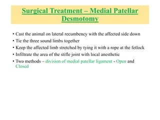
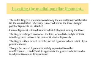
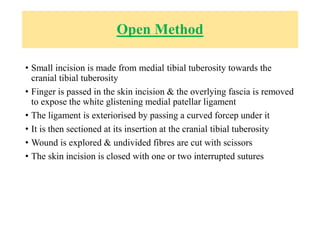
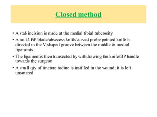
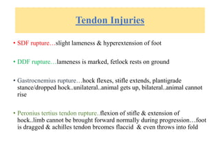
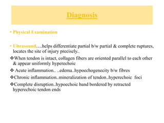
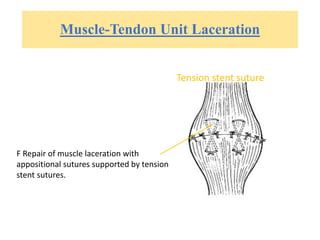
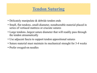
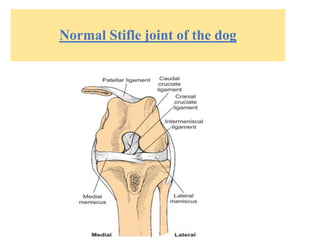
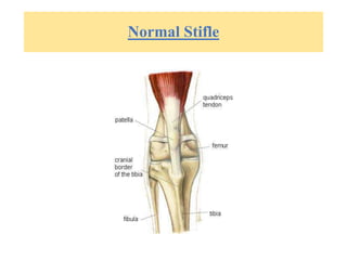
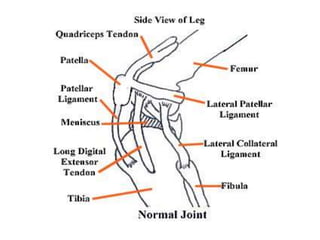
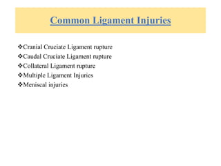
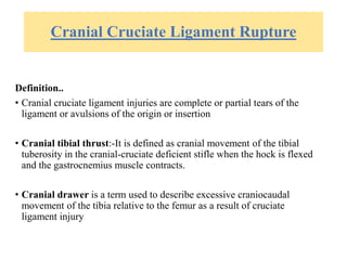
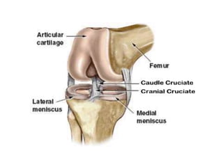
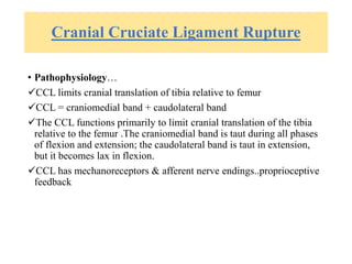
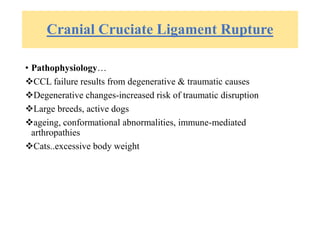
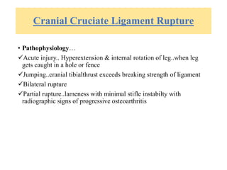
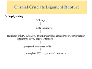
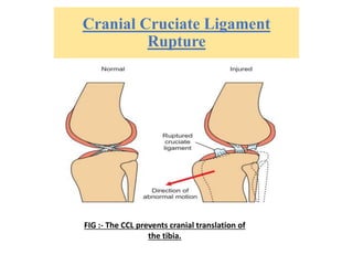
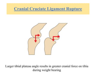
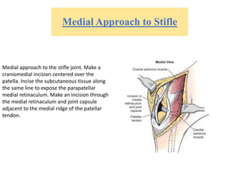
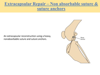
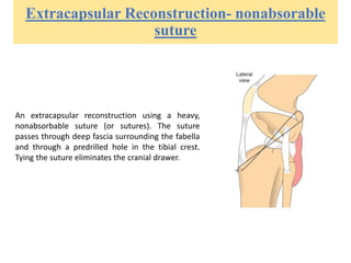
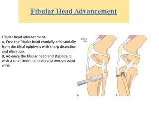
Various Affections Of Ligaments And
Tendons And Their Treatment
ICAR-IVRI Division Of VETERINARY SURGERY AND RADIOLOGY
Presented by
Dr. Rekha Pathak and Dr Anmol Dhiman
Ligament Injuries
Normal Stifle joint of the dog
Normal Stifle
Common Ligament Injuries
Cranial Cruciate Ligament rupture
Caudal Cruciate Ligament rupture
Collateral Ligament rupture
Multiple Ligament Injuries
Meniscal injuries
Cranial Cruciate Ligament Rupture
Definition..
• Cranial cruciate ligament injuries are complete or partial tears of the
ligament or avulsions of the origin or insertion
• Cranial tibial thrust:-It is defined as cranial movement of the tibial
tuberosity in the cranial-cruciate deficient stifle when the hock is flexed
and the gastrocnemius muscle contracts.
• Cranial drawer is a term used to describe excessive craniocaudal
movement of the tibia relative to the femur as a result of cruciate
ligament injury
Cranial Cruciate Ligament Rupture
• Translation:-it is defined as movement of a bone parallel to an axis
or a plane
• Pivot shift:-it is the cranial movement of the tibia combined with
internal rotation of the tibia
Cranial Cruciate Ligament Rupture
• Tibial plateau angle (TPA):-it is the angle between a line
perpendicular to the long axis of the tibia and a line parallel to the
tibial plateau
• Medial Butress:-it is a palpable thickening at the medial aspect of the
stifle
Cranial Cruciate Ligament Rupture
• Imbrication:-is defined as the tightening of a structure
• Isometry:-is defined as maintaining equal distance or tension
throughout a range of motion
GENERAL CONSIDERATIONS AND
CLINICALLY RELEVANT PATHOPHYSIOLOGY
• The CCL is divided into
• craniomedial and caudolateral bands,
• CCL functions primarily to limit cranial translation of the tibia
relative to the femur .
• The CCL also limits internal rotation of the tibia; as the stifle is
flexed, the cranial and caudal cruciate ligaments twist on each other,
limiting the degree of internal rotation of the tibia relative to the
femur.
• Interaction of the cranial and caudal cruciate ligaments during flexion
also provides a limited degree of varus-valgus support to the flexed
stifle joint.
• NOTE - The craniomedial band of the CCL is the primary check
against craniocaudal drawer motion.
• Acute injury is most commonly associated with the hyperextension and
internal rotation of the leg that occurs when a dog’s foot becomes caught in
a hole or fence.
• Jumping can also cause cruciate ligament rupture if the force of the cranial
tibial thrust exceeds the breaking strength of the ligament.
• ligament degeneration, even repetitive normal activities can cause
progressive rupturing of the ligament.
• Partial rupture of the CCL results in lameness with minimal detectable stifle
instability and progressive radiographic signs of osteoarthritis.
• Partial rupture generally proceeds to complete ligament rupture with time.
• CCL injury with stifle instability is part of a cascade of events that
include progressive osteoarthritis and medial meniscus injury. Stifle
instability results in synovitis, articular cartilage degeneration,
periarticular osteophyte development, and capsular fibrosis.
DIAGNOSIS
• Clinical Presentation
• History. Acute injury, chronic injury, and partial tears are three
clinical presentations associated with CCL injury.
• Patients with acute tears show a sudden onset of non weight-bearing
or partial-weight-bearing lameness.
• Lameness usually decreases somewhat within 3 to 6 weeks after
injury without treatment, particularly in patients that weigh less than
10 kg.
• An exception is dogs with associated meniscal injury. These dogs
usually maintain a minimally weight bearing or non-weight-bearing
lameness until surgical intervention takes place.
Physical Examination Findings
• A positive tibial compression test may be easier to elicit than a
positive drawer test.
• Patients with chronic tears may have thigh muscle atrophy.
• When the joint is extended from a flexed position, a clicking or
popping may be heard and felt; this is commonly associated with a
meniscal tear. However, the absence of joint noise does not eliminate
the possibility of meniscal injury. An enlargement along the medial
joint surface (medial buttress) often can be palpated and is caused by
osteophyte formation along the trochlear ridges.
• In chronic case this popping sound is absent as there is fibrosis.
Cranial Cruciate Ligament Rupture
• Pathophysiology…
CCL limits cranial translation of tibia relative to femur
CCL = craniomedial band + caudolateral band
The CCL functions primarily to limit cranial translation of the tibia
relative to the femur .The craniomedial band is taut during all phases
of flexion and extension; the caudolateral band is taut in extension,
but it becomes lax in flexion.
CCL has mechanoreceptors & afferent nerve endings..proprioceptive
feedback
Cranial Cruciate Ligament Rupture
• Pathophysiology…
CCL failure results from degenerative & traumatic causes
Degenerative changes-increased risk of traumatic disruption
Large breeds, active dogs
ageing, conformational abnormalities, immune-mediated
arthropathies
Cats..excessive body weight
Cranial Cruciate Ligament Rupture
• Pathophysiology…
Acute injury.. Hyperextension & internal rotation of leg..when leg
gets caught in a hole or fence
Jumping..cranial tibialthrust exceeds breaking strength of ligament
Bilateral rupture
Partial rupture..lameness with minimal stifle instabilty with
radiographic signs of progressive osteoarthritis
Cranial Cruciate Ligament Rupture
• Pathophysiology…
CCL injury
stifle instability
meniscus injury, synovitis, articular cartilage degeneration, periarticular
osteophyte devp, capsular fibrosis
progressive osteoarthritis
complete CCL rupture and lameness
Cranial Cruciate Ligament
Rupture
FIG :- The CCL prevents cranial translation of
the tibia.
Cranial Cruciate Ligament Rupture
Larger tibial plateau angle results in greater cranial force on tibia
during weight bearing
Cranial Cruciate Ligament Rupture
• Diagnosis
no specific gender, age or breed prediliction
young, active, large breeds
history..acute injury, chronic injury, partial
Acute tears..sudden onset of non-weight-bearing or partial-weight-
bearing lameness
Lameness usually improves 3-6 weeks after injury without treatment
(<10kg)
Cranial Cruciate Ligament Rupture
• Diagnosis…
 History..
 Chronic injury:-prolonged weight bearing lameness
 Difficulty in rising and sitting, dog sits with affected limb out to
the side of the body
 Lameness is typically worse after exercise or after sleeping
Cranial Cruciate Ligament Rupture
• Diagnosis…
History..
partial tears.. Mild weight-bearing lameness that resolves with
rest..stage persists for months
ligament continues to wear..degeneration..progressive
osteoarthritis..pronounced lameness not resolving with rest
Cranial Drawer Test
To examine for cruciate ligament
injury, place the thumb of one hand
over the lateral fabella and the index
finger over the patella. Stabilize the
femur with this hand. Place the
thumb of the opposite hand caudal to
the fibular head with the index finger
on the tibial tuberosity. With the stifle
flexed and then extended, attempt to
move the tibia cranially and distally to
the femur.
• The examiner must test for signs of instability with the stifle joint in
extension, in the normal standing angle, and in 90 degrees of flexion.
• If the degree of movement is questionable, comparison with the
opposite limb is helpful.
• A positive test result is craniocaudal movement beyond the 0 to 2 mm
found in normal stifle joints. In younger patients, normal craniocaudal
translation may be as great as 4 to 5 mm, but ligament rupture is
confirmed by the absence of an abrupt stop at the cranial extent of
movement.
• If a partial tear is present, the cranial drawer sign may reveal only 2
to 3 mm of instability when the test is done with the stifle flexed and
no instability with the stifle in extension
Tibial Compression Test
 Performed with the patient standing or in
lateral recumbency.
 To perform a tibial compression test, grasp the
distal quadriceps with one hand from the
cranial surface so that the index finger can be
extended down over the patella and the tip of
the finger is on the tibial crest.
 Use the second hand to grasp the foot at the
metatarsal region from the plantar surface.
With the limb in moderate extension, flex the
hock with the lower hand and prevent stifle
flexion with the upper hand.
 Feel for cranial movement of the tibia, which is
an indication of cruciate ligament damage.
 The technique should be repeated in different
degrees of stifle flexion to test for a partial CCL
rupture.
Diagnostic Imaging
Lateral radiograph of a dog
with a chronic cruciate
ligament rupture. Note the
loss of fat pad definition and
distention of the caudal
joint capsule. Also note the
osteophyte formation along
the trochlear ridge and the
subchondral bone sclerosis
of the tibial plateau.
Arthroscopy
• A large percentage of the surface of the cruciate ligament can be
examined arthroscopically for gross tears, fibrillation, or discoloration
associated with cruciate damage.
The caudal cruciate ligament (CdCL) is intact. A small cranial medial
band is all that remains of the cranial cruciate ligament (CaCL).
Laboratory Findings
• If joint palpation and radiographs are inconclusive synovial fluid
examination are helpful.
• Increased amounts of joint fluid and a two- to threefold increase in
cell numbers (6000 to 9000 WBC/µl— primarily mononuclear cells)
indicate secondary DJD
Cranial Cruciate Ligament Rupture
• Differential Diagnosis
Mild joint sprains, muscle strains, patellar luxation, caudal cruciate
ligament injury, long digital extensor tendon avulsion, primary or
secondary arthritis, immune-mediated arthritis
Medical management conservative treatment is tolerated in dogs less
than 10 kg in larger than 10 kg, lameness improves, but pre-injury
activity does not return.
Lameness often decreases within 6 weeks in small patients managed
conservatively (i.e., rest and antiinflammatory drugs).
Cranial Cruciate Ligament Rupture
• SURGICAL TREATMENT…
Intracapsular Reconstruction:-
Extracapsular Reconstruction
Imbrication techniques
Fibular Head Advancement technique
Tibial Plateau Leveling Osteotomy (TPLO)
Tibial Wedge Osteotomy (TWO)
Tibial Tuberosity Advancement (TTA)
• Intracapsular reconstruction consists of passing autogenous tissue
through the joint using the “over-the-top” method or passing the
tissue through predrilled holes in the femur or tibia or both.
• The most commonly used material is autogenous fascia lata.
• An advantage is that they most closely mimic the original position.
• Disadvantages of these techniques are their invasiveness and the
tendency of the graft to stretch or fail.
Lateral Approach to Stifle -1
• Extracapsular reconstruction involves placement of sutures outside
the joint or redirection of the lateral collateral ligament.
• Extracapsular reconstruction with sutures is often incorrectly referred
to as imbrication sutures
• Extracapsular sutures may also be secured from bone anchors or bone
tunnels.
• Materials used in extracapsular sutures include monofilament nylon
or fishing or leader line, manufactured orthopedic wire, or braided
orthopedic suture.
• Polyethylene sutures are stronger, stiffer and elongate less than nylon
leader.
• Imbrication for cruciate ligament rupture is performed by tightening
the fascia lata either through an advancement technique using a vest-
over-pants suture pattern or by partial excision and closure.
• The fibular head advancement technique advances the insertion of the
lateral collateral ligament to prevent abnormal drawer and internal
rotation of the tibia.
• Lateral approach to the stifle joint. A, Make a craniolateral skin
incision centered over the patella. Incise the subcutaneous tissue
along the same line to visualize the septum between the superficial
leaf of the fascia lata and the biceps femoris muscle proximally and
the lateral retinaculum distally.
Lateral Appproach to Stifle -2
Make an incision through the fascia lata proximally and carry the
incision through the fascia lata and lateral retinaculum distally.
Lateral Approach to Stifle -3
Incise the joint capsule and continue the
incision proximally adjacent to the patellar
tendon. Then incise along the border of the
vastus lateralis toward the fabella. Displace the
patella medially to expose the cranial surface
of the joint.
Medial Approach to Stifle
Medial approach to the stifle joint. Make a
craniomedial incision centered over the
patella. Incise the subcutaneous tissue along
the same line to expose the parapatellar
medial retinaculum. Make an incision through
the medial retinaculum and joint capsule
adjacent to the medial ridge of the patellar
tendon.
Extracapsular Repair – Non absorbable suture &
suture anchors
An extracapsular reconstruction using a heavy,
nonabsorbable suture and suture anchors.
Extracapsular Reconstruction- nonabsorable
suture
An extracapsular reconstruction using a heavy,
nonabsorbable suture (or sutures). The suture
passes through deep fascia surrounding the fabella
and through a predrilled hole in the tibial crest.
Tying the suture eliminates the cranial drawer.
Fibular Head Advancement
Fibular head advancement.
A, Free the fibular head cranially and caudally
from the tibial epiphysis with sharp dissection
and elevation.
B, Advance the fibular head and stabilize it
with a small Steinmann pin and tension band
wire.
Tibial Plateau Leveling
Osteotomy
Positioning for radiography before TPLO or
TTA surgery.
Measuring the TPA
To measure the TPA: On the lateral
radiograph, mark the center of the
trochlea of the talus and the center of
the intercondylar eminence of the tibial
plateau. Connect these two points with
a line (line a). Draw a second line to
estimate the tibial plateau (line b).
Where these two lines intersect, draw a
third line (line c) perpendicular to the
first line. Measure the angle between
lines b and c. This is the TPA.
A, Preoperative lateral radiograph of a dog showing measurement of the
tibial slope for a TPLO.
B, Postoperative lateral radiograph showing leveling of the tibial slope (5
degrees) for active stabilization of the cranial cruciate–deficient stifle joint.
Medial approach for TPLO
Medial approach to the proximal tibia. A, Make a medial skin incision centered at
the level of the proximal tibia. Begin the incision 3 cm proximal to the tibial
plateau and continue it distally 5 cm below the level of the tibial crest. Incise the
subcutaneous tissue along the same line sharply or with electrocautery to visualize
the insertion of the cranial head of the sartorius muscle. B, Incise the insertion of
the sartorius muscle and reflect it caudally to visualize the medial collateral
ligament and the caudal aspect of the proximal tibia.
C, Sharply incise the origin of the popliteus muscle from the caudomedial
aspect of the tibia. Bluntly dissect the origin of the muscle from the caudal
aspect of the tibia to the medial border. Once elevated, place a moistened
sponge in between the muscle and the bone to protect the muscle and
popliteal artery and vein during the osteotomy.
TPLO – the procedure
To perform a TPLO: A and B, Position the jig perpendicular to the
long axis of the tibia. C, Perform the osteotomy to a depth of one
third of the bone, keeping the saw parallel to the jig pins.
TPLO – the procedure
D, Mark the bone for rotation. E, Rotate the proximal segment to align the marks.
F, Secure the osteotomy with the appropriate-sized bone plate.
TPLO – post-operative radiograph
Instrumentation
Biradial saw
Tibial Wedge Osteotomy
– measurements
Radiographic measurement for TWO.
On the lateral radiograph, mark the center of the trochlea of the talus and the center
of the intercondylar eminence of the tibial plateau. Connect these two points with a
line (line a).
Draw a second line to estimate the tibial plateau (line b). Where these two lines
intersect, draw a third line perpendicular to the first line (line c).
Measure the angle between lines b and c. This is the tibial plateau angle.
Make a line perpendicular to line a at the base of the flare of the tibial crest (line d).
Make another line that intersects line d at the caudal aspect of the tibia and creates
an angle equal to the tibial plateau angle minus 6 degrees.
TWO – the technique
Technique for TWO. Using a straight saw blade,
mark both the lines of the osteotomy.
Alternating between the two cuts, complete
the osteotomy and remove the wedge of bone.
Reduce the bone segments and apply an
appropriate-sized bone plate.
Tibial Tuberosity
Advancement – radiograph
of tibia and overlying TTA
template for determining the
amount of advancement
TTA – the technique
Technique for TTA. A, Position the proper-sized plate on the tibial crest to assess its
selection. B, Place the fork template over the crest and drill the holes beginning with
the proximal pilot hole, the most distal hole, and then the remaining holes. C, Perform
a transverse partial crest osteotomy, leaving the lateral cortex intact.
TTA – the technique
D, Seat the plate into the tibial crest, then complete the osteotomy. E, Open the osteotomy
gap and insert the cage at the level of the proximal aspect of the osteotomy and secure with a
screw through the caudal cage ear. F, Insert the screws through the plate beginning with the
most distal screw. G, Insert the cranial cage ear screw, fill the gap with bone graft, and close
the surgical site.
TTA – post-operative radiograph
Caudal Cruciate Ligament Rupture
• Definition…
• Caudal cruciate ligament tear may be complete or partial, with
resultant instability causing progressive DJD
Caudal Cruciate Ligament Rupture
• Pathophysiology…
caudal cruciate ligament = large cranial portion + smaller caudal
portion
Large cranial…taut in flexion, lax in extension
Small caudal…taut in extension, lax in flexion
cranial and caudal bands prevent caudal translation of tibia during
flexion
Works with CCL to provide rotational stability in flexion & varus-
valgus stability in extension
Caudal Cruciate Ligament Rupture
• Pathophysiology…
rare because..
caudal cruciate lig is positioned in the joint such that loads that
commonly cause lig injury are directed toward the CCL
caudal cruciate ligament is stronger than CCL
the types of accidents that might cause caudal cruciate ligament
rupture are seldom encountered
Caudal Cruciate Ligament Rupture
• Pathophysiology…
can occur due to cranial-to-caudal blows directed against proximal
tibia…automobile accident, falling on the limb with stifle flexed
asso with severe derrangement of stifle joint
Caudal Cruciate Ligament Rupture
• Diagnosis…
- No specific age, gender or breed predicliction
- Large-breeds
- Cats..caudal cruciate and medial collateral occur simultaneously
• History…
- Isolated caudal cruciate ligament rupture..initially have non-weight-
bearing lameness…lameness progressively improves, seldom regains
athletic status………animal normal while walking, limps during
streneous activity..loss of joint stability during flexion
Caudal Cruciate Ligament Rupture
• History…
- During walking flexion occurs in the swing phase of the gait; during
running and turning, the kneeis more flexed in the stance (weight-
bearing) phase of the gait
- Non-athletic dogs usually function normally with caudal cruciate
tears
Caudal Cruciate Ligament Rupture
• Physical Examination...differentiation b/w CCL & caudal cruciate
rupture
1. When the joint is held in extension, the degree of palpable instabilty
is less with caudal cruciate ligament tears than with CCL tears
2. With patient in dorsal recumbency & the limb positioned such that the
stifle is flexed & the tibia is paralled to the ground, the tibial
tuberosity forms a distinct prominence cranial to the patella. If a
caudal cruciate ligament injury is is present, the weight of the limb
causes a caudal “sag” of the tibia, resulting in the loss of the
tuberosity prominence
Caudal Cruciate Ligament Rupture
• Physical Examination...differentiation b/w CCL & caudal cruciate
rupture
3. When the tibia is moved forward, there is aa distinct end-point to the
cranial movement when the caudal cruciate ligament is ruptured
4. With tibia in an extended position, there is a distinct caudal
subluxation of the tibia when the stifle joint is flexed & internally
rotated if the caudal cruciate ligament is torn
Caudal Cruciate Ligament Rupture
• Diagnosis…
- Radiographs may be helpful
- Small bone opacities asso with ligament avulsion (lat projection)
caudal & distal to femoral condyles
- Tibial plateau may be seen to be caudally displaced relative to the
femoral condyles
Caudal Cruciate Ligament Rupture
• Differential Diagnosis…
• CCL injury & Multiple ligament injury
• Medical Management…
• activity restricted to leash walking for 8 weeks..for small dogs
and cats..inactive lives
Caudal Cruciate Ligament Rupture
• Surgical Treatment…
- Exploratory Arthrotomy
- Suture Stabilization
- Use of Autogenous tissue
- Entrapment/Tendodesis of the Popliteal tendon
Extracapsular sutures
For caudal cruciate ligament
tears, extracapsular sutures are
placed from the patellar tendon
just distal to the patella to the
caudal tibia (medially) and
fibular head (laterally).
Redirection of medial collateral ligament
The medial collateral ligament may be
redirected caudally to stabilize caudal cruciate
ligament tears. The ventral surface of the
ligament is freed, and the ligament is secured
caudally with a bone screw.
Tendodesis of Popliteal Tendon
Tenodesis of the popliteal tendon may be
performed to stabilize caudal cruciate
ligament tears. Entrap the popliteal
tendon with a screw as it passes caudal
and proximal to the fibular head.
Collateral Ligament Injuries
• Definition…
Collateral ligament injury is a complete or partial tear of the
medial or lateral collateral ligament
Collateral Ligament Injuries
• Pathophysiology…
Medial & lateral collateral ligaments function in concert to limit
varus-valgus motion of the stifle joint
When stifle is extended both medial & lateral ligaments are taut
When stifle flexes..medial collateral ligament remains tight but lateral
collateral relaxes to allow internal tibial rotation..this motion allows
the foot to turn inward beneath the body during ambulation
When stifle extends the lateral collateral also becomes taut to assist in
the external rotation of tibia, plus the foot attains the right position for
weight-bearing
Collateral Ligament Injuries
• Pathophysiology…
- Isolated tears of medial or lateral collateral are rare
- These injuries occur in conjunction with injury to other primary or
secondary restraints of stifle joint
- Ususally a part of multiple ligament injury of the stifle
Collateral Ligament Injuries
• Diagnosis…
- No specific age, sex, breed prediliction
- May occur during exercise (w/o history of trauma) or Severe trauma
- Physical Examination..
- Valgus test…tibial abduction..+ if medial collateral, joint capsule or
peripheral meniscal ligaments are torn *
Varus test…tibial adduction..+ if lateral collateral, joint capsule,
peripheral meniscal ligaments are torn(stifle in extention)
Collateral Ligament Injuries
• Radiography..
To see bone fragments asso with ligament damage
To confirm presence or absence of bony avulsions
Stress radiographs are useful seeing increase in medial or lateral joint
space
FIG Stress radiograph of a cat with a medial
collateral ligament injury. Note the severity of
joint opening when a valgus stress is applied to
the joint.
Collateral Ligament Injuries
• Differential Diagnosis…
Muscle strains, cranial or caudal cruciate tears, nondisplaced physeal
fractures in immature animals
• Medical management…
• Degree of injury to the ligament & secondary joint restraints decides
(palp & rad)
• Minimal swelling, slight opening of joint space…fiberglass cast applied
for 2 weeks + controlled activity for 6 more weeks
Surgery-Medial collateral injury
FIG Medial and lateral views of the stifle showing soft tissue structures
surrounding the collateral ligaments.
Repair of Medial Restraint Injury
Repair of a medial restraint injury.
A, Incise the insertion of the caudal head of
the sartorius muscle and deep fascia along
the craniomedial border of the proximal
tibia.
B, Replace the collateral ligament in its
anatomic site and secure with a screw and
polyacetyl spiked washer. As an alternative,
use suture anchors.
C, If the ligament injury is an intrasubstance
tear, perform primary repair by suturing the
ligament ends with a locking-loop suture
pattern. Supplement the primary repair with
screws and figure-eight support.
Multiple Ligament Injuries
• Definitions…
Multiple ligament injuries -are injuries in which the cranial or caudal
cruciate ligaments & lateral collateral ligaments are damaged
simultaneously
Varus angulation -is an inward rotation of the leg(towards midline of the
body)
Valgus angulation -is an outward rotation of the of the leg(away from the
midline of the body)
Multiple Ligament Injuries
• Definitions…
Primary stifle restraints -these include the collateral and crucaiate
ligaments
Secondary stifle restraints -these include the joint capsule, meniscus,
tendon and muscle
FIG Structures commonly injured with multiple ligament derangement of the
stifle joint. Note the loss of the cranial and caudal cruciate ligaments and the
disruption of the medial restraints.
Multiple Ligament Injuries
• Pathophysiology…
Automobile accidents, etc
Common traid = cranial/cuadal tear + pri/sec restraint failure +
peripheral medial meniscal tears
Multiple Ligament Injuries
• Diagnosis…
No specific age, sex, breed predisposition
- History…
acute, non-weight-bearing lameness
trauma..+/-
- Physical examination..
cranial + caudal rupture..marked craniocaudal movement
varus & valgus test..collateral injury
Multiple Ligament Injuries
• Diagnosis…
- Radiographs..craniocaudal + lateromedial..to note bone fragments
asso with ligament injuries
Multiple Ligament Injuries
• Differential diagnosis…
Distal femoral and proximal tibial metaphyseal fractures
• Medical management…
Conservative treatment with external co-aptation is not effective in
maintaining alignment of the stifle joint and leads to severe djd.
Surgical intervention is a must.
Multiple Ligament Injuries
• Surgical Treatment…
Involves careful reconstruction of cruciate ligaments, collateral
ligaments and the menisci
 Collateral complex is repaired first, followed by construction of
cruciate ligaments
Meniscal Injury
• Definitions…
Meniscal injury -occurs when excessive crushing or shearing forces asso
with stifle injury result in meniscocapsular detachment or separation in
the substance of the meniscus
Radial tears -are those that run in axial to abaxial direction
Circumferential tears -are those that follow the curvature of the meniscus
Meniscal Injury
• Definitions…
Bucket Handle tears -are circumferential or transverse tears with the
separation of the meniscus at the site of the tear
Meniscal release -it is a midbody or meniscotibial incision of the
medial meniscus intended to prevent future meniscal impingement
and damage
Meniscal Injury
• Pathophysiology…
Functions of menisci…
1. Load transmission
2. Energy absorption
3. Help provide rotational and varus-valgus stability
4. Lubricate the joint
5. Render joint surfaces congruent
Meniscal Injury
• Isolated meniscal injuries..rare but occur during a fall if leg is twisted
• Most MT occur in conjunction with CCL ruptures..caudal body of
medial meniscus
CCL injury
Craniocaudal instabilty of stifle
Displacement of medial femoral condyle caudally during flexion
Caudal body of medial meniscus gets wedged between the femur and tibia
Gets crushed upon weight-bearing and joint extension
Forms of Meniscal Injury
FIG Meniscal tears in dogs. Bucket handle tears occur most often.
Meniscal Injury
• Bucket-handle tear is most common..circumferential or transverse
tear in the caudal body of medial meniscus..free portion gets folded
forward
• Peripheral meniscal tears are second most common..occur in response
to sever trauma or multiple ligament injuries
• Rupture of medial meniscocapsular ligament commonly
occurs..allowing entire meniscal body to fold forward
Meniscal Injury
• Diagnosis…
No specific age, gender, breed predisposition
Rare in cats
- History..
CCL rupture
more pain if both CCL injury & meniscal tear occur simlt
long-standing lameness asso with CCL suddenly worsens -tearing of
meniscus must be suspected
popping sound when dog walks
popping sound when stifle is examined-caused by movement of
‘free’ section of bucket-handle tear
Meniscal Injury
• Diagnosis…
- Physical examination..
more pain is elicited upon exm,
meniscal click at later stage of joint flexion
meniscal click may not be audible or palpable all the time
- Diagnostic Imaging..
radiography.-not useful
Ultrasound has been tried out
MRI
Arthroscopy
Meniscal Injury
• Differential Diagnosis
cruciate ligament injury must be suspected in animal with meniscal
tear
sprain of medial restraints
• Medical management…
not recommended as continued back and forth sliding of the torn
meniscus causes severe pain & leads to DJD
Surgical Treatment
• Meniscal trauma…
partial meniscectomy
Use of Hohmann retractor to improve
visualization of caudomedial
compartment :
To improve visualization of the
caudomedial compartment (where most
meniscal tears occur), place a small
Hohmann retractor to force the tibia
forward and down.
Meniscal Injury
• Meniscal Release
Locations of release of the medial meniscus.
Transection of the meniscotibial ligament (line
A).
Transection of the midbody of the meniscus
(line B).
• Definition…
• Medial patellar luxation is a displacement of the patella from the
trochlear sulcus
Medial Patellar Luxation
Pathophysiology…
Small breeds>large breeds
Occurs in conjunction with other deformities like..medial displacment
of quadriceps group, lateral torsion/bowing of the distal femur,
femoral epiphyseal dysplasia, rotational instabilty of the stifle joint,
tibial deformity
Medial Patellar Luxation
• Medial patellar luxation
• Medial deviation of quadriceps
• Increased pressure on medial side & decreased pressure on
lateral side of distal femoral physis
• Growth of medial side is suppresed & that of lateral side
continues
• Lateral bowing of the distal femoral physis
• Medial displacement of tibial tuberosity + medial bowing of
proximal tibia + lateral torsion of distal tibia
Medial Patellar Luxation
• Articulation of patella within the trochlear groove exerts a physiologic
pressure on articular cartilage that retards cartilage growth
• Continued pressure-adequate & normal depth of trochlear groove
• When this is absent, trochlea fail to gain proper depth
Medial Patellar Luxation
• Mild luxations..minimal loss of depth
• Severe luxations..no trochlear groove
• May occur secondary to hip luxation or femoral head ostectomy…but
ressolves with time
Grades of Patellar Luxation
• GRADE 1 :
- Patella can be luxated but spontaneous luxation of the patella during
normal joint motion rarely occurs
- Manual luxation of patella may be accomplished during physical
examination but patella reduces when pressure is released
- Flexion & extension of the joint are normal
Grades of Patellar Luxation
• GRADE 2 :
• Angular & torsional deformities of the femur may be present to a mild
degree
• Patella may be manually displaced with the lateral pressure or may be
displaced with flexion of the stifle joint
• Patella remains luxated until it is reduced by the examiner or is
spontaneously reduced when the animal extends or derotates the tibia
Grades of Patellar Luxation
• GRADE 3 :
- Patella remains luxated medially most of the time, but may be manually
reduced with stifle in extension
- However after manual reduction, flexion & extension of the stifle results
in reluxation of the patella
- There is a medial displacement of the quadriceps muscle group
- Abnormalities of the supporting structures of the stifle joint &
deformities of distal femur & tibia may be present
Grades of Patellar Luxation
• GRADE 4 :
- There may be 80- to 90-degree medial rotation of the proximal tibial
plateau
- The patella is permanently luxated & cannot be manually repositioned
- The femoral trochlear groove is shallow or absent
- There is medial displacement of quadriceps muscle group
- Abnormalities of stifle & deformities of distal femur & tibia are
notable
Medial Patellar Luxation
• Diagnosis…
- No specific age or sex prediliction, small & toy breeds are frq affected
- Medial patellar luxations are more common than lateral luxations in
both large & small breeds, but large breeds have lateral luxations
more than small ones
Medial Patellar Luxation
• Dignosis
- History..
Intermittent weight-bearing lameness
dog occasionally holds the leg in a flexed position for one or two
steps
dogs with grade 4, have severe lameness and & gait abnormalities
Medial Patellar Luxation
• Diagnosis…
- Physical Examination..
- elicitation of patellar luxation
Grade 1- no lameness, accidental finding
Grade 2- occasional skipping while walking or running,
stretching of lateral retinacular structures, non-weight-bearing
lameness
Grade 3- occasional skip to weight-bearing lameness
Grade 4- walk with rear quarters in crouched position because
stifle cannot be completely extended, patella is hypoplastic,
found medially displaced alongside femoral condyle
Medial Patellar Luxation
• Diagnostic Imaging…
- Radiography.
Grade 1 & 2 – patella may or may not be displaced from trochlear
groove
Grade 3 & 4 – patella displaced medially (AP & lat)
Full-limb – varus, valgus or torsional deformities of tibia & femur
for ostectomies..CT
Medial Patellar Luxation
• Differential Diagnosis…
Avascular necrosis of femoral head
Coxofemoral luxation
Ligamentous sprain of stifle
Muscle strain
Hip dysplacia
Hip luxation
Medial Patellar Luxation
• Medical management…
- Recommended in old, asymptomatic patients
- Not in young, lame animals
Medial Patellar Luxation
• Surgical treatment…
It is recommended in symptomatic immature or young adult patients
because intermediate patellar luxations may prematurely wear the
cartilage of the patella
Must be performed in patients of any age showing lameness
Strongly advised in those with active growth plates as skeletal deformity
may worsen rapidly
Surgical Techniques
• Trochlear wedge recession
• Trochlear block recession
• Trochlear groove deepening
• Tibial tuberosity transposition
• Medial restraint release
• Lateral restraint reinforcement/imbrication
• Femoral osteotomy
• Tibial osteotomy
• Antirotational sutures
• Tranposition of origin of rectus femoris
A combination of techniques is used maximum surgical success
Trochlear Wedge Recession
Trochlear wedge recession. A, Resect an osteochondral wedge from the patellar
groove. B, Remove bone from the sides of the incised groove to deepen the sulcus. C,
Replace the osteochondral wedge.
Trochlear Block Recession
Trochlear block recession.
A, Use a thin saw blade to make two
parallel cuts just axial to both trochlear
ridges.
B, Use an osteotome from proximal and
distal to elevate an osteochondral block
from the patellar groove.
C, Remove bone from the bottom of the
incised block to deepen the sulcus. D,
Replace the osteochondral block.
Trochlear Resection
A, Resect an
osteochondral wedge
from the patellar groove.
B, Remove bone from
the sides of the incised
groove to deepen the
sulcus.
C, Replace the
osteochondral wedge.
Tibial Tuberosity
Transposition
Medial patellar luxations.
A, Transpose the tibial crest laterally.
Make a lateral parapatellar incision
through the fascia lata and carry the
incision distally onto the tibial tuberosity
below the joint line. Reflect the cranialis
tibialis muscle from the lateral tibial
tuberosity and tibial plateau to the level
of the long digital extensor tendon.
B, Position an osteotome beneath the
patellar ligament and partly ostectomize
the tibial crest. Do not transect the distal
periosteal attachment.
C, Stabilize the tibial tuberosity in its new
location with one or two small Kirschner
wires.
Lateral Imbrication
Lateral reinforcement of the
retinaculum may be performed by
placing a polyester suture through
the femoral-fabellar ligament and
lateral parapatellar fibrocartilage.
Lateral Patellar Luxation
• Definition…
it is an intermittent or permanent displacement of the patella
from the trochlear sulcus in the lateral direction
Lateral Patellar Luxation
• Anteversion.. It is excessive
external rotation of proximal femur relative to the distal femur
• Coxa vulga.. It is an
abnormal increase in the angle formed by the femoral neck and shaft
in the frontal plane
Lateral Patellar Luxation
• Pathophysiology…
- Seen often in large breeds; does not occur in small/toy breeds
- Anteversion or Coxa valga
- Shift of line of pull of quadriceps lateral to the longitudinal axis of
the trochlear groove
- Abnormal lateral force pulls patella out from the trochlear groove
- Abnormal & unequal forces on the growth plates
- Skeletal deformities
Lateral Patellar Luxation
• Diagnosis…
No gender, age prediliction
Seen more often in large breeds
- History...
Intermittent weight-bearing lameness
animal occasionally holds the leg in flexed position for one or two
steps
Lateral Patellar Luxation
• Diagnosis…
- Physical Examination..
- elicitation of lateral luxation of patella
Grade 1- no lameness, incidental finding
Grade 2- occ skipping while walking or running; occ
stretch medial retinacular structures..nin-weight-
bearing lameness
Grade 3- occ skip to weight-bearing lameness
Grade 4- a’ walks with rear quarters in crouched
position because of inability to completely extend the
stifle
Lateral Patellar Luxation
• Diagnosis..
- Imaging..
Grade 1 & 2..patella may or may not be the trochlear groove
Grade 3 & 4..patella is laterally displaced
varying degree of osteoarthrosis
Lateral Patellar Luxation
• Differential Diagnosis..
- Hip Dysplacia
- Osteochondritis of stifle or tarsal joints
- Panosteitis
- Hypertrophic osteodystrophy
- Capital physeal injury
- CCLR
- Muscle strain
Lateral Patellar Luxation
• Medical management…
- Recommended in old, asymptomatic patients
- Not in young, lame animals
Surgical Techniques
• Trochlear wedge recession
• Trochlear block recession
• Trochlear groove deepening
• Tibial tuberosity transposition
• Lateral restraint release
• Medial restraint reinforcement/imbrication
• Femoral osteotomy
• Tibial osteotomy
• Antirotational sutures
• Tranposition of origin of rectus femoris
A combination of techniques is used maximum surgical success
Tibial Tubercle Transposition
For lateral
patellar luxations, transpose the tibial crest
medially.
A, Make a parapatellar incision through the
fascia lata and carry the incision distally onto
the tibial tuberosity below the joint line.
Position an osteotome beneath the patellar
ligament and partly ostectomize the tibial
crest. Do not transect the distal periosteal
attachment.
B, Stabilize the tibial tuberosity in its new
location with one or two Kirschner wires and a
figure-8 wire or a screw.
C, Reinforce the medial retinaculum with
suture placed from the fabella to the
parapatellar fibrocartilage.
Upward Fixation of Patella
• Definition…
• impaired patellar function characterised by jerky movements during
flexion to complete immobilization of joint
• ‘Limb in extension’ as patella glides up over the trochlea to its
maximum height on the articular surface…recurrent or permanent
upward fixation of patella
• Extension followed by flexion..ruminants
• Common non-inflammatory condition of stifle jt of cattle, buffaloes &
camel
Normal Stifle joint of cattle
Upward Fixation of Patella
•Pathophysiology..
• Ligament laxity
• Relaxed ligaments allow patella to glide freely on trochlea
• If limb is over-extended, patellar apex gets jammed between the trochlear
ridges by hooking the medial fibrocartilage over the medial ridge
• Complete extension of limb
• Signs are more severe in winter & draught purpose animals
Upward Fixation of Patella
• Clinical signs…
- Posture of animal normal while at rest
- Attempt to move animal backward is resisted
- Animal shows jerky flexions during movement or drags the affected
limb with flexed pastern
- Affected limb is brought forward with jerky flexion on every step
- These signs disappear with a few steps but come back after rest
Upward Fixation of Patella
• Clinical Signs…
- If both limbs are affected, signs are shown alternatively in both limbs
- Constant dragging..wear & tear of toes, bleeding
- A’ keeps the limb in extension during progression, raises the affected
hindquarter & moves by swinging the limb outward & forward
- Due to complete extension of the limbs, the animal becomes unable to
move
- While sitting, animal lies down on the sound side & keeps the
affected limb stretched
- Symptoms are exaggerated in advanced pregnancy are ligaments are
relaxed
Surgical Treatment – Medial Patellar
Desmotomy
• Cast the animal on lateral recumbency with the affected side down
• Tie the three sound limbs together
• Keep the affected limb stretched by tying it with a rope at the fetlock
• Infiltrate the area of the stifle joint with local anesthetic
• Two methods – division of medial patellar ligament - Open and
Closed
Locating the medial patellar ligament..
• The index finger is moved upward along the cranial border of the tibia
till the cranial tibial tuberosity is reached where the three straight
patellar ligaments are attached
• Cranial ligament is traced as a broadest & thickest among the three
• The finger is slipped inwards at the level of medial condyle of tibia
into the groove between the cranial & medial ligaments
• The finger is then moved over the medial ligament which is felt like a
prominent cord
• Though the medial ligament is widely separated from the
middle/cranial, it is difficult to appreciate the groove in between due
to adipose tissue and fibrous tissue
Open Method
• Small incision is made from medial tibial tuberosity towards the
cranial tibial tuberosity
• Finger is passed in the skin incision & the overlying fascia is removed
to expose the white glistening medial patellar ligament
• The ligament is exteriorised by passing a curved forcep under it
• It is then sectioned at its insertion at the cranial tibial tuberosity
• Wound is explored & undivided fibres are cut with scissors
• The skin incision is closed with one or two interrupted sutures
Closed method
• A stab incision is made at the medial tibial tuberosity
• A no.12 BP blade/absecess knife/curved probe pointed knife is
directed in the V-shaped groove between the middle & medial
ligaments
• The ligamentis then transected by withdrawing the knife/BP handle
towards the surgeon
• A small qty of tincture iodine is instilled in the wound; it is left
unsutured
Tendon Injuries
Healing of Tendon
• Tendon is a band of dense connective tissue that attaches a muscle to
bone
• Collagen fibers embedded in the matrix are arranged in parallel
bundles & surrounded by loose areolar connective tissue
• Endotenon, Epitenon
• Peritenon
• Tendon sheath , Bursa
Healing of Tendon
• Tendon is supplied by vessels entering at the musculotendinous
junction & insertion of tendon, extrinsic vessels in the paratenon or
synovial sheaths & intinsic vessels between the tendon bundles
• At tendon insertion, interstial tissue of the tendon becomes continuous
with periosteum & collagen fibers are inserted into cortical bone
• Thus rupture of collagenous fibers at their insertion separates
periosteum from the surface of the bone and leads to exostoses
Healing of Tendon
• Extrinsic (peritenon, tendon sheath) & Intrinsic components
(endotenon, epitenon)…~…capillary & fibroblastic growth
• ‘One-wound-one-scar’ theorey…as adhesions are formed with the
adjacent tissues
• Fibroblasts & capillary buds migrate from surrounding connective
tissue into the wound & between the tendon ends
Healing of Tendon
• Fibroblasts synthesize the collagen & mucoplysaccherides
• Collgen polymerises into fibrils that are randomly deposited in &
around the wound
• With time, as remodelling phase ensues, collagen fibres become
longitudinally oriented & continue with the fibres of the tendon
• Adhesions are weakened & remodelled as scar tissue matures
Principles of Tendon Surgery
• Strict asepsis
• Atraumtic handling
• Use of non-reactive suture material
• Adequate immobilization during healing
• Skin incision should be placed away from the affected tendon to avoid
adhesions between the healing skin & tendon
• Devitalised tissue/contaminated paratenon & subcut tissue must be
debrided & site must be lavaged copiously with warm sterile saline
• Hemorrhage should be checked
• Use of instruments to grasp tendons must be discouraged
Principles of Tendon surgery
• Tissues should be kept moist with saline throughout the surgical
procedure
• Suture material should be strong & should readily pass thru the
tendon
• Bunnell, Bunnell-Mayer, far-near-near-far, Locking-loop patterns may
be used
• Antibiotics, analgesics, tetanus prophylaxis
• Exercise restriction, physiotherapy post-surgery
Suture patterns
Suture patterns used to appose tendon ends.
Suture Patterns
Tendon Injuries
• Common in draught & racing purpose animals
• Mischief/Intentional
• Lacerations, rupture, luxation…aquired anomalies
• Contracted tendons, Spastic paresis…congenital
Tendon Injuries
• Achilles tendon rupture…bullocks, dogs
• Flexor & Extensor tendons below carpus/hock
• Complete Rupture…discontinuity can be palpated as the two cut ends
are drawn apart by muscle contraction
• Partial ruptures/strained tendons
• Rupture occurs when tendon is suddenly over-extended beyond its
elastic limit
Tendon Injuries
• Violent jumping, falling, kicking
• Supervening tendinitis, neurectomy..decrease strength of tendon
• Gastrocnemius muscle rupture..bovines
• Tendon rupture..spontaneous, traumatic, necrotic
• Mounting, post-partum paralysis
• Necrosis..DDF tendon..puncture wounds of foot, foot rot,
pododermatitis
Tendon Injuries
• SDF rupture…slight lameness & hyperextension of foot
• DDF rupture…lameness is marked, fetlock rests on ground
• Gastrocnemius rupture…hock flexes, stifle extends, plantigrade
stance/dropped hock..unilateral..animal gets up, bilateral..animal cannot
rise
• Peronius tertius tendon rupture..flexion of stifle & extension of
hock..limb cannot be brought forward normally during progression…foot
is dragged & achilles tendon brcomes flaccid & even throws into fold
Diagnosis
• Physical Examination
• Ultrasound….helps differentiate partial b/w partial & complete ruptures,
locates the site of injury precisely..
When tendon is intact, collagen fibers are oriented parallel to each other
& appear uniformly hyperechoic
 Acute inflammation.. ..edema..hypoechogenecity b/w fibres
Chronic inflammation..mineralization of tendon..hyperechoic foci
Complete disruption..hypoechoic band bordered by retracted
hyperechoic tendon ends
Muscle-Tendon Unit Laceration
Tension stent suture
F Repair of muscle laceration with
appositional sutures supported by tension
stent sutures.
Tendon Suturing
• Delicately manipulate & debride tendon ends
• Small, flat tendons..small-diameter, nonabsorable material placed in
series of vertiacal mattress or cruciate sutures
• Large tendons..largest suture diameter that will readily pass through
the tendon atraumatically
• Use adjacent fascia to support tendon appositional sutures
• Suture material must maintain its mechanical strenght for 3-4 weeks
• Prefer swaged-on needles
Tendon Plating
FTendon anastomosis supported with a small bone plate. The plate
serves to neutralize forces acting on the anastomosis.
Post-operative care
• After tendon repair, limb shd be immobilised for 3 weeks…external
co-aptation with joint positioned to release stress on repaired tendon
• After splint is removed, limb must be semirigidly immobilised for
another 3 weeks… heavy padded bandage or half cast
• Muscle repair..immobilize for 5 days, followed by 4-6 weeks of
protected activity
Post-operative care
Hock-bandaging after repair
Achilles tendon rupture in
the dog
Post-operative care
• During immobilization…Pulsed 3-MHz therapeutic ultrasound…aids
in collagen repair
• After immobilization..physical rehabilitation is vital to reverse effects
of immb on other joints & muscles
• Gradual return to normal activity
Achilles Tendon Avulsion –methods to
repair
1. Treated by suturing the ruptured tendon & then securing to the
calcaneus by passing the suture through holes drilled in proximal
calcaneus
2. Transfixation pins..distal tibial diaphysis & calcaneus…connected
together with bilateral external fixator bands
3. Place a raised, threaded transfixation pin thru the free end of
calcaneus into distal tibia; cut thru the pin shaft to leave the pin
just below the skin surface; apply fiberglass cast
Contracted Tendons
• Knuckling of fetlock joint, rarely carpal joint..calves
• Shortening of digital flexors
• Degree of knuckling varies from slight knuckling to complete flexion
of the pastern & fetlock joints which rest on the ground
• Symptoms seen within first 1-2 weeks of birth
• Usually in forelimbs, rarely in hindlimbs
• Heredity, abnormal posture in uterus
Contracted Tendons
Flexed fetlocks
Contracted tendons
• Clinical Signs…
- Slight fetlock flexion…animal is able to walk on toes without
touching its heels on the ground
- Advanced cases..fetlock cannot be extended, a’ stumbles or falls
down when made to walk
- Very severe cases..a’ rests dorsal aspect of the pastern or fetlock on
the ground; skin excoriation..~..joint infection
Treatment
• Mild cases..Rx not req..deformity resolves by daily normal walking
exercise; OR place the affected joint in extension with a splint or plaster
cast..inverse myotactic reflex
• Splint placement…
place on caudal aspect on limb
must extend from foot upto elbow or hock
proper padding
must be removed every 1 or 3 days for massage, then reapplied
Tenotomy
• Lateral recumbency with sedation
• Affected limb down
• Local an_ at site
• Site – mid-metacarpal/metatarsal region..tendon is devoid of tendon
sheath
• Both tendons are identified
• Affected tendon is transected while forcibly extending the fetlock
joint
• Skin wound then sutured routinely
Z-tenotomy..lengthens contracted tendon
Z-tenotomy is performed by making
overlapping incisions, spaced some distance
apart, from opposite sides of the tendon.
Thank you

Ligament Injuries.pptx

  • 1.
    Various Affections OfLigaments And Tendons And Their Treatment ICAR-IVRI Division Of VETERINARY SURGERY AND RADIOLOGY Presented by Dr. Rekha Pathak and Dr Anmol Dhiman
  • 2.
  • 3.
  • 4.
  • 6.
    Common Ligament Injuries CranialCruciate Ligament rupture Caudal Cruciate Ligament rupture Collateral Ligament rupture Multiple Ligament Injuries Meniscal injuries
  • 7.
    Cranial Cruciate LigamentRupture Definition.. • Cranial cruciate ligament injuries are complete or partial tears of the ligament or avulsions of the origin or insertion • Cranial tibial thrust:-It is defined as cranial movement of the tibial tuberosity in the cranial-cruciate deficient stifle when the hock is flexed and the gastrocnemius muscle contracts. • Cranial drawer is a term used to describe excessive craniocaudal movement of the tibia relative to the femur as a result of cruciate ligament injury
  • 8.
    Cranial Cruciate LigamentRupture • Translation:-it is defined as movement of a bone parallel to an axis or a plane • Pivot shift:-it is the cranial movement of the tibia combined with internal rotation of the tibia
  • 9.
    Cranial Cruciate LigamentRupture • Tibial plateau angle (TPA):-it is the angle between a line perpendicular to the long axis of the tibia and a line parallel to the tibial plateau • Medial Butress:-it is a palpable thickening at the medial aspect of the stifle
  • 10.
    Cranial Cruciate LigamentRupture • Imbrication:-is defined as the tightening of a structure • Isometry:-is defined as maintaining equal distance or tension throughout a range of motion
  • 11.
    GENERAL CONSIDERATIONS AND CLINICALLYRELEVANT PATHOPHYSIOLOGY • The CCL is divided into • craniomedial and caudolateral bands, • CCL functions primarily to limit cranial translation of the tibia relative to the femur . • The CCL also limits internal rotation of the tibia; as the stifle is flexed, the cranial and caudal cruciate ligaments twist on each other, limiting the degree of internal rotation of the tibia relative to the femur. • Interaction of the cranial and caudal cruciate ligaments during flexion also provides a limited degree of varus-valgus support to the flexed stifle joint.
  • 12.
    • NOTE -The craniomedial band of the CCL is the primary check against craniocaudal drawer motion. • Acute injury is most commonly associated with the hyperextension and internal rotation of the leg that occurs when a dog’s foot becomes caught in a hole or fence. • Jumping can also cause cruciate ligament rupture if the force of the cranial tibial thrust exceeds the breaking strength of the ligament. • ligament degeneration, even repetitive normal activities can cause progressive rupturing of the ligament. • Partial rupture of the CCL results in lameness with minimal detectable stifle instability and progressive radiographic signs of osteoarthritis. • Partial rupture generally proceeds to complete ligament rupture with time.
  • 13.
    • CCL injurywith stifle instability is part of a cascade of events that include progressive osteoarthritis and medial meniscus injury. Stifle instability results in synovitis, articular cartilage degeneration, periarticular osteophyte development, and capsular fibrosis.
  • 14.
    DIAGNOSIS • Clinical Presentation •History. Acute injury, chronic injury, and partial tears are three clinical presentations associated with CCL injury. • Patients with acute tears show a sudden onset of non weight-bearing or partial-weight-bearing lameness. • Lameness usually decreases somewhat within 3 to 6 weeks after injury without treatment, particularly in patients that weigh less than 10 kg. • An exception is dogs with associated meniscal injury. These dogs usually maintain a minimally weight bearing or non-weight-bearing lameness until surgical intervention takes place.
  • 15.
    Physical Examination Findings •A positive tibial compression test may be easier to elicit than a positive drawer test. • Patients with chronic tears may have thigh muscle atrophy. • When the joint is extended from a flexed position, a clicking or popping may be heard and felt; this is commonly associated with a meniscal tear. However, the absence of joint noise does not eliminate the possibility of meniscal injury. An enlargement along the medial joint surface (medial buttress) often can be palpated and is caused by osteophyte formation along the trochlear ridges. • In chronic case this popping sound is absent as there is fibrosis.
  • 17.
    Cranial Cruciate LigamentRupture • Pathophysiology… CCL limits cranial translation of tibia relative to femur CCL = craniomedial band + caudolateral band The CCL functions primarily to limit cranial translation of the tibia relative to the femur .The craniomedial band is taut during all phases of flexion and extension; the caudolateral band is taut in extension, but it becomes lax in flexion. CCL has mechanoreceptors & afferent nerve endings..proprioceptive feedback
  • 18.
    Cranial Cruciate LigamentRupture • Pathophysiology… CCL failure results from degenerative & traumatic causes Degenerative changes-increased risk of traumatic disruption Large breeds, active dogs ageing, conformational abnormalities, immune-mediated arthropathies Cats..excessive body weight
  • 19.
    Cranial Cruciate LigamentRupture • Pathophysiology… Acute injury.. Hyperextension & internal rotation of leg..when leg gets caught in a hole or fence Jumping..cranial tibialthrust exceeds breaking strength of ligament Bilateral rupture Partial rupture..lameness with minimal stifle instabilty with radiographic signs of progressive osteoarthritis
  • 20.
    Cranial Cruciate LigamentRupture • Pathophysiology… CCL injury stifle instability meniscus injury, synovitis, articular cartilage degeneration, periarticular osteophyte devp, capsular fibrosis progressive osteoarthritis complete CCL rupture and lameness
  • 21.
    Cranial Cruciate Ligament Rupture FIG:- The CCL prevents cranial translation of the tibia.
  • 22.
    Cranial Cruciate LigamentRupture Larger tibial plateau angle results in greater cranial force on tibia during weight bearing
  • 23.
    Cranial Cruciate LigamentRupture • Diagnosis no specific gender, age or breed prediliction young, active, large breeds history..acute injury, chronic injury, partial Acute tears..sudden onset of non-weight-bearing or partial-weight- bearing lameness Lameness usually improves 3-6 weeks after injury without treatment (<10kg)
  • 24.
    Cranial Cruciate LigamentRupture • Diagnosis…  History..  Chronic injury:-prolonged weight bearing lameness  Difficulty in rising and sitting, dog sits with affected limb out to the side of the body  Lameness is typically worse after exercise or after sleeping
  • 25.
    Cranial Cruciate LigamentRupture • Diagnosis… History.. partial tears.. Mild weight-bearing lameness that resolves with rest..stage persists for months ligament continues to wear..degeneration..progressive osteoarthritis..pronounced lameness not resolving with rest
  • 26.
    Cranial Drawer Test Toexamine for cruciate ligament injury, place the thumb of one hand over the lateral fabella and the index finger over the patella. Stabilize the femur with this hand. Place the thumb of the opposite hand caudal to the fibular head with the index finger on the tibial tuberosity. With the stifle flexed and then extended, attempt to move the tibia cranially and distally to the femur.
  • 27.
    • The examinermust test for signs of instability with the stifle joint in extension, in the normal standing angle, and in 90 degrees of flexion. • If the degree of movement is questionable, comparison with the opposite limb is helpful. • A positive test result is craniocaudal movement beyond the 0 to 2 mm found in normal stifle joints. In younger patients, normal craniocaudal translation may be as great as 4 to 5 mm, but ligament rupture is confirmed by the absence of an abrupt stop at the cranial extent of movement. • If a partial tear is present, the cranial drawer sign may reveal only 2 to 3 mm of instability when the test is done with the stifle flexed and no instability with the stifle in extension
  • 28.
    Tibial Compression Test Performed with the patient standing or in lateral recumbency.  To perform a tibial compression test, grasp the distal quadriceps with one hand from the cranial surface so that the index finger can be extended down over the patella and the tip of the finger is on the tibial crest.  Use the second hand to grasp the foot at the metatarsal region from the plantar surface. With the limb in moderate extension, flex the hock with the lower hand and prevent stifle flexion with the upper hand.  Feel for cranial movement of the tibia, which is an indication of cruciate ligament damage.  The technique should be repeated in different degrees of stifle flexion to test for a partial CCL rupture.
  • 29.
    Diagnostic Imaging Lateral radiographof a dog with a chronic cruciate ligament rupture. Note the loss of fat pad definition and distention of the caudal joint capsule. Also note the osteophyte formation along the trochlear ridge and the subchondral bone sclerosis of the tibial plateau.
  • 30.
    Arthroscopy • A largepercentage of the surface of the cruciate ligament can be examined arthroscopically for gross tears, fibrillation, or discoloration associated with cruciate damage. The caudal cruciate ligament (CdCL) is intact. A small cranial medial band is all that remains of the cranial cruciate ligament (CaCL).
  • 31.
    Laboratory Findings • Ifjoint palpation and radiographs are inconclusive synovial fluid examination are helpful. • Increased amounts of joint fluid and a two- to threefold increase in cell numbers (6000 to 9000 WBC/µl— primarily mononuclear cells) indicate secondary DJD
  • 32.
    Cranial Cruciate LigamentRupture • Differential Diagnosis Mild joint sprains, muscle strains, patellar luxation, caudal cruciate ligament injury, long digital extensor tendon avulsion, primary or secondary arthritis, immune-mediated arthritis Medical management conservative treatment is tolerated in dogs less than 10 kg in larger than 10 kg, lameness improves, but pre-injury activity does not return. Lameness often decreases within 6 weeks in small patients managed conservatively (i.e., rest and antiinflammatory drugs).
  • 33.
    Cranial Cruciate LigamentRupture • SURGICAL TREATMENT… Intracapsular Reconstruction:- Extracapsular Reconstruction Imbrication techniques Fibular Head Advancement technique Tibial Plateau Leveling Osteotomy (TPLO) Tibial Wedge Osteotomy (TWO) Tibial Tuberosity Advancement (TTA)
  • 34.
    • Intracapsular reconstructionconsists of passing autogenous tissue through the joint using the “over-the-top” method or passing the tissue through predrilled holes in the femur or tibia or both. • The most commonly used material is autogenous fascia lata. • An advantage is that they most closely mimic the original position. • Disadvantages of these techniques are their invasiveness and the tendency of the graft to stretch or fail.
  • 35.
  • 36.
    • Extracapsular reconstructioninvolves placement of sutures outside the joint or redirection of the lateral collateral ligament. • Extracapsular reconstruction with sutures is often incorrectly referred to as imbrication sutures • Extracapsular sutures may also be secured from bone anchors or bone tunnels. • Materials used in extracapsular sutures include monofilament nylon or fishing or leader line, manufactured orthopedic wire, or braided orthopedic suture. • Polyethylene sutures are stronger, stiffer and elongate less than nylon leader.
  • 37.
    • Imbrication forcruciate ligament rupture is performed by tightening the fascia lata either through an advancement technique using a vest- over-pants suture pattern or by partial excision and closure. • The fibular head advancement technique advances the insertion of the lateral collateral ligament to prevent abnormal drawer and internal rotation of the tibia.
  • 38.
    • Lateral approachto the stifle joint. A, Make a craniolateral skin incision centered over the patella. Incise the subcutaneous tissue along the same line to visualize the septum between the superficial leaf of the fascia lata and the biceps femoris muscle proximally and the lateral retinaculum distally.
  • 39.
    Lateral Appproach toStifle -2 Make an incision through the fascia lata proximally and carry the incision through the fascia lata and lateral retinaculum distally.
  • 40.
    Lateral Approach toStifle -3 Incise the joint capsule and continue the incision proximally adjacent to the patellar tendon. Then incise along the border of the vastus lateralis toward the fabella. Displace the patella medially to expose the cranial surface of the joint.
  • 41.
    Medial Approach toStifle Medial approach to the stifle joint. Make a craniomedial incision centered over the patella. Incise the subcutaneous tissue along the same line to expose the parapatellar medial retinaculum. Make an incision through the medial retinaculum and joint capsule adjacent to the medial ridge of the patellar tendon.
  • 42.
    Extracapsular Repair –Non absorbable suture & suture anchors An extracapsular reconstruction using a heavy, nonabsorbable suture and suture anchors.
  • 43.
    Extracapsular Reconstruction- nonabsorable suture Anextracapsular reconstruction using a heavy, nonabsorbable suture (or sutures). The suture passes through deep fascia surrounding the fabella and through a predrilled hole in the tibial crest. Tying the suture eliminates the cranial drawer.
  • 44.
    Fibular Head Advancement Fibularhead advancement. A, Free the fibular head cranially and caudally from the tibial epiphysis with sharp dissection and elevation. B, Advance the fibular head and stabilize it with a small Steinmann pin and tension band wire.
  • 45.
    Tibial Plateau Leveling Osteotomy Positioningfor radiography before TPLO or TTA surgery.
  • 46.
    Measuring the TPA Tomeasure the TPA: On the lateral radiograph, mark the center of the trochlea of the talus and the center of the intercondylar eminence of the tibial plateau. Connect these two points with a line (line a). Draw a second line to estimate the tibial plateau (line b). Where these two lines intersect, draw a third line (line c) perpendicular to the first line. Measure the angle between lines b and c. This is the TPA.
  • 47.
    A, Preoperative lateralradiograph of a dog showing measurement of the tibial slope for a TPLO. B, Postoperative lateral radiograph showing leveling of the tibial slope (5 degrees) for active stabilization of the cranial cruciate–deficient stifle joint.
  • 48.
    Medial approach forTPLO Medial approach to the proximal tibia. A, Make a medial skin incision centered at the level of the proximal tibia. Begin the incision 3 cm proximal to the tibial plateau and continue it distally 5 cm below the level of the tibial crest. Incise the subcutaneous tissue along the same line sharply or with electrocautery to visualize the insertion of the cranial head of the sartorius muscle. B, Incise the insertion of the sartorius muscle and reflect it caudally to visualize the medial collateral ligament and the caudal aspect of the proximal tibia.
  • 49.
    C, Sharply incisethe origin of the popliteus muscle from the caudomedial aspect of the tibia. Bluntly dissect the origin of the muscle from the caudal aspect of the tibia to the medial border. Once elevated, place a moistened sponge in between the muscle and the bone to protect the muscle and popliteal artery and vein during the osteotomy.
  • 50.
    TPLO – theprocedure To perform a TPLO: A and B, Position the jig perpendicular to the long axis of the tibia. C, Perform the osteotomy to a depth of one third of the bone, keeping the saw parallel to the jig pins.
  • 51.
    TPLO – theprocedure D, Mark the bone for rotation. E, Rotate the proximal segment to align the marks. F, Secure the osteotomy with the appropriate-sized bone plate.
  • 52.
  • 53.
  • 54.
    Tibial Wedge Osteotomy –measurements Radiographic measurement for TWO. On the lateral radiograph, mark the center of the trochlea of the talus and the center of the intercondylar eminence of the tibial plateau. Connect these two points with a line (line a). Draw a second line to estimate the tibial plateau (line b). Where these two lines intersect, draw a third line perpendicular to the first line (line c). Measure the angle between lines b and c. This is the tibial plateau angle. Make a line perpendicular to line a at the base of the flare of the tibial crest (line d). Make another line that intersects line d at the caudal aspect of the tibia and creates an angle equal to the tibial plateau angle minus 6 degrees.
  • 55.
    TWO – thetechnique Technique for TWO. Using a straight saw blade, mark both the lines of the osteotomy. Alternating between the two cuts, complete the osteotomy and remove the wedge of bone. Reduce the bone segments and apply an appropriate-sized bone plate.
  • 56.
    Tibial Tuberosity Advancement –radiograph of tibia and overlying TTA template for determining the amount of advancement
  • 57.
    TTA – thetechnique Technique for TTA. A, Position the proper-sized plate on the tibial crest to assess its selection. B, Place the fork template over the crest and drill the holes beginning with the proximal pilot hole, the most distal hole, and then the remaining holes. C, Perform a transverse partial crest osteotomy, leaving the lateral cortex intact.
  • 58.
    TTA – thetechnique D, Seat the plate into the tibial crest, then complete the osteotomy. E, Open the osteotomy gap and insert the cage at the level of the proximal aspect of the osteotomy and secure with a screw through the caudal cage ear. F, Insert the screws through the plate beginning with the most distal screw. G, Insert the cranial cage ear screw, fill the gap with bone graft, and close the surgical site.
  • 59.
  • 60.
    Caudal Cruciate LigamentRupture • Definition… • Caudal cruciate ligament tear may be complete or partial, with resultant instability causing progressive DJD
  • 61.
    Caudal Cruciate LigamentRupture • Pathophysiology… caudal cruciate ligament = large cranial portion + smaller caudal portion Large cranial…taut in flexion, lax in extension Small caudal…taut in extension, lax in flexion cranial and caudal bands prevent caudal translation of tibia during flexion Works with CCL to provide rotational stability in flexion & varus- valgus stability in extension
  • 62.
    Caudal Cruciate LigamentRupture • Pathophysiology… rare because.. caudal cruciate lig is positioned in the joint such that loads that commonly cause lig injury are directed toward the CCL caudal cruciate ligament is stronger than CCL the types of accidents that might cause caudal cruciate ligament rupture are seldom encountered
  • 63.
    Caudal Cruciate LigamentRupture • Pathophysiology… can occur due to cranial-to-caudal blows directed against proximal tibia…automobile accident, falling on the limb with stifle flexed asso with severe derrangement of stifle joint
  • 64.
    Caudal Cruciate LigamentRupture • Diagnosis… - No specific age, gender or breed predicliction - Large-breeds - Cats..caudal cruciate and medial collateral occur simultaneously • History… - Isolated caudal cruciate ligament rupture..initially have non-weight- bearing lameness…lameness progressively improves, seldom regains athletic status………animal normal while walking, limps during streneous activity..loss of joint stability during flexion
  • 65.
    Caudal Cruciate LigamentRupture • History… - During walking flexion occurs in the swing phase of the gait; during running and turning, the kneeis more flexed in the stance (weight- bearing) phase of the gait - Non-athletic dogs usually function normally with caudal cruciate tears
  • 66.
    Caudal Cruciate LigamentRupture • Physical Examination...differentiation b/w CCL & caudal cruciate rupture 1. When the joint is held in extension, the degree of palpable instabilty is less with caudal cruciate ligament tears than with CCL tears 2. With patient in dorsal recumbency & the limb positioned such that the stifle is flexed & the tibia is paralled to the ground, the tibial tuberosity forms a distinct prominence cranial to the patella. If a caudal cruciate ligament injury is is present, the weight of the limb causes a caudal “sag” of the tibia, resulting in the loss of the tuberosity prominence
  • 67.
    Caudal Cruciate LigamentRupture • Physical Examination...differentiation b/w CCL & caudal cruciate rupture 3. When the tibia is moved forward, there is aa distinct end-point to the cranial movement when the caudal cruciate ligament is ruptured 4. With tibia in an extended position, there is a distinct caudal subluxation of the tibia when the stifle joint is flexed & internally rotated if the caudal cruciate ligament is torn
  • 68.
    Caudal Cruciate LigamentRupture • Diagnosis… - Radiographs may be helpful - Small bone opacities asso with ligament avulsion (lat projection) caudal & distal to femoral condyles - Tibial plateau may be seen to be caudally displaced relative to the femoral condyles
  • 69.
    Caudal Cruciate LigamentRupture • Differential Diagnosis… • CCL injury & Multiple ligament injury • Medical Management… • activity restricted to leash walking for 8 weeks..for small dogs and cats..inactive lives
  • 70.
    Caudal Cruciate LigamentRupture • Surgical Treatment… - Exploratory Arthrotomy - Suture Stabilization - Use of Autogenous tissue - Entrapment/Tendodesis of the Popliteal tendon
  • 71.
    Extracapsular sutures For caudalcruciate ligament tears, extracapsular sutures are placed from the patellar tendon just distal to the patella to the caudal tibia (medially) and fibular head (laterally).
  • 72.
    Redirection of medialcollateral ligament The medial collateral ligament may be redirected caudally to stabilize caudal cruciate ligament tears. The ventral surface of the ligament is freed, and the ligament is secured caudally with a bone screw.
  • 73.
    Tendodesis of PoplitealTendon Tenodesis of the popliteal tendon may be performed to stabilize caudal cruciate ligament tears. Entrap the popliteal tendon with a screw as it passes caudal and proximal to the fibular head.
  • 74.
    Collateral Ligament Injuries •Definition… Collateral ligament injury is a complete or partial tear of the medial or lateral collateral ligament
  • 75.
    Collateral Ligament Injuries •Pathophysiology… Medial & lateral collateral ligaments function in concert to limit varus-valgus motion of the stifle joint When stifle is extended both medial & lateral ligaments are taut When stifle flexes..medial collateral ligament remains tight but lateral collateral relaxes to allow internal tibial rotation..this motion allows the foot to turn inward beneath the body during ambulation When stifle extends the lateral collateral also becomes taut to assist in the external rotation of tibia, plus the foot attains the right position for weight-bearing
  • 76.
    Collateral Ligament Injuries •Pathophysiology… - Isolated tears of medial or lateral collateral are rare - These injuries occur in conjunction with injury to other primary or secondary restraints of stifle joint - Ususally a part of multiple ligament injury of the stifle
  • 77.
    Collateral Ligament Injuries •Diagnosis… - No specific age, sex, breed prediliction - May occur during exercise (w/o history of trauma) or Severe trauma - Physical Examination.. - Valgus test…tibial abduction..+ if medial collateral, joint capsule or peripheral meniscal ligaments are torn * Varus test…tibial adduction..+ if lateral collateral, joint capsule, peripheral meniscal ligaments are torn(stifle in extention)
  • 78.
    Collateral Ligament Injuries •Radiography.. To see bone fragments asso with ligament damage To confirm presence or absence of bony avulsions Stress radiographs are useful seeing increase in medial or lateral joint space
  • 79.
    FIG Stress radiographof a cat with a medial collateral ligament injury. Note the severity of joint opening when a valgus stress is applied to the joint.
  • 80.
    Collateral Ligament Injuries •Differential Diagnosis… Muscle strains, cranial or caudal cruciate tears, nondisplaced physeal fractures in immature animals • Medical management… • Degree of injury to the ligament & secondary joint restraints decides (palp & rad) • Minimal swelling, slight opening of joint space…fiberglass cast applied for 2 weeks + controlled activity for 6 more weeks
  • 81.
    Surgery-Medial collateral injury FIGMedial and lateral views of the stifle showing soft tissue structures surrounding the collateral ligaments.
  • 82.
    Repair of MedialRestraint Injury Repair of a medial restraint injury. A, Incise the insertion of the caudal head of the sartorius muscle and deep fascia along the craniomedial border of the proximal tibia. B, Replace the collateral ligament in its anatomic site and secure with a screw and polyacetyl spiked washer. As an alternative, use suture anchors. C, If the ligament injury is an intrasubstance tear, perform primary repair by suturing the ligament ends with a locking-loop suture pattern. Supplement the primary repair with screws and figure-eight support.
  • 83.
    Multiple Ligament Injuries •Definitions… Multiple ligament injuries -are injuries in which the cranial or caudal cruciate ligaments & lateral collateral ligaments are damaged simultaneously Varus angulation -is an inward rotation of the leg(towards midline of the body) Valgus angulation -is an outward rotation of the of the leg(away from the midline of the body)
  • 84.
    Multiple Ligament Injuries •Definitions… Primary stifle restraints -these include the collateral and crucaiate ligaments Secondary stifle restraints -these include the joint capsule, meniscus, tendon and muscle
  • 85.
    FIG Structures commonlyinjured with multiple ligament derangement of the stifle joint. Note the loss of the cranial and caudal cruciate ligaments and the disruption of the medial restraints.
  • 86.
    Multiple Ligament Injuries •Pathophysiology… Automobile accidents, etc Common traid = cranial/cuadal tear + pri/sec restraint failure + peripheral medial meniscal tears
  • 87.
    Multiple Ligament Injuries •Diagnosis… No specific age, sex, breed predisposition - History… acute, non-weight-bearing lameness trauma..+/- - Physical examination.. cranial + caudal rupture..marked craniocaudal movement varus & valgus test..collateral injury
  • 88.
    Multiple Ligament Injuries •Diagnosis… - Radiographs..craniocaudal + lateromedial..to note bone fragments asso with ligament injuries
  • 89.
    Multiple Ligament Injuries •Differential diagnosis… Distal femoral and proximal tibial metaphyseal fractures • Medical management… Conservative treatment with external co-aptation is not effective in maintaining alignment of the stifle joint and leads to severe djd. Surgical intervention is a must.
  • 90.
    Multiple Ligament Injuries •Surgical Treatment… Involves careful reconstruction of cruciate ligaments, collateral ligaments and the menisci  Collateral complex is repaired first, followed by construction of cruciate ligaments
  • 91.
    Meniscal Injury • Definitions… Meniscalinjury -occurs when excessive crushing or shearing forces asso with stifle injury result in meniscocapsular detachment or separation in the substance of the meniscus Radial tears -are those that run in axial to abaxial direction Circumferential tears -are those that follow the curvature of the meniscus
  • 92.
    Meniscal Injury • Definitions… BucketHandle tears -are circumferential or transverse tears with the separation of the meniscus at the site of the tear Meniscal release -it is a midbody or meniscotibial incision of the medial meniscus intended to prevent future meniscal impingement and damage
  • 93.
    Meniscal Injury • Pathophysiology… Functionsof menisci… 1. Load transmission 2. Energy absorption 3. Help provide rotational and varus-valgus stability 4. Lubricate the joint 5. Render joint surfaces congruent
  • 94.
    Meniscal Injury • Isolatedmeniscal injuries..rare but occur during a fall if leg is twisted • Most MT occur in conjunction with CCL ruptures..caudal body of medial meniscus CCL injury Craniocaudal instabilty of stifle Displacement of medial femoral condyle caudally during flexion Caudal body of medial meniscus gets wedged between the femur and tibia Gets crushed upon weight-bearing and joint extension
  • 95.
    Forms of MeniscalInjury FIG Meniscal tears in dogs. Bucket handle tears occur most often.
  • 96.
    Meniscal Injury • Bucket-handletear is most common..circumferential or transverse tear in the caudal body of medial meniscus..free portion gets folded forward • Peripheral meniscal tears are second most common..occur in response to sever trauma or multiple ligament injuries • Rupture of medial meniscocapsular ligament commonly occurs..allowing entire meniscal body to fold forward
  • 97.
    Meniscal Injury • Diagnosis… Nospecific age, gender, breed predisposition Rare in cats - History.. CCL rupture more pain if both CCL injury & meniscal tear occur simlt long-standing lameness asso with CCL suddenly worsens -tearing of meniscus must be suspected popping sound when dog walks popping sound when stifle is examined-caused by movement of ‘free’ section of bucket-handle tear
  • 98.
    Meniscal Injury • Diagnosis… -Physical examination.. more pain is elicited upon exm, meniscal click at later stage of joint flexion meniscal click may not be audible or palpable all the time - Diagnostic Imaging.. radiography.-not useful Ultrasound has been tried out MRI Arthroscopy
  • 99.
    Meniscal Injury • DifferentialDiagnosis cruciate ligament injury must be suspected in animal with meniscal tear sprain of medial restraints • Medical management… not recommended as continued back and forth sliding of the torn meniscus causes severe pain & leads to DJD
  • 100.
    Surgical Treatment • Meniscaltrauma… partial meniscectomy Use of Hohmann retractor to improve visualization of caudomedial compartment : To improve visualization of the caudomedial compartment (where most meniscal tears occur), place a small Hohmann retractor to force the tibia forward and down.
  • 101.
    Meniscal Injury • MeniscalRelease Locations of release of the medial meniscus. Transection of the meniscotibial ligament (line A). Transection of the midbody of the meniscus (line B).
  • 102.
    • Definition… • Medialpatellar luxation is a displacement of the patella from the trochlear sulcus Medial Patellar Luxation
  • 103.
    Medial Patellar Luxation Pathophysiology… Smallbreeds>large breeds Occurs in conjunction with other deformities like..medial displacment of quadriceps group, lateral torsion/bowing of the distal femur, femoral epiphyseal dysplasia, rotational instabilty of the stifle joint, tibial deformity
  • 104.
    Medial Patellar Luxation •Medial patellar luxation • Medial deviation of quadriceps • Increased pressure on medial side & decreased pressure on lateral side of distal femoral physis • Growth of medial side is suppresed & that of lateral side continues • Lateral bowing of the distal femoral physis • Medial displacement of tibial tuberosity + medial bowing of proximal tibia + lateral torsion of distal tibia
  • 105.
    Medial Patellar Luxation •Articulation of patella within the trochlear groove exerts a physiologic pressure on articular cartilage that retards cartilage growth • Continued pressure-adequate & normal depth of trochlear groove • When this is absent, trochlea fail to gain proper depth
  • 106.
    Medial Patellar Luxation •Mild luxations..minimal loss of depth • Severe luxations..no trochlear groove • May occur secondary to hip luxation or femoral head ostectomy…but ressolves with time
  • 107.
    Grades of PatellarLuxation • GRADE 1 : - Patella can be luxated but spontaneous luxation of the patella during normal joint motion rarely occurs - Manual luxation of patella may be accomplished during physical examination but patella reduces when pressure is released - Flexion & extension of the joint are normal
  • 108.
    Grades of PatellarLuxation • GRADE 2 : • Angular & torsional deformities of the femur may be present to a mild degree • Patella may be manually displaced with the lateral pressure or may be displaced with flexion of the stifle joint • Patella remains luxated until it is reduced by the examiner or is spontaneously reduced when the animal extends or derotates the tibia
  • 109.
    Grades of PatellarLuxation • GRADE 3 : - Patella remains luxated medially most of the time, but may be manually reduced with stifle in extension - However after manual reduction, flexion & extension of the stifle results in reluxation of the patella - There is a medial displacement of the quadriceps muscle group - Abnormalities of the supporting structures of the stifle joint & deformities of distal femur & tibia may be present
  • 110.
    Grades of PatellarLuxation • GRADE 4 : - There may be 80- to 90-degree medial rotation of the proximal tibial plateau - The patella is permanently luxated & cannot be manually repositioned - The femoral trochlear groove is shallow or absent - There is medial displacement of quadriceps muscle group - Abnormalities of stifle & deformities of distal femur & tibia are notable
  • 111.
    Medial Patellar Luxation •Diagnosis… - No specific age or sex prediliction, small & toy breeds are frq affected - Medial patellar luxations are more common than lateral luxations in both large & small breeds, but large breeds have lateral luxations more than small ones
  • 112.
    Medial Patellar Luxation •Dignosis - History.. Intermittent weight-bearing lameness dog occasionally holds the leg in a flexed position for one or two steps dogs with grade 4, have severe lameness and & gait abnormalities
  • 113.
    Medial Patellar Luxation •Diagnosis… - Physical Examination.. - elicitation of patellar luxation Grade 1- no lameness, accidental finding Grade 2- occasional skipping while walking or running, stretching of lateral retinacular structures, non-weight-bearing lameness Grade 3- occasional skip to weight-bearing lameness Grade 4- walk with rear quarters in crouched position because stifle cannot be completely extended, patella is hypoplastic, found medially displaced alongside femoral condyle
  • 114.
    Medial Patellar Luxation •Diagnostic Imaging… - Radiography. Grade 1 & 2 – patella may or may not be displaced from trochlear groove Grade 3 & 4 – patella displaced medially (AP & lat) Full-limb – varus, valgus or torsional deformities of tibia & femur for ostectomies..CT
  • 115.
    Medial Patellar Luxation •Differential Diagnosis… Avascular necrosis of femoral head Coxofemoral luxation Ligamentous sprain of stifle Muscle strain Hip dysplacia Hip luxation
  • 116.
    Medial Patellar Luxation •Medical management… - Recommended in old, asymptomatic patients - Not in young, lame animals
  • 117.
    Medial Patellar Luxation •Surgical treatment… It is recommended in symptomatic immature or young adult patients because intermediate patellar luxations may prematurely wear the cartilage of the patella Must be performed in patients of any age showing lameness Strongly advised in those with active growth plates as skeletal deformity may worsen rapidly
  • 118.
    Surgical Techniques • Trochlearwedge recession • Trochlear block recession • Trochlear groove deepening • Tibial tuberosity transposition • Medial restraint release • Lateral restraint reinforcement/imbrication • Femoral osteotomy • Tibial osteotomy • Antirotational sutures • Tranposition of origin of rectus femoris A combination of techniques is used maximum surgical success
  • 119.
    Trochlear Wedge Recession Trochlearwedge recession. A, Resect an osteochondral wedge from the patellar groove. B, Remove bone from the sides of the incised groove to deepen the sulcus. C, Replace the osteochondral wedge.
  • 120.
    Trochlear Block Recession Trochlearblock recession. A, Use a thin saw blade to make two parallel cuts just axial to both trochlear ridges. B, Use an osteotome from proximal and distal to elevate an osteochondral block from the patellar groove. C, Remove bone from the bottom of the incised block to deepen the sulcus. D, Replace the osteochondral block.
  • 121.
    Trochlear Resection A, Resectan osteochondral wedge from the patellar groove. B, Remove bone from the sides of the incised groove to deepen the sulcus. C, Replace the osteochondral wedge.
  • 122.
    Tibial Tuberosity Transposition Medial patellarluxations. A, Transpose the tibial crest laterally. Make a lateral parapatellar incision through the fascia lata and carry the incision distally onto the tibial tuberosity below the joint line. Reflect the cranialis tibialis muscle from the lateral tibial tuberosity and tibial plateau to the level of the long digital extensor tendon. B, Position an osteotome beneath the patellar ligament and partly ostectomize the tibial crest. Do not transect the distal periosteal attachment. C, Stabilize the tibial tuberosity in its new location with one or two small Kirschner wires.
  • 123.
    Lateral Imbrication Lateral reinforcementof the retinaculum may be performed by placing a polyester suture through the femoral-fabellar ligament and lateral parapatellar fibrocartilage.
  • 124.
    Lateral Patellar Luxation •Definition… it is an intermittent or permanent displacement of the patella from the trochlear sulcus in the lateral direction
  • 125.
    Lateral Patellar Luxation •Anteversion.. It is excessive external rotation of proximal femur relative to the distal femur • Coxa vulga.. It is an abnormal increase in the angle formed by the femoral neck and shaft in the frontal plane
  • 126.
    Lateral Patellar Luxation •Pathophysiology… - Seen often in large breeds; does not occur in small/toy breeds - Anteversion or Coxa valga - Shift of line of pull of quadriceps lateral to the longitudinal axis of the trochlear groove - Abnormal lateral force pulls patella out from the trochlear groove - Abnormal & unequal forces on the growth plates - Skeletal deformities
  • 127.
    Lateral Patellar Luxation •Diagnosis… No gender, age prediliction Seen more often in large breeds - History... Intermittent weight-bearing lameness animal occasionally holds the leg in flexed position for one or two steps
  • 128.
    Lateral Patellar Luxation •Diagnosis… - Physical Examination.. - elicitation of lateral luxation of patella Grade 1- no lameness, incidental finding Grade 2- occ skipping while walking or running; occ stretch medial retinacular structures..nin-weight- bearing lameness Grade 3- occ skip to weight-bearing lameness Grade 4- a’ walks with rear quarters in crouched position because of inability to completely extend the stifle
  • 129.
    Lateral Patellar Luxation •Diagnosis.. - Imaging.. Grade 1 & 2..patella may or may not be the trochlear groove Grade 3 & 4..patella is laterally displaced varying degree of osteoarthrosis
  • 130.
    Lateral Patellar Luxation •Differential Diagnosis.. - Hip Dysplacia - Osteochondritis of stifle or tarsal joints - Panosteitis - Hypertrophic osteodystrophy - Capital physeal injury - CCLR - Muscle strain
  • 131.
    Lateral Patellar Luxation •Medical management… - Recommended in old, asymptomatic patients - Not in young, lame animals
  • 132.
    Surgical Techniques • Trochlearwedge recession • Trochlear block recession • Trochlear groove deepening • Tibial tuberosity transposition • Lateral restraint release • Medial restraint reinforcement/imbrication • Femoral osteotomy • Tibial osteotomy • Antirotational sutures • Tranposition of origin of rectus femoris A combination of techniques is used maximum surgical success
  • 133.
    Tibial Tubercle Transposition Forlateral patellar luxations, transpose the tibial crest medially. A, Make a parapatellar incision through the fascia lata and carry the incision distally onto the tibial tuberosity below the joint line. Position an osteotome beneath the patellar ligament and partly ostectomize the tibial crest. Do not transect the distal periosteal attachment. B, Stabilize the tibial tuberosity in its new location with one or two Kirschner wires and a figure-8 wire or a screw. C, Reinforce the medial retinaculum with suture placed from the fabella to the parapatellar fibrocartilage.
  • 134.
  • 135.
    Upward Fixation ofPatella • Definition… • impaired patellar function characterised by jerky movements during flexion to complete immobilization of joint • ‘Limb in extension’ as patella glides up over the trochlea to its maximum height on the articular surface…recurrent or permanent upward fixation of patella • Extension followed by flexion..ruminants • Common non-inflammatory condition of stifle jt of cattle, buffaloes & camel
  • 136.
  • 137.
    Upward Fixation ofPatella •Pathophysiology.. • Ligament laxity • Relaxed ligaments allow patella to glide freely on trochlea • If limb is over-extended, patellar apex gets jammed between the trochlear ridges by hooking the medial fibrocartilage over the medial ridge • Complete extension of limb • Signs are more severe in winter & draught purpose animals
  • 138.
    Upward Fixation ofPatella • Clinical signs… - Posture of animal normal while at rest - Attempt to move animal backward is resisted - Animal shows jerky flexions during movement or drags the affected limb with flexed pastern - Affected limb is brought forward with jerky flexion on every step - These signs disappear with a few steps but come back after rest
  • 139.
    Upward Fixation ofPatella • Clinical Signs… - If both limbs are affected, signs are shown alternatively in both limbs - Constant dragging..wear & tear of toes, bleeding - A’ keeps the limb in extension during progression, raises the affected hindquarter & moves by swinging the limb outward & forward - Due to complete extension of the limbs, the animal becomes unable to move - While sitting, animal lies down on the sound side & keeps the affected limb stretched - Symptoms are exaggerated in advanced pregnancy are ligaments are relaxed
  • 140.
    Surgical Treatment –Medial Patellar Desmotomy • Cast the animal on lateral recumbency with the affected side down • Tie the three sound limbs together • Keep the affected limb stretched by tying it with a rope at the fetlock • Infiltrate the area of the stifle joint with local anesthetic • Two methods – division of medial patellar ligament - Open and Closed
  • 141.
    Locating the medialpatellar ligament.. • The index finger is moved upward along the cranial border of the tibia till the cranial tibial tuberosity is reached where the three straight patellar ligaments are attached • Cranial ligament is traced as a broadest & thickest among the three • The finger is slipped inwards at the level of medial condyle of tibia into the groove between the cranial & medial ligaments • The finger is then moved over the medial ligament which is felt like a prominent cord • Though the medial ligament is widely separated from the middle/cranial, it is difficult to appreciate the groove in between due to adipose tissue and fibrous tissue
  • 142.
    Open Method • Smallincision is made from medial tibial tuberosity towards the cranial tibial tuberosity • Finger is passed in the skin incision & the overlying fascia is removed to expose the white glistening medial patellar ligament • The ligament is exteriorised by passing a curved forcep under it • It is then sectioned at its insertion at the cranial tibial tuberosity • Wound is explored & undivided fibres are cut with scissors • The skin incision is closed with one or two interrupted sutures
  • 143.
    Closed method • Astab incision is made at the medial tibial tuberosity • A no.12 BP blade/absecess knife/curved probe pointed knife is directed in the V-shaped groove between the middle & medial ligaments • The ligamentis then transected by withdrawing the knife/BP handle towards the surgeon • A small qty of tincture iodine is instilled in the wound; it is left unsutured
  • 144.
  • 145.
    Healing of Tendon •Tendon is a band of dense connective tissue that attaches a muscle to bone • Collagen fibers embedded in the matrix are arranged in parallel bundles & surrounded by loose areolar connective tissue • Endotenon, Epitenon • Peritenon • Tendon sheath , Bursa
  • 146.
    Healing of Tendon •Tendon is supplied by vessels entering at the musculotendinous junction & insertion of tendon, extrinsic vessels in the paratenon or synovial sheaths & intinsic vessels between the tendon bundles • At tendon insertion, interstial tissue of the tendon becomes continuous with periosteum & collagen fibers are inserted into cortical bone • Thus rupture of collagenous fibers at their insertion separates periosteum from the surface of the bone and leads to exostoses
  • 147.
    Healing of Tendon •Extrinsic (peritenon, tendon sheath) & Intrinsic components (endotenon, epitenon)…~…capillary & fibroblastic growth • ‘One-wound-one-scar’ theorey…as adhesions are formed with the adjacent tissues • Fibroblasts & capillary buds migrate from surrounding connective tissue into the wound & between the tendon ends
  • 148.
    Healing of Tendon •Fibroblasts synthesize the collagen & mucoplysaccherides • Collgen polymerises into fibrils that are randomly deposited in & around the wound • With time, as remodelling phase ensues, collagen fibres become longitudinally oriented & continue with the fibres of the tendon • Adhesions are weakened & remodelled as scar tissue matures
  • 149.
    Principles of TendonSurgery • Strict asepsis • Atraumtic handling • Use of non-reactive suture material • Adequate immobilization during healing • Skin incision should be placed away from the affected tendon to avoid adhesions between the healing skin & tendon • Devitalised tissue/contaminated paratenon & subcut tissue must be debrided & site must be lavaged copiously with warm sterile saline • Hemorrhage should be checked • Use of instruments to grasp tendons must be discouraged
  • 150.
    Principles of Tendonsurgery • Tissues should be kept moist with saline throughout the surgical procedure • Suture material should be strong & should readily pass thru the tendon • Bunnell, Bunnell-Mayer, far-near-near-far, Locking-loop patterns may be used • Antibiotics, analgesics, tetanus prophylaxis • Exercise restriction, physiotherapy post-surgery
  • 151.
    Suture patterns Suture patternsused to appose tendon ends.
  • 152.
  • 153.
    Tendon Injuries • Commonin draught & racing purpose animals • Mischief/Intentional • Lacerations, rupture, luxation…aquired anomalies • Contracted tendons, Spastic paresis…congenital
  • 154.
    Tendon Injuries • Achillestendon rupture…bullocks, dogs • Flexor & Extensor tendons below carpus/hock • Complete Rupture…discontinuity can be palpated as the two cut ends are drawn apart by muscle contraction • Partial ruptures/strained tendons • Rupture occurs when tendon is suddenly over-extended beyond its elastic limit
  • 155.
    Tendon Injuries • Violentjumping, falling, kicking • Supervening tendinitis, neurectomy..decrease strength of tendon • Gastrocnemius muscle rupture..bovines • Tendon rupture..spontaneous, traumatic, necrotic • Mounting, post-partum paralysis • Necrosis..DDF tendon..puncture wounds of foot, foot rot, pododermatitis
  • 156.
    Tendon Injuries • SDFrupture…slight lameness & hyperextension of foot • DDF rupture…lameness is marked, fetlock rests on ground • Gastrocnemius rupture…hock flexes, stifle extends, plantigrade stance/dropped hock..unilateral..animal gets up, bilateral..animal cannot rise • Peronius tertius tendon rupture..flexion of stifle & extension of hock..limb cannot be brought forward normally during progression…foot is dragged & achilles tendon brcomes flaccid & even throws into fold
  • 157.
    Diagnosis • Physical Examination •Ultrasound….helps differentiate partial b/w partial & complete ruptures, locates the site of injury precisely.. When tendon is intact, collagen fibers are oriented parallel to each other & appear uniformly hyperechoic  Acute inflammation.. ..edema..hypoechogenecity b/w fibres Chronic inflammation..mineralization of tendon..hyperechoic foci Complete disruption..hypoechoic band bordered by retracted hyperechoic tendon ends
  • 158.
    Muscle-Tendon Unit Laceration Tensionstent suture F Repair of muscle laceration with appositional sutures supported by tension stent sutures.
  • 159.
    Tendon Suturing • Delicatelymanipulate & debride tendon ends • Small, flat tendons..small-diameter, nonabsorable material placed in series of vertiacal mattress or cruciate sutures • Large tendons..largest suture diameter that will readily pass through the tendon atraumatically • Use adjacent fascia to support tendon appositional sutures • Suture material must maintain its mechanical strenght for 3-4 weeks • Prefer swaged-on needles
  • 160.
    Tendon Plating FTendon anastomosissupported with a small bone plate. The plate serves to neutralize forces acting on the anastomosis.
  • 161.
    Post-operative care • Aftertendon repair, limb shd be immobilised for 3 weeks…external co-aptation with joint positioned to release stress on repaired tendon • After splint is removed, limb must be semirigidly immobilised for another 3 weeks… heavy padded bandage or half cast • Muscle repair..immobilize for 5 days, followed by 4-6 weeks of protected activity
  • 162.
    Post-operative care Hock-bandaging afterrepair Achilles tendon rupture in the dog
  • 163.
    Post-operative care • Duringimmobilization…Pulsed 3-MHz therapeutic ultrasound…aids in collagen repair • After immobilization..physical rehabilitation is vital to reverse effects of immb on other joints & muscles • Gradual return to normal activity
  • 164.
    Achilles Tendon Avulsion–methods to repair 1. Treated by suturing the ruptured tendon & then securing to the calcaneus by passing the suture through holes drilled in proximal calcaneus 2. Transfixation pins..distal tibial diaphysis & calcaneus…connected together with bilateral external fixator bands 3. Place a raised, threaded transfixation pin thru the free end of calcaneus into distal tibia; cut thru the pin shaft to leave the pin just below the skin surface; apply fiberglass cast
  • 165.
    Contracted Tendons • Knucklingof fetlock joint, rarely carpal joint..calves • Shortening of digital flexors • Degree of knuckling varies from slight knuckling to complete flexion of the pastern & fetlock joints which rest on the ground • Symptoms seen within first 1-2 weeks of birth • Usually in forelimbs, rarely in hindlimbs • Heredity, abnormal posture in uterus
  • 166.
  • 167.
    Contracted tendons • ClinicalSigns… - Slight fetlock flexion…animal is able to walk on toes without touching its heels on the ground - Advanced cases..fetlock cannot be extended, a’ stumbles or falls down when made to walk - Very severe cases..a’ rests dorsal aspect of the pastern or fetlock on the ground; skin excoriation..~..joint infection
  • 168.
    Treatment • Mild cases..Rxnot req..deformity resolves by daily normal walking exercise; OR place the affected joint in extension with a splint or plaster cast..inverse myotactic reflex • Splint placement… place on caudal aspect on limb must extend from foot upto elbow or hock proper padding must be removed every 1 or 3 days for massage, then reapplied
  • 169.
    Tenotomy • Lateral recumbencywith sedation • Affected limb down • Local an_ at site • Site – mid-metacarpal/metatarsal region..tendon is devoid of tendon sheath • Both tendons are identified • Affected tendon is transected while forcibly extending the fetlock joint • Skin wound then sutured routinely
  • 170.
    Z-tenotomy..lengthens contracted tendon Z-tenotomyis performed by making overlapping incisions, spaced some distance apart, from opposite sides of the tendon.
  • 171.