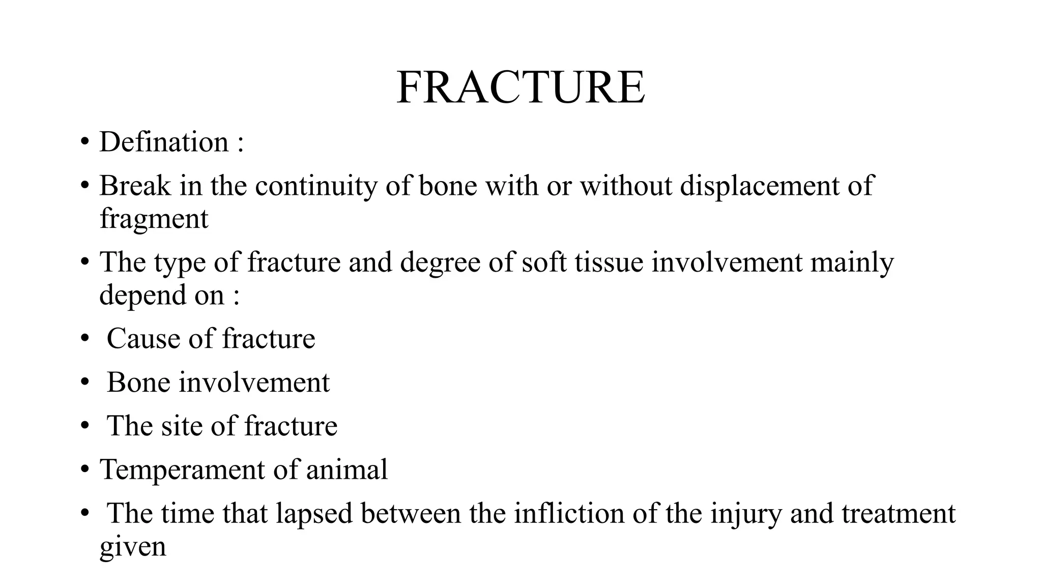
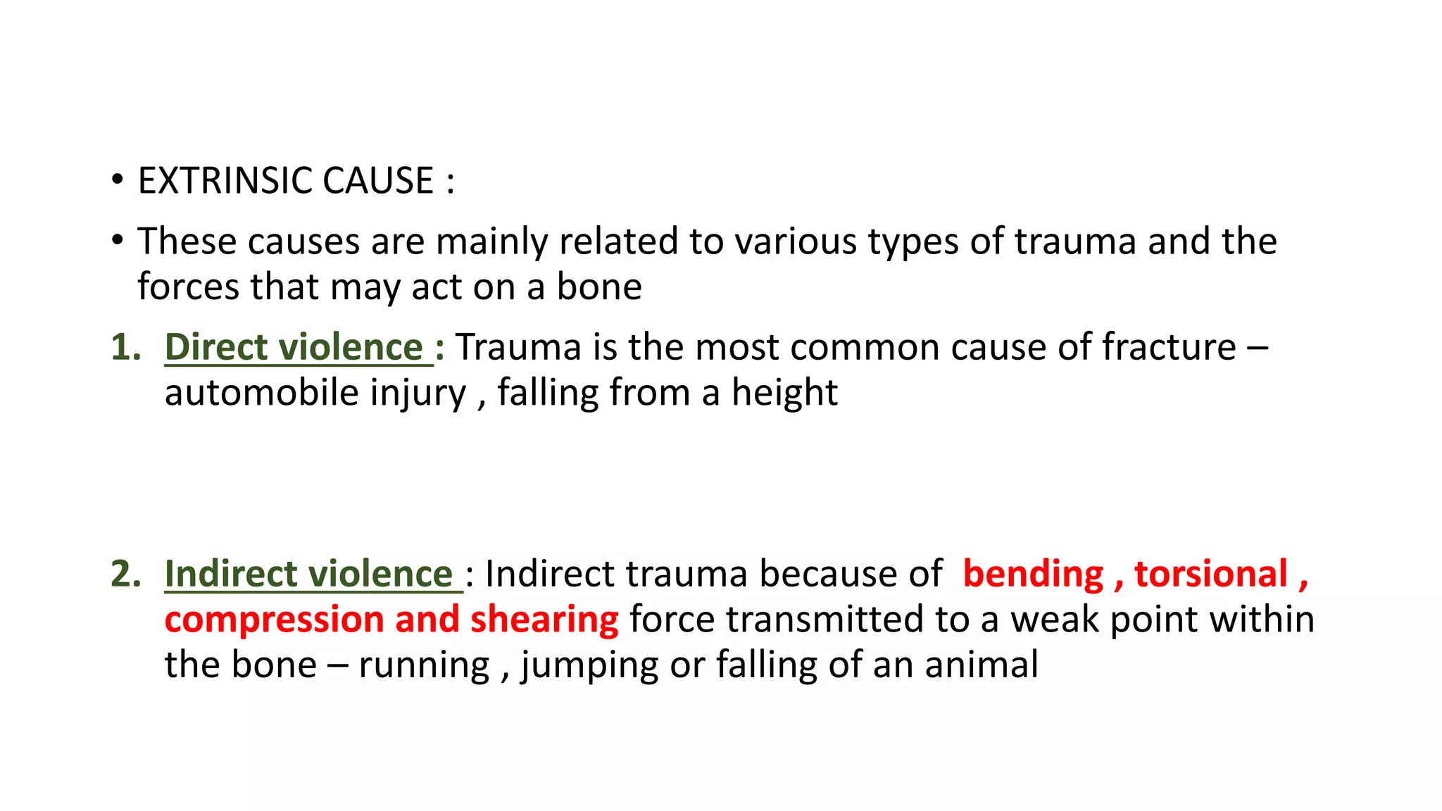
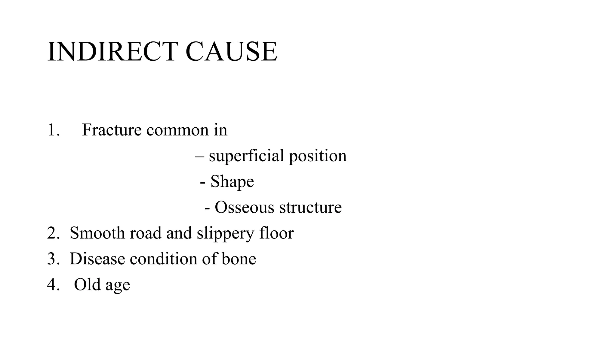
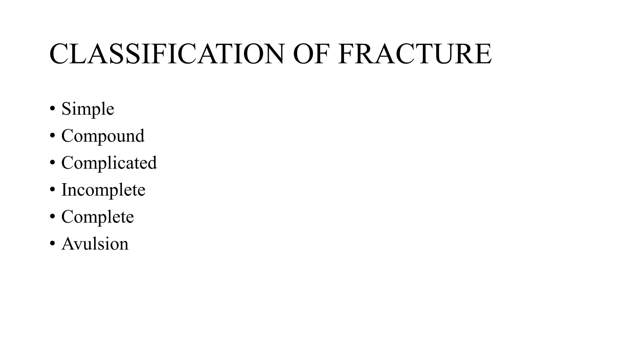
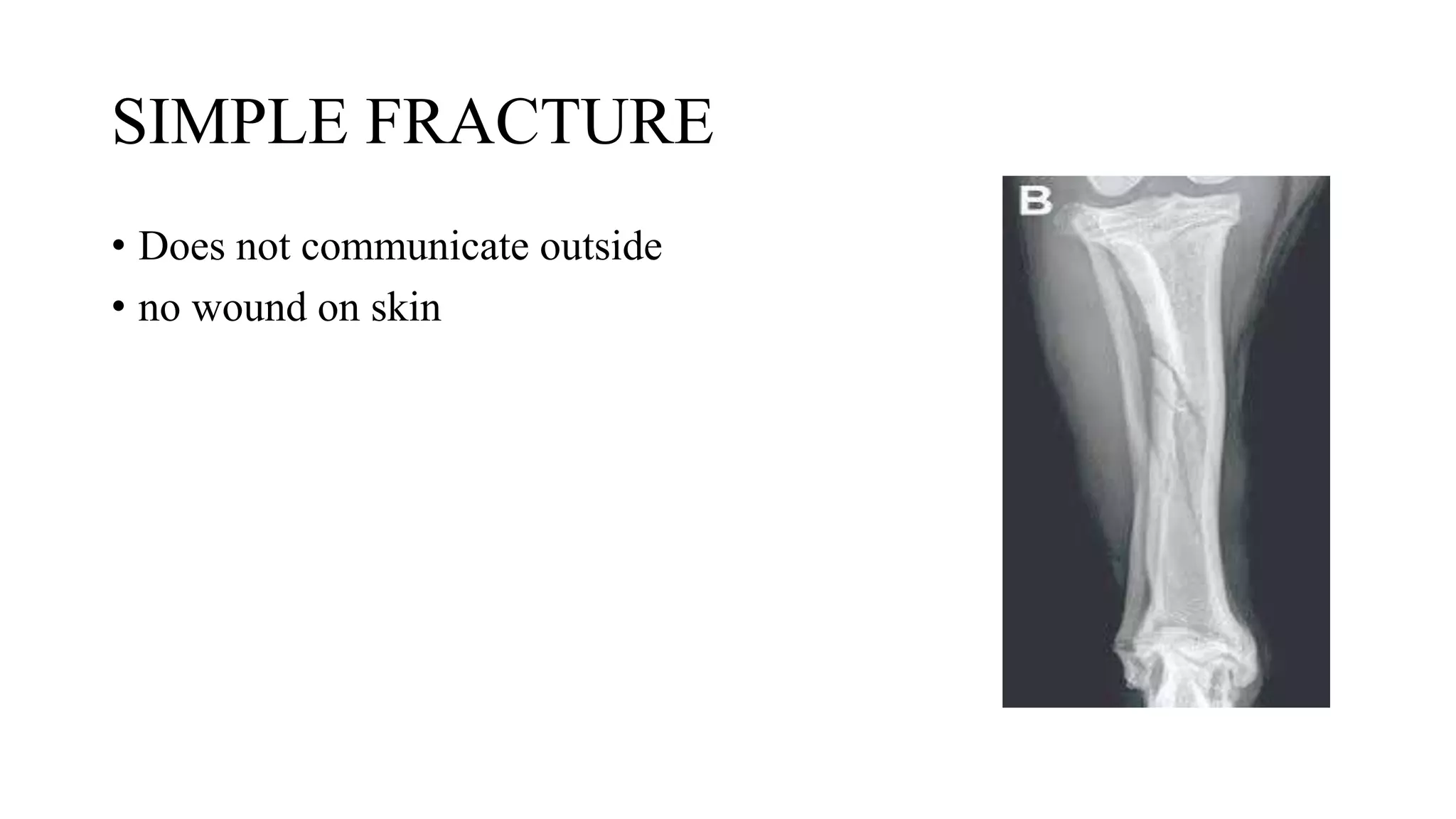
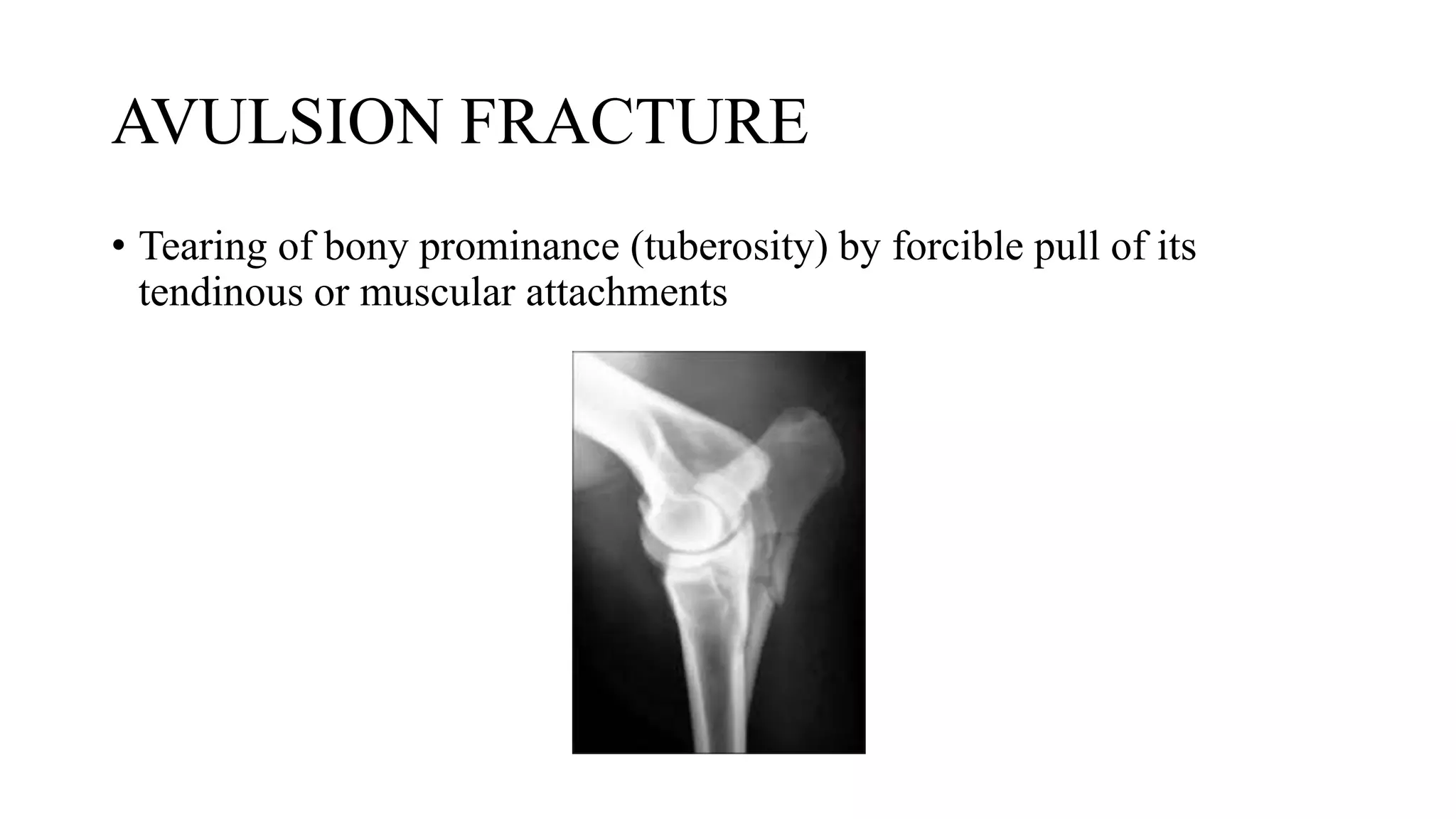
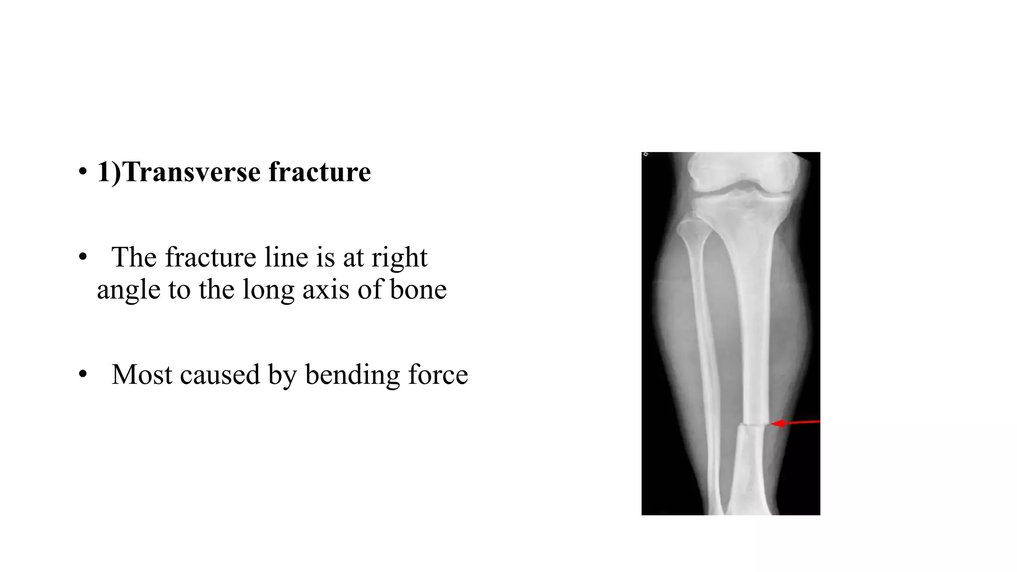
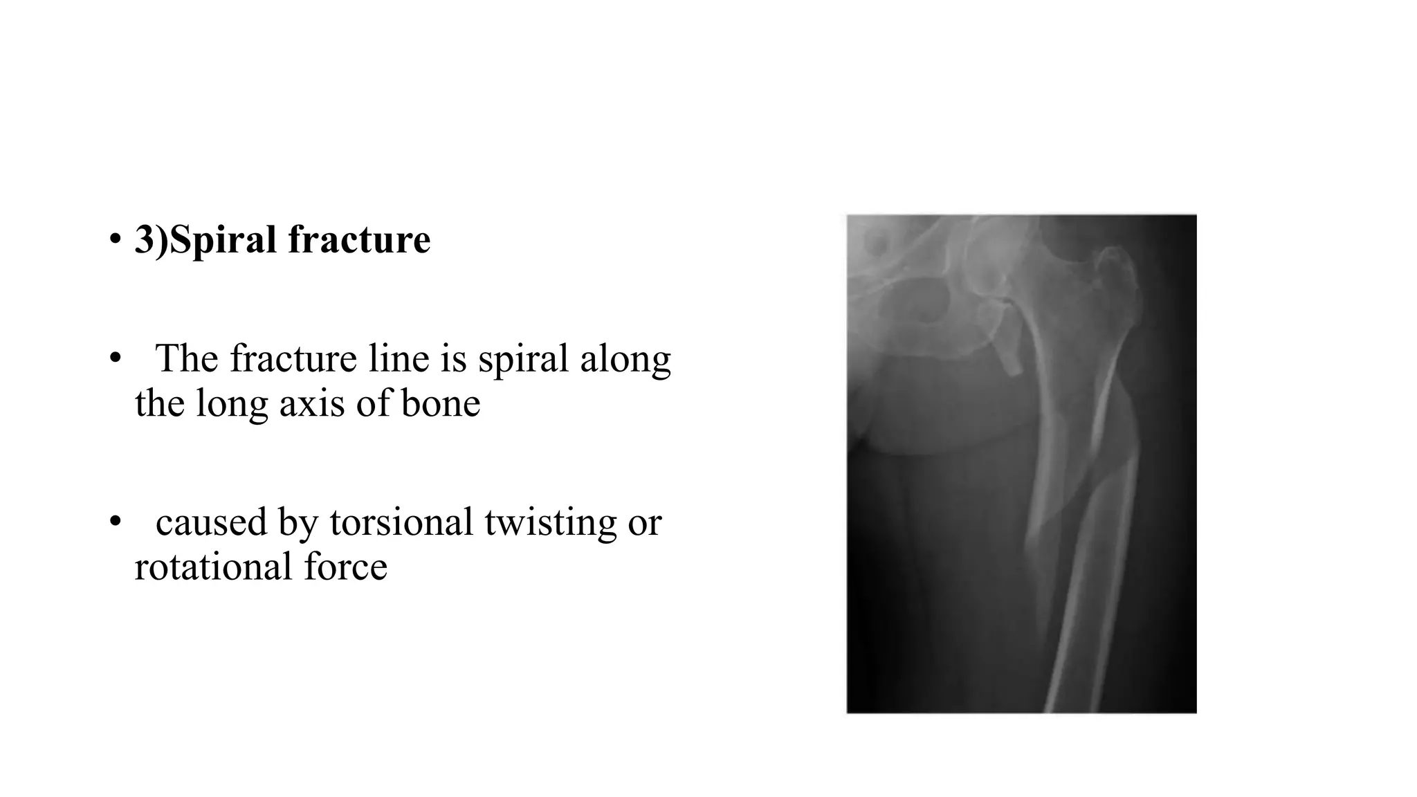
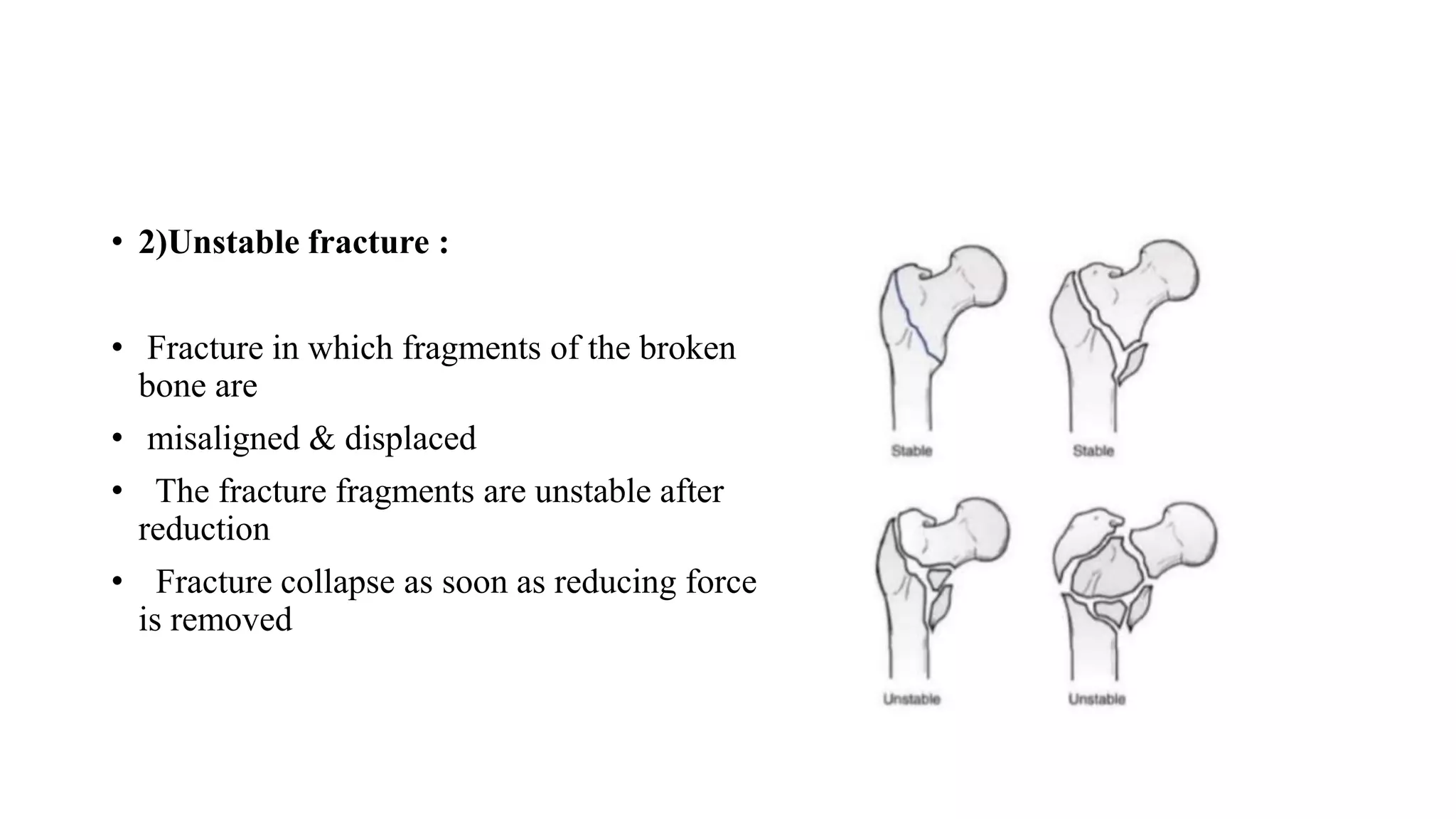
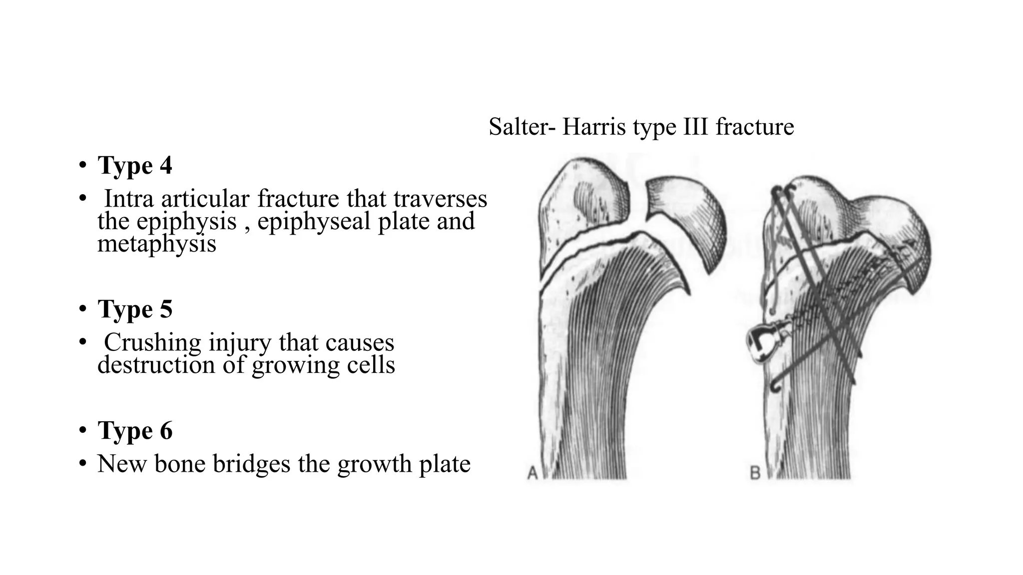
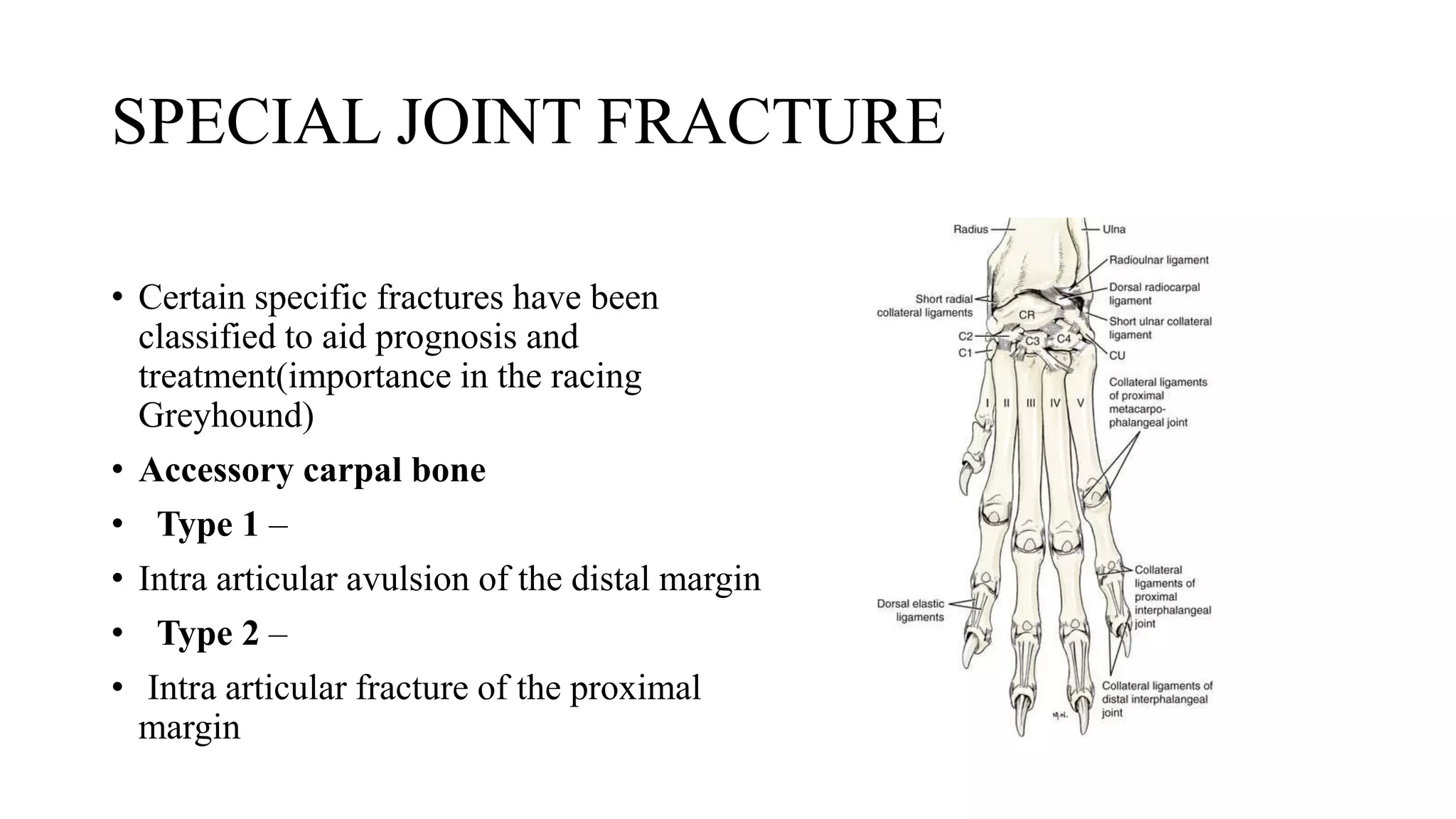
INDIAN VETERINARY RESEARCH INSTITUTE,
IZATNAGAR
CLASSIFICATION OF FRACTURE
FRACTURE HEALING
Dr Rekha Pathak and Prarthana R
Division of Veterinary Surgery & Radiology
IVRI
FRACTURE
• Defination :
• Break in the continuity of bone with or without displacement of
fragment
• The type of fracture and degree of soft tissue involvement mainly
depend on :
• Cause of fracture
• Bone involvement
• The site of fracture
• Temperament of animal
• The time that lapsed between the infliction of the injury and treatment
given
• ETIOLOGY
1) Direct cause
I. Intrinsic causes
II. Extrinsic causes
2) Indirect cause
DIRECT CAUSE
• INTRINSIC CAUSE:
• These are related to either violent muscle contractions and local or systemic
disease
• Muscular contraction :
1. Violent contraction of muscle cause avulsion fracture
2. Avulsion fracture : eg – Acromion ,greater humeral tubercle ,
olecranon, Ischial tuberosity, trochanter major, cranial tibia tuberosity and
calcaneous bone.
• EXTRINSIC CAUSE :
• These causes are mainly related to various types of trauma and the
forces that may act on a bone
1. Direct violence : Trauma is the most common cause of fracture –
automobile injury , falling from a height
2. Indirect violence : Indirect trauma because of bending , torsional ,
compression and shearing force transmitted to a weak point within
the bone – running , jumping or falling of an animal
INDIRECT CAUSE
1. Fracture common in
– superficial position
- Shape
- Osseous structure
2. Smooth road and slippery floor
3. Disease condition of bone
4. Old age
CLASSIFICATION OF FRACTURE
• Simple
• Compound
• Complicated
• Incomplete
• Complete
• Avulsion
SIMPLE FRACTURE
• Does not communicate outside
• no wound on skin
COMPOUND FRACTURE
• Communicate with open wound
on skin
INCOMPLETE FRACTURE
• Does not extend through complete thickness of bone
1. Green stick
2. Partial or Splintered
3. Fissured
4. Deferred
Green stick fracture
• It is characterized by partial loss of
continuity and displacement is minimal
• Part of the bone remain intact
• Green stick fracture
• Bone is partially broken like bent green
stick
• Young animals
FISSURED FRACTURE
• Fissure (crack) extending
through the bone without causing
any displacement of the
fragments
• Undisplaced fragments which
may displace at operation or
under stress
• Longitudinal, transverse or
oblique
DEFFERED FRACTURE
• Separation of fragments occur only after a varying period
after the incident
• Violence, strain or concussion
COMPLETE FRACTURE
• Bone is broken completely through its thickness
• Single
• Double
• Multiple
a)Single b)Double c) Communited
AVULSION FRACTURE
• Tearing of bony prominance (tuberosity) by forcible pull of its
tendinous or muscular attachments
COMPLICATED FRACTURE
• A closed fracture in which there is
considerable injury to important
neighboring vessels ,nerves or joint
involvement
• Accompanied by the opening of a
joint or visceral cavity
• Example : Complicated fracture of rib
Others
• Based on portion of bone involved-
1. Diphysary
2. Epiphysary
3. Supracondylar
4. Condylar
5. Transcondylar
6. Intercondylar
7. Articular
• Articular fracture
• Fracture involving subchondral bone
& articular cartilage
• Extra articular
• Not involving the joint surface
• Based on direcion of Fracture
1. Transverse
2. Longitudinal
3. Oblique
4. Spiral
• 1)Transverse fracture
• The fracture line is at right
angle to the long axis of bone
• Most caused by bending force
• 2)Oblique fracture
• The line of the fracture is
diagonal to the long axis of bone
• Caused by bending with axial
compression
• 3)Spiral fracture
• The fracture line is spiral along
the long axis of bone
• caused by torsional twisting or
rotational force
• 4)Comminuted fracture
• It comprises of at least three
fracture lines and all of them
meet at a common point
• Caused by high energy trauma
• 6)Avulsion fracture/
apophyseal
• A fragment of bone at the site of
muscle insertion is detached due
to its forceful contraction
• Pulled by tendon or ligament
Correction of Avulsion fracture
• Depending on relationship between fragments
1. Impacted
2. Distracted
3. Depressed
4. Over-riding
5. Compression
IMPACTED FRACTURE
• One end of broken bone is driven
into other end or one bone is
driven into the fracture site of
another
• Union is rapid but bone is
shortened
DISTRACTED FRACTURE
• A fracture resulting in increased overall bone length,is due
distraction of bone component
DEPRESSED FRACTURE
• The fragments are displaced and produce a cavity
• Especially part of the skull
a)Impacted c) Compression
b)Over riding
According to stability of fractured fragment
• Charnley classification
• Used to determine which fracture would respond to closed
reduction and reduction
1. Stable Fracture
2. Unstable Fracture
• 1) Stable after fracture :
• The fragments more or less interlock after reduction
• Maintain the approximate length of the segment
• Eg – simple fracture ; fractured pieces of bone are well aligned and
stable
• 2)Unstable fracture :
• Fracture in which fragments of the broken
bone are
• misaligned & displaced
• The fracture fragments are unstable after
reduction
• Fracture collapse as soon as reducing force
is removed
Epiphyseal fracture or growth plate fracture
• The most commonly used is the Salter- Harris system (salter &
Harris, 1963 )
• Fracture of epiphysis occurs in mature animal after closure of the
epiphyseal plate
• Type 1
• Complete , through the
hypertrophied cartilage cell zone
• Type 2
• Partially includes the metaphysis
• Type 3
• Intra articular fracture to the
hypertrophied zone and then along
the epiphyseal plate to the edge
Salter - Harris type I
• Type 4
• Intra articular fracture that traverses
the epiphysis , epiphyseal plate and
metaphysis
• Type 5
• Crushing injury that causes
destruction of growing cells
• Type 6
• New bone bridges the growth plate
Salter- Harris type III fracture
SPECIAL JOINT FRACTURE
• Certain specific fractures have been
classified to aid prognosis and
treatment(importance in the racing
Greyhound)
• Accessory carpal bone
• Type 1 –
• Intra articular avulsion of the distal margin
• Type 2 –
• Intra articular fracture of the proximal
margin
Type 3 –
• Extra articular avulsion of the
• distal margin
• Type 4 –
• Extra articular avulsion of the
insertion of flexor carpi ulnaris at
proximal palmer surface
• Type 5 – Comminuted fracture
of the body which may involve
the articular surface
• Central tarsal bone
• Type 1 –
• Small dorsal stab fracture with minimal
displacement
• Type 2 –
• Dorsal stab fracture with displacement
• Type 3 –
• One third to half of the bone fractured in
the median plane and displaced medially
or dorsally
• Type 4 –
• Combination of types 2 & 3
• Type 5 –
• Sever comminution
• Various combintions of fractures
of the tarsus have been described
as Triads ( Newton and
Nunamaker)
• A system for classification of the soft tissue injury ( muller et al.
1992)
• System is developed for humans
• System may be too complicated for animals – can be used with little
variation
• Integument closed (IC)
• IC1 No injury
• IC2 No laceration but contusion
• IC3 Circumscribed degloving
• IC4 Extensive closed degloving
• IC5 Necropsis from contusion
• Integument open (IO)
• IO1 Skin breakage from inside out
• IO2 Skin breakage from outside in > 5cm,devitalized edges
• IO3 Skin breakage from outside in < 5cm, devitalised edges,
• circumscribed degloving
• IO4 Full thickness contusion, abrasion, skin loss
• IO5 Extensive degloving
• Muscle tendon (MT)
• MT1 No injury
• MT 2 Circumscribed injury, one muscle group only
• MT3 Extensive injury, two or more muscle groups
• MT4 Avulsion or loss of entire muscle groups,
• tendon laceration
• MT5 Compartment syndrome/ crush syndrome
• Neurovascular (NV)
• NV1 No injury
• NV2 Isolated nerve injury
• NV3 Localized vascular injury
• NV 4 Combined neurovascular injury
• NV5 Subtotal / total amputation
• General classification of fractures was developed by by Muller and others of the
AO / ASIF group for human fractures .
• System using alphanumeric classifications combined with electronically stored X-
ray images
• AO - Arbeitsgemeinschaftn fur Osteosynthesefragen
• ASIF - Association for the study of internal fixation
• This has been modified to use in small animals and horse
Two systems for small animals
• 1) Prieur ( Prier et al., 1990)
• 2) Unger (unger et al., 1990)
• These fractures describes the bone , the location and the type of fracture
• Each of the proposed system creates a 4 digit record in a similar wat to the
human AO system
• The Prieur & Unger fracture classification system used only for long bones .
• Not involving the skull , vertebral coloumn , pelvis or small limb bones.
• Doesn’t involve soft tissue problems
• The prieur system
• This is simplest system – slightly less information
• Digits are allocated under each fields
• (Bone ; location ; fracture area ; fragment number )
• The location Zones of each bone are determined by drawing a square
around the ends , of length and width equal to the widest dimension of
the bone end
Field Number
•Bone Humerus 1
Radius & Ulna 2
Femur 3
Tibia 4
•Location Proximal segment 1
Middle segment 2
Distal Segment 3
•Fracture area( % of bone length )
< 5% (& or not involving articular cartilage) 1
5–25%(Specific fractures of femur neck) 2
> 25%(&/or involving articular surfaces) 3
•Number of fragments
Two 2
Example
• The unger system
• Simliar to Prieur system - Records more data by attempting to
identify reducible or non reducible wedges or the direction of the
fracture line.
• Charts of both letters and numbers for each bone and codes allocated
for various fractures
Clinical Signs
• Pain
• Dysfunction
• Local trauma
• Abnormal posture
• Crepitus
Diagnosis
• Based on history and clinical examination- Pain, Dysfunction , Local
trauma , Abnormal posture,Crepitus
• Radiography
• Bone scan
• Computed tomography scan(CT)
• Micro CT
• Magnetic resonance imaging (MRI)
• Positron emission tomography(PET)
Treatment option for open fracture
K wire Intramedullary pin Interlocking nail
Orthopedic wire Screw & plate Plate rod
FRACTURE HEALING
• Indirect bone healing - callus formation present
• Direct bone healing- without callus formation
• Others-
• Intramembranous bone healing- (Mesenchyme---- Osteoblast)
• Trabecular bone healing
• Physeal bone healing
STAGES OF HEALING OF FRACTURE
• Formation of hematoma ( within 24 hours)
• Formation of soft callus (10-21 days)
• Formation of primary bone callus (21 days -6 weeks)
• Formation of secondary bone callus (6-8 weeks)
RADIOGRAPHIC VIEW
THANKYOU

FRACTURE PPT.pptx

  • 1.
    INDIAN VETERINARY RESEARCHINSTITUTE, IZATNAGAR CLASSIFICATION OF FRACTURE FRACTURE HEALING Dr Rekha Pathak and Prarthana R Division of Veterinary Surgery & Radiology IVRI
  • 2.
    FRACTURE • Defination : •Break in the continuity of bone with or without displacement of fragment • The type of fracture and degree of soft tissue involvement mainly depend on : • Cause of fracture • Bone involvement • The site of fracture • Temperament of animal • The time that lapsed between the infliction of the injury and treatment given
  • 3.
    • ETIOLOGY 1) Directcause I. Intrinsic causes II. Extrinsic causes 2) Indirect cause
  • 4.
    DIRECT CAUSE • INTRINSICCAUSE: • These are related to either violent muscle contractions and local or systemic disease • Muscular contraction : 1. Violent contraction of muscle cause avulsion fracture 2. Avulsion fracture : eg – Acromion ,greater humeral tubercle , olecranon, Ischial tuberosity, trochanter major, cranial tibia tuberosity and calcaneous bone.
  • 5.
    • EXTRINSIC CAUSE: • These causes are mainly related to various types of trauma and the forces that may act on a bone 1. Direct violence : Trauma is the most common cause of fracture – automobile injury , falling from a height 2. Indirect violence : Indirect trauma because of bending , torsional , compression and shearing force transmitted to a weak point within the bone – running , jumping or falling of an animal
  • 6.
    INDIRECT CAUSE 1. Fracturecommon in – superficial position - Shape - Osseous structure 2. Smooth road and slippery floor 3. Disease condition of bone 4. Old age
  • 7.
    CLASSIFICATION OF FRACTURE •Simple • Compound • Complicated • Incomplete • Complete • Avulsion
  • 8.
    SIMPLE FRACTURE • Doesnot communicate outside • no wound on skin
  • 9.
    COMPOUND FRACTURE • Communicatewith open wound on skin
  • 10.
    INCOMPLETE FRACTURE • Doesnot extend through complete thickness of bone 1. Green stick 2. Partial or Splintered 3. Fissured 4. Deferred
  • 11.
    Green stick fracture •It is characterized by partial loss of continuity and displacement is minimal • Part of the bone remain intact • Green stick fracture • Bone is partially broken like bent green stick • Young animals
  • 12.
    FISSURED FRACTURE • Fissure(crack) extending through the bone without causing any displacement of the fragments • Undisplaced fragments which may displace at operation or under stress • Longitudinal, transverse or oblique
  • 13.
    DEFFERED FRACTURE • Separationof fragments occur only after a varying period after the incident • Violence, strain or concussion
  • 14.
    COMPLETE FRACTURE • Boneis broken completely through its thickness • Single • Double • Multiple
  • 15.
  • 16.
    AVULSION FRACTURE • Tearingof bony prominance (tuberosity) by forcible pull of its tendinous or muscular attachments
  • 18.
    COMPLICATED FRACTURE • Aclosed fracture in which there is considerable injury to important neighboring vessels ,nerves or joint involvement • Accompanied by the opening of a joint or visceral cavity • Example : Complicated fracture of rib
  • 19.
    Others • Based onportion of bone involved- 1. Diphysary 2. Epiphysary 3. Supracondylar 4. Condylar 5. Transcondylar 6. Intercondylar 7. Articular
  • 20.
    • Articular fracture •Fracture involving subchondral bone & articular cartilage • Extra articular • Not involving the joint surface
  • 21.
    • Based ondirecion of Fracture 1. Transverse 2. Longitudinal 3. Oblique 4. Spiral
  • 22.
    • 1)Transverse fracture •The fracture line is at right angle to the long axis of bone • Most caused by bending force
  • 23.
    • 2)Oblique fracture •The line of the fracture is diagonal to the long axis of bone • Caused by bending with axial compression
  • 24.
    • 3)Spiral fracture •The fracture line is spiral along the long axis of bone • caused by torsional twisting or rotational force
  • 25.
    • 4)Comminuted fracture •It comprises of at least three fracture lines and all of them meet at a common point • Caused by high energy trauma
  • 26.
    • 6)Avulsion fracture/ apophyseal •A fragment of bone at the site of muscle insertion is detached due to its forceful contraction • Pulled by tendon or ligament
  • 27.
  • 29.
    • Depending onrelationship between fragments 1. Impacted 2. Distracted 3. Depressed 4. Over-riding 5. Compression
  • 30.
    IMPACTED FRACTURE • Oneend of broken bone is driven into other end or one bone is driven into the fracture site of another • Union is rapid but bone is shortened
  • 31.
    DISTRACTED FRACTURE • Afracture resulting in increased overall bone length,is due distraction of bone component
  • 32.
    DEPRESSED FRACTURE • Thefragments are displaced and produce a cavity • Especially part of the skull
  • 33.
  • 34.
    According to stabilityof fractured fragment • Charnley classification • Used to determine which fracture would respond to closed reduction and reduction 1. Stable Fracture 2. Unstable Fracture
  • 35.
    • 1) Stableafter fracture : • The fragments more or less interlock after reduction • Maintain the approximate length of the segment • Eg – simple fracture ; fractured pieces of bone are well aligned and stable
  • 36.
    • 2)Unstable fracture: • Fracture in which fragments of the broken bone are • misaligned & displaced • The fracture fragments are unstable after reduction • Fracture collapse as soon as reducing force is removed
  • 37.
    Epiphyseal fracture orgrowth plate fracture • The most commonly used is the Salter- Harris system (salter & Harris, 1963 ) • Fracture of epiphysis occurs in mature animal after closure of the epiphyseal plate
  • 38.
    • Type 1 •Complete , through the hypertrophied cartilage cell zone • Type 2 • Partially includes the metaphysis • Type 3 • Intra articular fracture to the hypertrophied zone and then along the epiphyseal plate to the edge Salter - Harris type I
  • 39.
    • Type 4 •Intra articular fracture that traverses the epiphysis , epiphyseal plate and metaphysis • Type 5 • Crushing injury that causes destruction of growing cells • Type 6 • New bone bridges the growth plate Salter- Harris type III fracture
  • 40.
    SPECIAL JOINT FRACTURE •Certain specific fractures have been classified to aid prognosis and treatment(importance in the racing Greyhound) • Accessory carpal bone • Type 1 – • Intra articular avulsion of the distal margin • Type 2 – • Intra articular fracture of the proximal margin
  • 41.
    Type 3 – •Extra articular avulsion of the • distal margin
  • 42.
    • Type 4– • Extra articular avulsion of the insertion of flexor carpi ulnaris at proximal palmer surface • Type 5 – Comminuted fracture of the body which may involve the articular surface
  • 43.
    • Central tarsalbone • Type 1 – • Small dorsal stab fracture with minimal displacement • Type 2 – • Dorsal stab fracture with displacement • Type 3 – • One third to half of the bone fractured in the median plane and displaced medially or dorsally
  • 44.
    • Type 4– • Combination of types 2 & 3 • Type 5 – • Sever comminution • Various combintions of fractures of the tarsus have been described as Triads ( Newton and Nunamaker)
  • 45.
    • A systemfor classification of the soft tissue injury ( muller et al. 1992) • System is developed for humans • System may be too complicated for animals – can be used with little variation
  • 46.
    • Integument closed(IC) • IC1 No injury • IC2 No laceration but contusion • IC3 Circumscribed degloving • IC4 Extensive closed degloving • IC5 Necropsis from contusion • Integument open (IO) • IO1 Skin breakage from inside out • IO2 Skin breakage from outside in > 5cm,devitalized edges • IO3 Skin breakage from outside in < 5cm, devitalised edges, • circumscribed degloving • IO4 Full thickness contusion, abrasion, skin loss • IO5 Extensive degloving
  • 47.
    • Muscle tendon(MT) • MT1 No injury • MT 2 Circumscribed injury, one muscle group only • MT3 Extensive injury, two or more muscle groups • MT4 Avulsion or loss of entire muscle groups, • tendon laceration • MT5 Compartment syndrome/ crush syndrome • Neurovascular (NV) • NV1 No injury • NV2 Isolated nerve injury • NV3 Localized vascular injury • NV 4 Combined neurovascular injury • NV5 Subtotal / total amputation
  • 48.
    • General classificationof fractures was developed by by Muller and others of the AO / ASIF group for human fractures . • System using alphanumeric classifications combined with electronically stored X- ray images • AO - Arbeitsgemeinschaftn fur Osteosynthesefragen • ASIF - Association for the study of internal fixation • This has been modified to use in small animals and horse Two systems for small animals • 1) Prieur ( Prier et al., 1990) • 2) Unger (unger et al., 1990)
  • 49.
    • These fracturesdescribes the bone , the location and the type of fracture • Each of the proposed system creates a 4 digit record in a similar wat to the human AO system • The Prieur & Unger fracture classification system used only for long bones . • Not involving the skull , vertebral coloumn , pelvis or small limb bones. • Doesn’t involve soft tissue problems
  • 50.
    • The prieursystem • This is simplest system – slightly less information • Digits are allocated under each fields • (Bone ; location ; fracture area ; fragment number ) • The location Zones of each bone are determined by drawing a square around the ends , of length and width equal to the widest dimension of the bone end
  • 51.
    Field Number •Bone Humerus1 Radius & Ulna 2 Femur 3 Tibia 4 •Location Proximal segment 1 Middle segment 2 Distal Segment 3 •Fracture area( % of bone length ) < 5% (& or not involving articular cartilage) 1 5–25%(Specific fractures of femur neck) 2 > 25%(&/or involving articular surfaces) 3 •Number of fragments Two 2
  • 52.
  • 53.
    • The ungersystem • Simliar to Prieur system - Records more data by attempting to identify reducible or non reducible wedges or the direction of the fracture line. • Charts of both letters and numbers for each bone and codes allocated for various fractures
  • 54.
    Clinical Signs • Pain •Dysfunction • Local trauma • Abnormal posture • Crepitus
  • 55.
    Diagnosis • Based onhistory and clinical examination- Pain, Dysfunction , Local trauma , Abnormal posture,Crepitus • Radiography • Bone scan • Computed tomography scan(CT) • Micro CT • Magnetic resonance imaging (MRI) • Positron emission tomography(PET)
  • 56.
    Treatment option foropen fracture K wire Intramedullary pin Interlocking nail Orthopedic wire Screw & plate Plate rod
  • 57.
    FRACTURE HEALING • Indirectbone healing - callus formation present • Direct bone healing- without callus formation • Others- • Intramembranous bone healing- (Mesenchyme---- Osteoblast) • Trabecular bone healing • Physeal bone healing
  • 58.
    STAGES OF HEALINGOF FRACTURE • Formation of hematoma ( within 24 hours) • Formation of soft callus (10-21 days) • Formation of primary bone callus (21 days -6 weeks) • Formation of secondary bone callus (6-8 weeks)
  • 60.
  • 61.