Downloaded 240 times


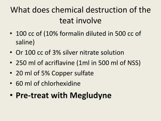

Chemical destruction of the teat involves injecting solutions such as diluted formalin, silver nitrate, acriflavine, copper sulfate, or chlorhexidine into the teat to destroy the teat tissue. This is done as a last resort when the teat cannot otherwise be salvaged due to extensive damage or infection. The solutions work to kill the teat tissue over the course of a few days through their disinfectant and caustic properties. Pretreating with a local anesthetic like megludyne can help reduce pain from the chemical destruction process.