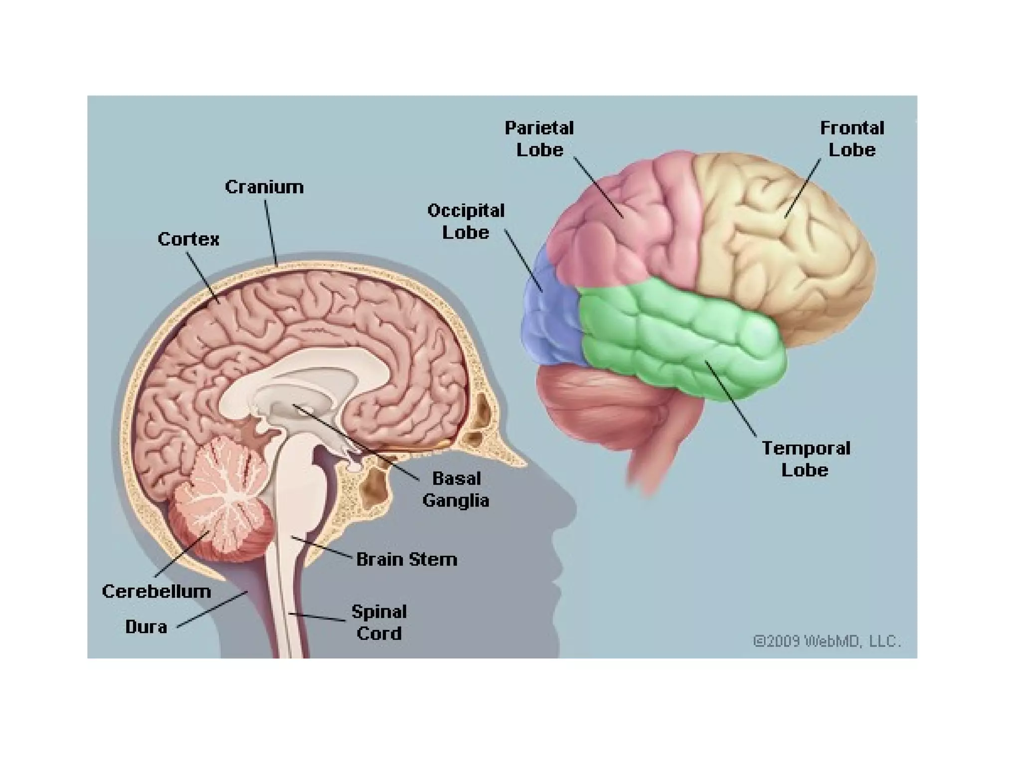
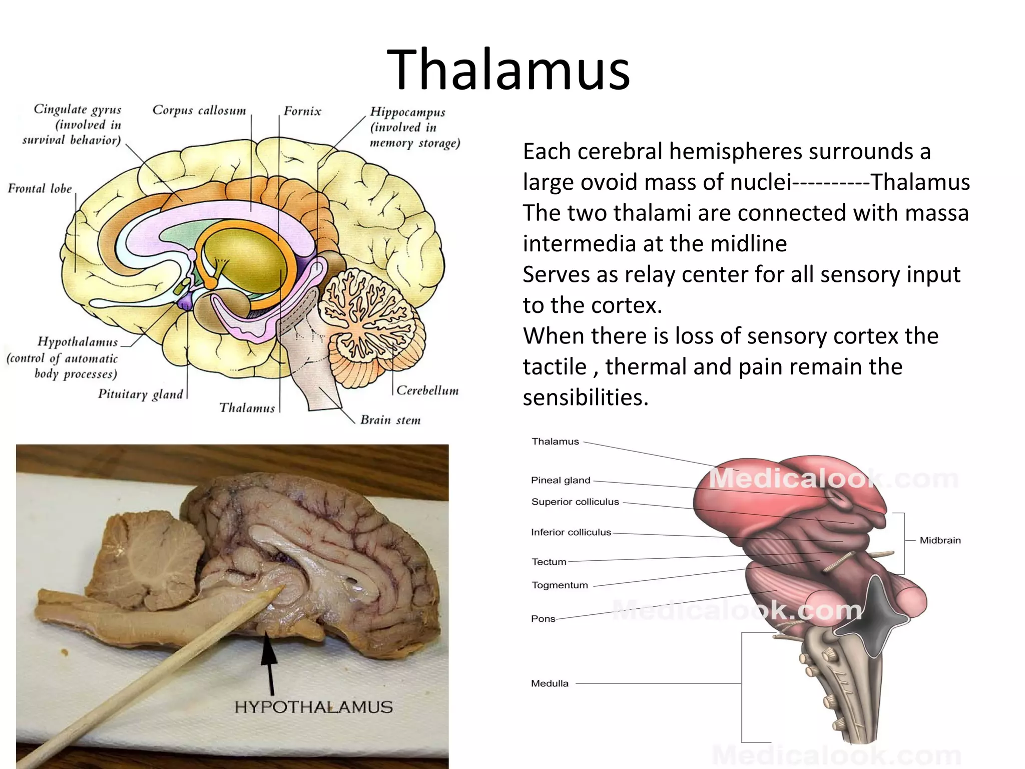
Corpus callosum
•The bridge of nerve fibres that
connect the two hemispheres-
Corpus callosum
•Agenesis: split brain; lack of
communication between the two
halves (ACC: agenesis of corpus
callosum)
•Gyri
•Sulci
•Falx cerebri: the extension of dura
mater within these two
hemispheres
•Outer layer of the cerebrum
---cerebral cortex
• Not as important as in primates
• Experimental removal of cortex still continues carrying
out many major motor functions, run walk, maintain
equilibrium and right itself.
• Cannot seek out and eat food but if put inside the mouth
they can eat
• Intellectual functions are lost
• Electrical stimulation of specific area and surgical
removal of specific area enables to understand the
function of that area
• Some areas of cerebral cortex is sensory and
some is motor
• Temporal lobe has auditory and memory functions
• Occipital lobe houses the visual perception and visual
interpretaton areas
• Removal of occipital area----total blindness
• Frontal area: controls the psychic level of dog and very
important area . The destuctive lesions of this area cause
marked changes in personality
• Animal fail to recognize the owner and unable to perform
learned tasks
• Impulsive and ill tempered
Thalamus
Each cerebral hemispheres surrounds a
large ovoid mass of nuclei----------Thalamus
The two thalami are connected with massa
intermedia at the midline
Serves as relay center for all sensory input
to the cortex.
When there is loss of sensory cortex the
tactile , thermal and pain remain the
sensibilities.
Thalamus
All the pain stimuli pass through
thalami---------------perception of pain
Whereas cortical areas-----------help in
localization of pain
Damage to thalamic areas lead to
overeaction to minimal stimulation of pain
receptiors called as thalamic syndrome.
This is due to block in the thelamogeniculate
branch of the posterior cerebral artery.
Hypothalamus
Collection of nuclei ventral
to the thalmus and
forming the floor of lateral
walls of the third ventricle.
Hypothalamus
It has supraoptic nuclei, mammilary bodies MB, tuber cinereum
TC and the infundibulum I
Structure of pituitary
There are various nuclei in the hypothalamus which are
responsible for various releasing factors
Pituitary releases the hormones in response
•The anterior lobe of pituitary
•Thyroid Stimulating Hormone (TSH)
•Follicle-Stimulating Hormone (FSH)
•Luteinizing Hormone (LH)
•Prolactin (PRL)
•Growth Hormone (GH)
•ACTH — the adrenocorticotropic hormone
•Alpha Melanocyte-Stimulating Hormone (α-MSH)
Posterior lobe
•Releases Vasopressin and oxytocin -------synthesised in
hypothalamus
•So only stored in posterior lobe of pituitary
•Hypothalamic regulatory mechanisms;
•Temperature regulation
•Neuroendocrine control of catecholamines
•Appetite behaviour
•Thirst
•Hunger
•Sexual behaviour
•Defensive reaction
Midbrain
•Short portion between the Pons and the diencephlon
•Four corpora quadrigemina
•Anterior colliculi are associated with the optic and posterior
colliculi are associated with the auditory system
•The roots of trochlear , oculomotor and trigeminal roots are
located on the midrain and the lesions are associated with the
dysfunction of these nerves
Pons
•Just below the midbrain lies
the pons---------------
composed or fiber tracts and
number of nuclei
•The fibres run transversely
across the pons and through
the middle cerebellar
peduncle into the
cerebellum
•5, 6, and 7 and 8th cranial
nerves are located in the
upper part of the pons
•Pneumotaxic center: control of respiration
•Apneustic center:The apneustic center of pons sends signals
to the dorsal respiratory center in the medulla to delay the
'switch off' signal of the inspiratory ramp provided by the
pneumotaxic center of pons. It controls the intensity of
breathing. The apneustic center is inhibited by pulmonary
stretch receptors. However, it gives positive impulses to the
inspiratory (I) neurons.
•Lesions of the pons : increased sensitivity to touch-----called
as trigeminal neualgia
Medulla oblongata
•Separated from the pons by
a transverse groove
•8th
, 9th,
10th,
11th
and 12th
cranial nerves project from
medulla
•Nuclei of autonomic reflex
centers like respiratory
centre, cardiac and
vasomotor are present in
medulla.(they are called vital
centres)
•The reflex function like
Vomiting , coughing sneezing,
hicoughing and swallowing
functions are regulated

Brain structure

  • 2.
    Corpus callosum •The bridgeof nerve fibres that connect the two hemispheres- Corpus callosum •Agenesis: split brain; lack of communication between the two halves (ACC: agenesis of corpus callosum) •Gyri •Sulci •Falx cerebri: the extension of dura mater within these two hemispheres •Outer layer of the cerebrum ---cerebral cortex
  • 3.
    • Not asimportant as in primates • Experimental removal of cortex still continues carrying out many major motor functions, run walk, maintain equilibrium and right itself. • Cannot seek out and eat food but if put inside the mouth they can eat • Intellectual functions are lost • Electrical stimulation of specific area and surgical removal of specific area enables to understand the function of that area
  • 4.
    • Some areasof cerebral cortex is sensory and some is motor
  • 5.
    • Temporal lobehas auditory and memory functions • Occipital lobe houses the visual perception and visual interpretaton areas • Removal of occipital area----total blindness • Frontal area: controls the psychic level of dog and very important area . The destuctive lesions of this area cause marked changes in personality • Animal fail to recognize the owner and unable to perform learned tasks • Impulsive and ill tempered
  • 6.
    Thalamus Each cerebral hemispheressurrounds a large ovoid mass of nuclei----------Thalamus The two thalami are connected with massa intermedia at the midline Serves as relay center for all sensory input to the cortex. When there is loss of sensory cortex the tactile , thermal and pain remain the sensibilities.
  • 7.
    Thalamus All the painstimuli pass through thalami---------------perception of pain Whereas cortical areas-----------help in localization of pain Damage to thalamic areas lead to overeaction to minimal stimulation of pain receptiors called as thalamic syndrome. This is due to block in the thelamogeniculate branch of the posterior cerebral artery.
  • 8.
    Hypothalamus Collection of nucleiventral to the thalmus and forming the floor of lateral walls of the third ventricle.
  • 9.
    Hypothalamus It has supraopticnuclei, mammilary bodies MB, tuber cinereum TC and the infundibulum I
  • 10.
  • 11.
    There are variousnuclei in the hypothalamus which are responsible for various releasing factors Pituitary releases the hormones in response •The anterior lobe of pituitary •Thyroid Stimulating Hormone (TSH) •Follicle-Stimulating Hormone (FSH) •Luteinizing Hormone (LH) •Prolactin (PRL) •Growth Hormone (GH) •ACTH — the adrenocorticotropic hormone •Alpha Melanocyte-Stimulating Hormone (α-MSH)
  • 12.
    Posterior lobe •Releases Vasopressinand oxytocin -------synthesised in hypothalamus •So only stored in posterior lobe of pituitary •Hypothalamic regulatory mechanisms; •Temperature regulation •Neuroendocrine control of catecholamines •Appetite behaviour •Thirst •Hunger •Sexual behaviour •Defensive reaction
  • 13.
    Midbrain •Short portion betweenthe Pons and the diencephlon •Four corpora quadrigemina •Anterior colliculi are associated with the optic and posterior colliculi are associated with the auditory system •The roots of trochlear , oculomotor and trigeminal roots are located on the midrain and the lesions are associated with the dysfunction of these nerves
  • 15.
    Pons •Just below themidbrain lies the pons--------------- composed or fiber tracts and number of nuclei •The fibres run transversely across the pons and through the middle cerebellar peduncle into the cerebellum •5, 6, and 7 and 8th cranial nerves are located in the upper part of the pons
  • 16.
    •Pneumotaxic center: controlof respiration •Apneustic center:The apneustic center of pons sends signals to the dorsal respiratory center in the medulla to delay the 'switch off' signal of the inspiratory ramp provided by the pneumotaxic center of pons. It controls the intensity of breathing. The apneustic center is inhibited by pulmonary stretch receptors. However, it gives positive impulses to the inspiratory (I) neurons. •Lesions of the pons : increased sensitivity to touch-----called as trigeminal neualgia
  • 17.
    Medulla oblongata •Separated fromthe pons by a transverse groove •8th , 9th, 10th, 11th and 12th cranial nerves project from medulla •Nuclei of autonomic reflex centers like respiratory centre, cardiac and vasomotor are present in medulla.(they are called vital centres) •The reflex function like Vomiting , coughing sneezing, hicoughing and swallowing functions are regulated