INTRODUCTION
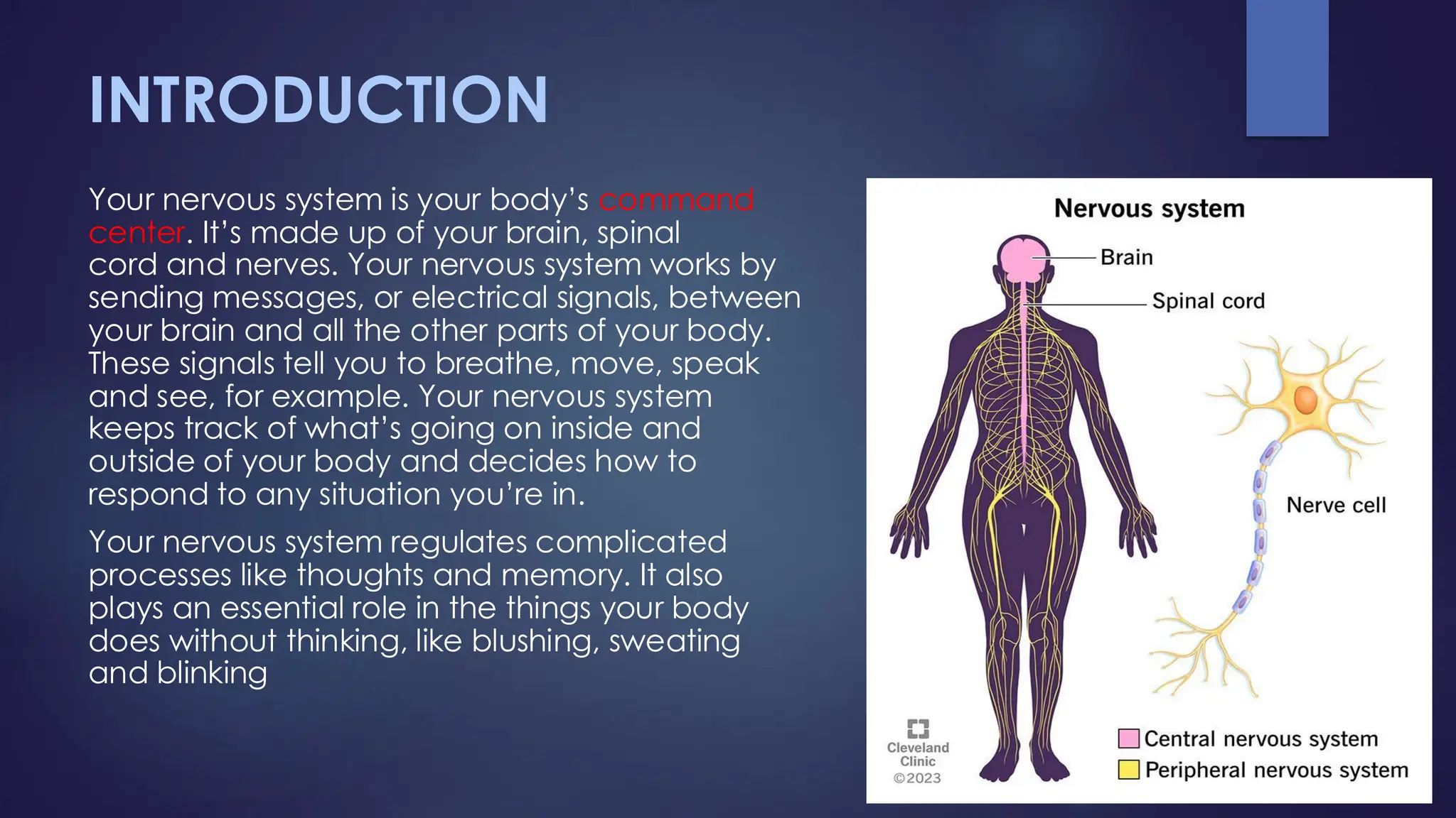
Your nervous systemis your body’s command
center. It’s made up of your brain, spinal
cord and nerves. Your nervous system works by
sending messages, or electrical signals, between
your brain and all the other parts of your body.
These signals tell you to breathe, move, speak
and see, for example. Your nervous system
keeps track of what’s going on inside and
outside of your body and decides how to
respond to any situation you’re in.
Your nervous system regulates complicated
processes like thoughts and memory. It also
plays an essential role in the things your body
does without thinking, like blushing, sweating
and blinking
3.
FUNCTIONS OF NERVOUSSYSTEM
Your nervous system’s main function is to send messages from various parts of your
body to your brain, and from your brain back out to your body to tell your body
what to do. These messages regulate your:
❑ Thoughts, memory, learning and feelings.
❑ Movements (balance and coordination).
❑ Senses (how your brain interprets what you see, hear, taste, touch and feel).
❑ Wound healing.
❑ Sleep.
❑ Heartbeat and breathing patterns.
❑ Response to stressful situations, including sweat production.
❑ Digestion.
❑ Body processes, such as puberty and aging.
4.
WORKING MECHANISM
Your nervoussystem uses nerve cells called neurons to send signals, or
messages, all over your body. These electrical signals travel among your
brain, skin, organs, glands and muscles.
The messages help you move your limbs and feel sensations, like pain.
Your eyes, ears, tongue, nose and the nerves all over your body take in
information about your environment. Then, nerves carry that data to and
from your brain.
There are different types of neurons. Each type of neuron has a different
job:
1) Motor neurons take signals from your brain and spinal cord to your
muscles. They help you move. They also assist with breathing,
swallowing and speaking.
2) Sensory neurons take information from your senses (what you see,
touch, taste, etc.) to your brain.
3) Interneurons communicate between motor and sensory neurons.
These neurons regulate your movement in response to sensory
information (like moving away from a hot surface) and play a role in
how you learn, think and remember.
5.
PARTS OF NERVOUSSYSTEM
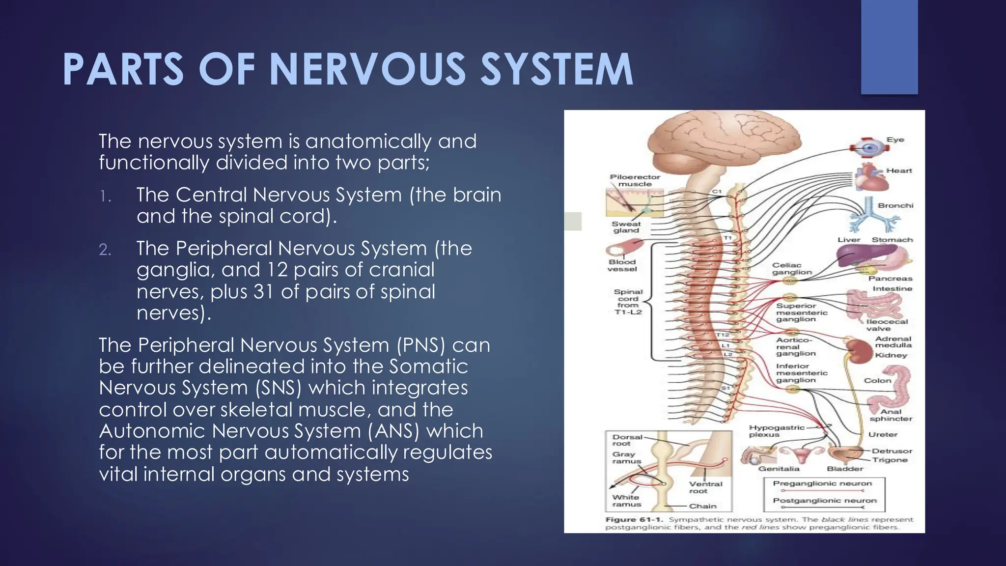
The nervous system is anatomically and
functionally divided into two parts;
1. The Central Nervous System (the brain
and the spinal cord).
2. The Peripheral Nervous System (the
ganglia, and 12 pairs of cranial
nerves, plus 31 of pairs of spinal
nerves).
The Peripheral Nervous System (PNS) can
be further delineated into the Somatic
Nervous System (SNS) which integrates
control over skeletal muscle, and the
Autonomic Nervous System (ANS) which
for the most part automatically regulates
vital internal organs and systems
6.
THE CENTRAL NERVOUSSYSTEM –
THE BRAIN
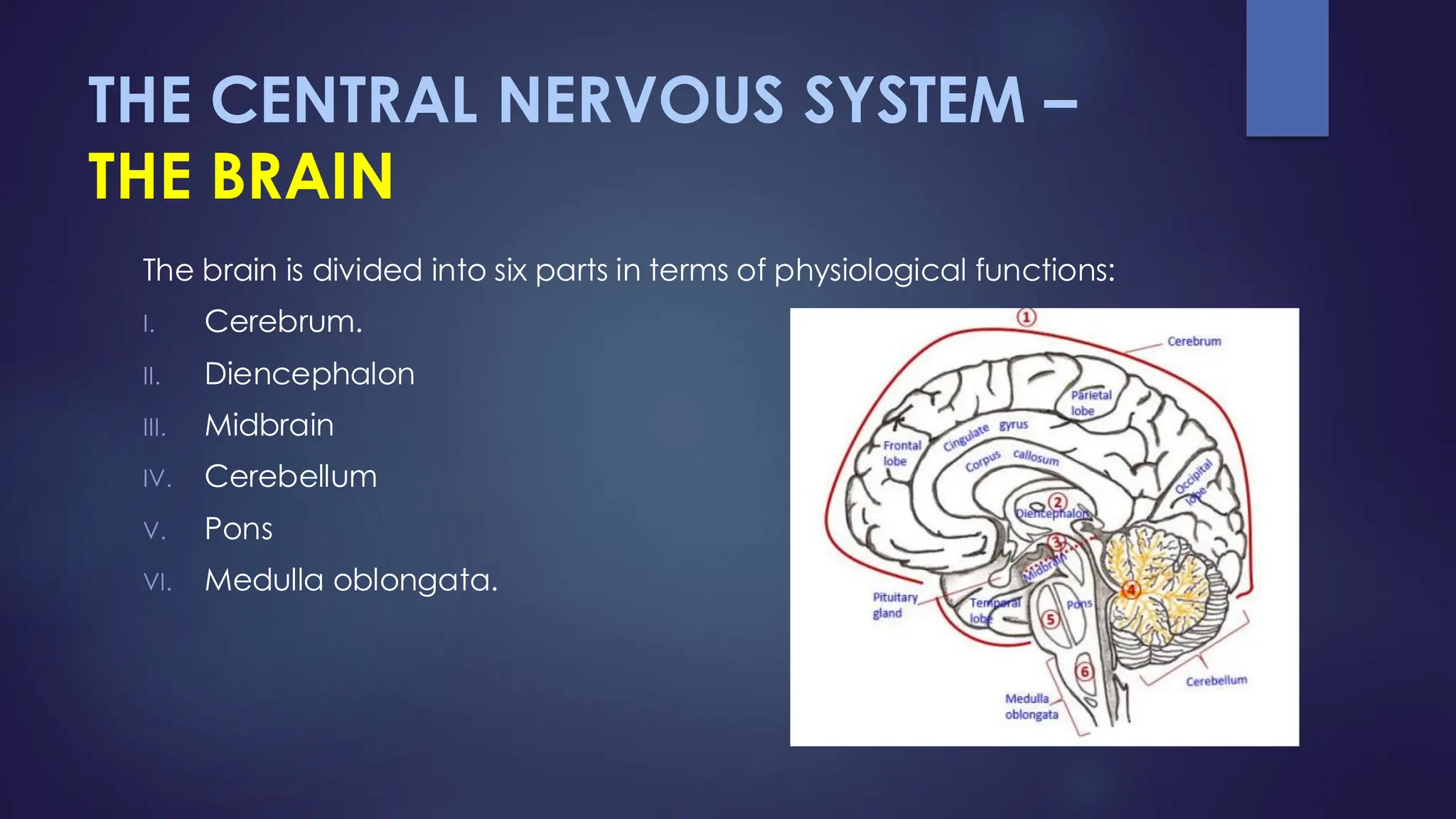
The brain is divided into six parts in terms of physiological functions:
I. Cerebrum.
II. Diencephalon
III. Midbrain
IV. Cerebellum
V. Pons
VI. Medulla oblongata.
7.
CEREBRUM
The cerebrum isthe largest most developed area of the human brain
and is considered to be the center of the highest functions. Its major
functions include: Awareness of sensory perception, voluntary control
of movement (regulation of skeletal muscle movement), language;
personality traits, sophisticated mental activities such as thinking,
memory, decision making, predictive ability, creativity and self-
consciousness.
The cerebrum is composed of 5 lobes, here is some basic information
about them:
1. The Frontal Lobe: it is concerned with higher intellectual functions
and is involved in the many behavioral aspects of humans (controls
the movement of the body).
2. The Parietal Lobe: . It is involved in body senses (touch, vibration
and position sense of the body).
8.
3. The TemporalLobe: The temporal lobe contains the auditory cortex
for the reception and interpretation of sound information, and the
olfactory cortex for the sense of smell. It also houses the language
cortex in the dominant hemisphere (usually the left hemisphere)
and participates in recognition and interpretation of language.
Parts of the limbic system (the amygdala and hippocampus) are
connected to the temporal lobe and aid in memory formation
related to emotions, the sense of smell and sound.
4. The Occipital Lobe: The occipital lobe is involved in functions
including: Visual perception, color recognition, reading and
reading comprehension, depth perception, recognition of object
movement.
5. The Insula Lobe: A key role is visceral perception, that is, conscious
awareness of internal organs and various bodily states (e.g. heart
beating awareness, bladder state etc.).
9.
THE LIMBIC SYSTEM:
Itis more of a functional system than an anatomical one. The
limbic system is the "emotional brain", participating in the
creation of emotional states such as fear, pleasure, anger,
affection, arousal, etc. and processing vivid memories
associated with those states.
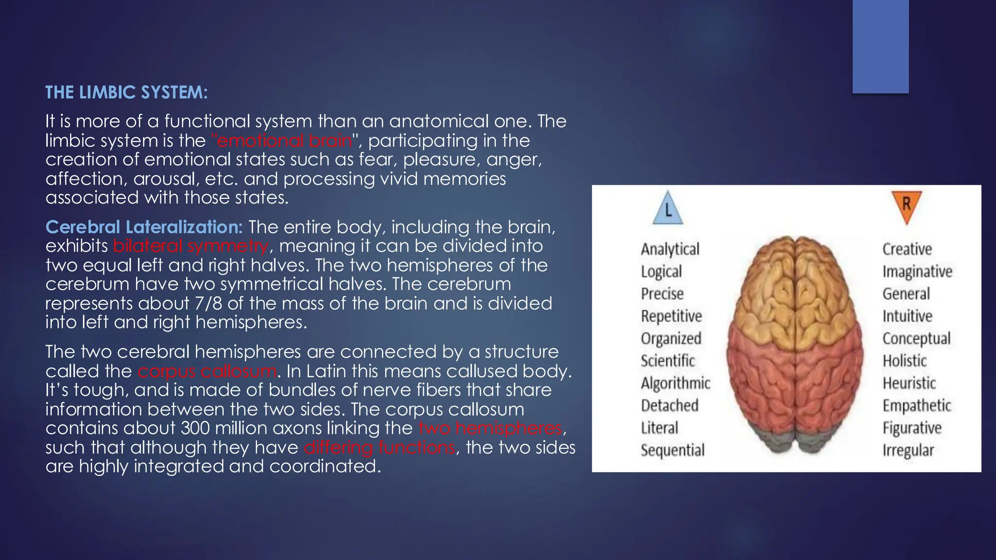
Cerebral Lateralization: The entire body, including the brain,
exhibits bilateral symmetry, meaning it can be divided into
two equal left and right halves. The two hemispheres of the
cerebrum have two symmetrical halves. The cerebrum
represents about 7/8 of the mass of the brain and is divided
into left and right hemispheres.
The two cerebral hemispheres are connected by a structure
called the corpus callosum. In Latin this means callused body.
It’s tough, and is made of bundles of nerve fibers that share
information between the two sides. The corpus callosum
contains about 300 million axons linking the two hemispheres,
such that although they have differing functions, the two sides
are highly integrated and coordinated.
10.
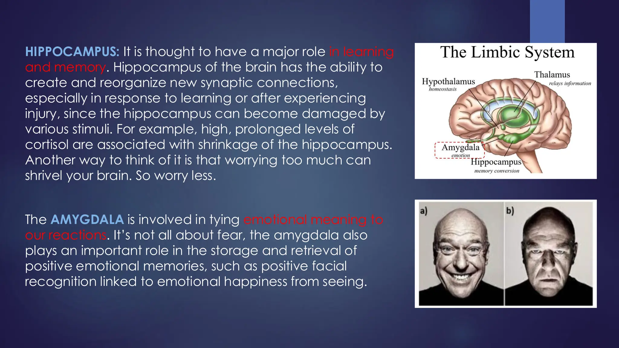
HIPPOCAMPUS: It isthought to have a major role in learning
and memory. Hippocampus of the brain has the ability to
create and reorganize new synaptic connections,
especially in response to learning or after experiencing
injury, since the hippocampus can become damaged by
various stimuli. For example, high, prolonged levels of
cortisol are associated with shrinkage of the hippocampus.
Another way to think of it is that worrying too much can
shrivel your brain. So worry less.
The AMYGDALA is involved in tying emotional meaning to
our reactions. It’s not all about fear, the amygdala also
plays an important role in the storage and retrieval of
positive emotional memories, such as positive facial
recognition linked to emotional happiness from seeing.
11.
DIENCEPHALON
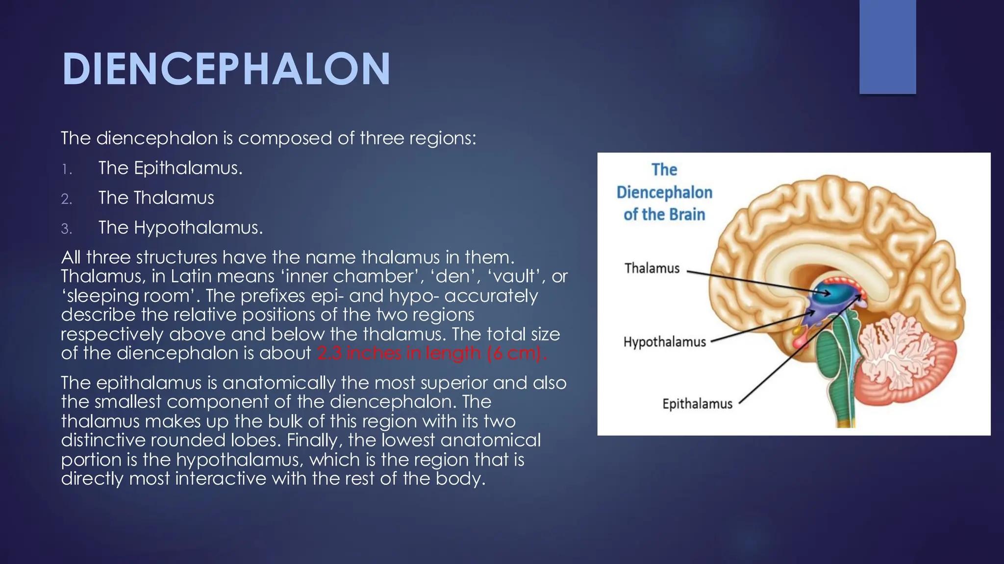
The diencephalon iscomposed of three regions:
1. The Epithalamus.
2. The Thalamus
3. The Hypothalamus.
All three structures have the name thalamus in them.
Thalamus, in Latin means ‘inner chamber’, ‘den’, ‘vault’, or
‘sleeping room’. The prefixes epi- and hypo- accurately
describe the relative positions of the two regions
respectively above and below the thalamus. The total size
of the diencephalon is about 2.3 inches in length (6 cm).
The epithalamus is anatomically the most superior and also
the smallest component of the diencephalon. The
thalamus makes up the bulk of this region with its two
distinctive rounded lobes. Finally, the lowest anatomical
portion is the hypothalamus, which is the region that is
directly most interactive with the rest of the body.
12.
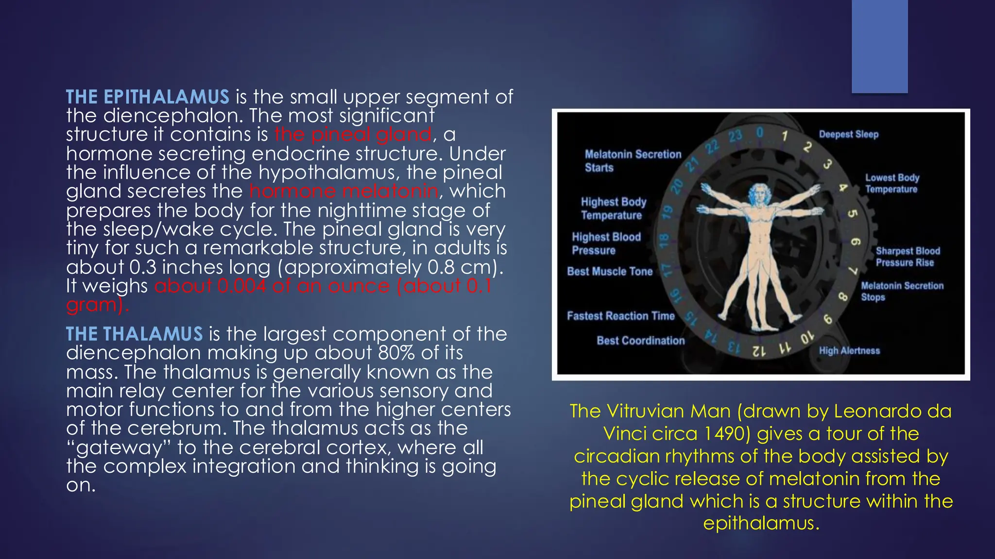
THE EPITHALAMUS isthe small upper segment of
the diencephalon. The most significant
structure it contains is the pineal gland, a
hormone secreting endocrine structure. Under
the influence of the hypothalamus, the pineal
gland secretes the hormone melatonin, which
prepares the body for the nighttime stage of
the sleep/wake cycle. The pineal gland is very
tiny for such a remarkable structure, in adults is
about 0.3 inches long (approximately 0.8 cm).
It weighs about 0.004 of an ounce (about 0.1
gram).
THE THALAMUS is the largest component of the
diencephalon making up about 80% of its
mass. The thalamus is generally known as the
main relay center for the various sensory and
motor functions to and from the higher centers
of the cerebrum. The thalamus acts as the
“gateway” to the cerebral cortex, where all
the complex integration and thinking is going
on.
The Vitruvian Man (drawn by Leonardo da
Vinci circa 1490) gives a tour of the
circadian rhythms of the body assisted by
the cyclic release of melatonin from the
pineal gland which is a structure within the
epithalamus.
13.
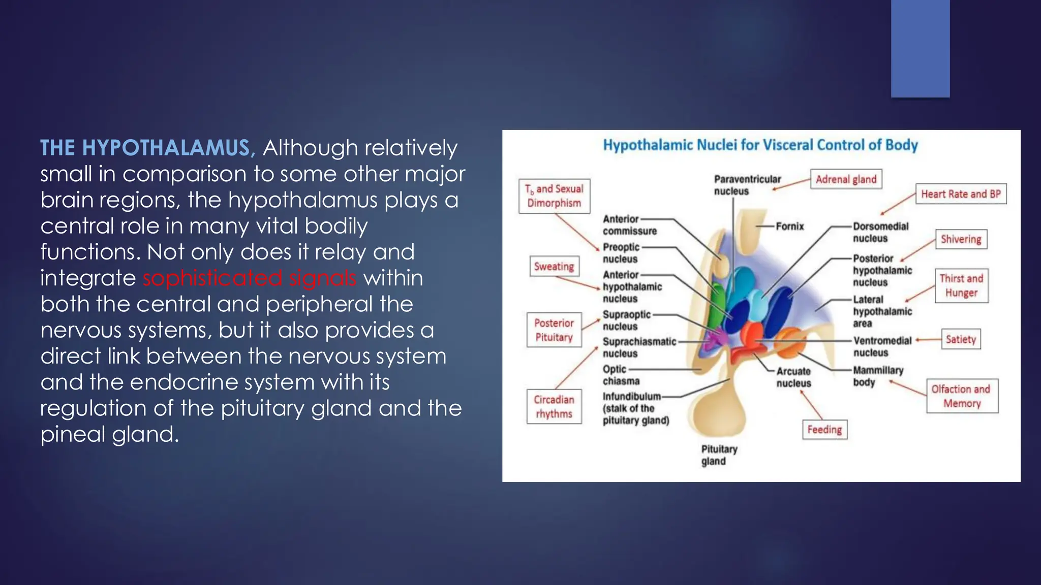
THE HYPOTHALAMUS, Althoughrelatively
small in comparison to some other major
brain regions, the hypothalamus plays a
central role in many vital bodily
functions. Not only does it relay and
integrate sophisticated signals within
both the central and peripheral the
nervous systems, but it also provides a
direct link between the nervous system
and the endocrine system with its
regulation of the pituitary gland and the
pineal gland.
14.
Following are thelist of Hormones regulated by Hypothalamus;
❑ PREOPTIC NUCLEUS modulates the secretion of Gonadotropin Releasing
Hormone (GnRH), necessary for sexual maturity and reproduction. It
sends GnRH to the adenohypophysis (or anterior pituitary gland) which
responds by releasing follicular stimulating hormone (FSH) and luteinizing
hormone (LH) which promotes the development of the gametes(germ
cell) and sex hormones in both sexes.
❑ SUPRAOPTIC NUCLEI, contains about 3,000 neurons and regulates
osmolality of blood and body fluids via the production of antidiuretic
hormone (ADH), and aids in birthing by the action of oxytocin on the
uterus.
❑ ARCUATE NUCLEUS, Responsible for producing growth hormone-
releasing hormone (GHRH), and involved in the regulation of feeding
and metabolism. Dopamine of this region inhibits lactation by inhibiting
the release of prolactin (PRL).
❑ MAMMILLARY BODY, An important area for recollective memory
(recalling a specific episode from the past), understood to be
integrated with olfaction. Damage can to spatial memory deficit.
15.
Functions regulates byHypothalamus are as follows;
❑ Control of Autonomic Nervous System (heart rate, B.P, G.I tract
activity).
❑ Control of emotional responses ( fear, rage, pleasure & sex drive).
❑ Regulation of body temperature.
❑ Regulation of hunger & thirst sensations.
16.
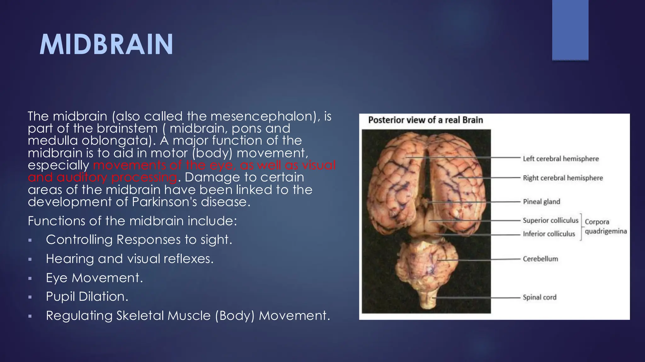
MIDBRAIN
The midbrain (alsocalled the mesencephalon), is
part of the brainstem ( midbrain, pons and
medulla oblongata). A major function of the
midbrain is to aid in motor (body) movement,
especially movements of the eye, as well as visual
and auditory processing. Damage to certain
areas of the midbrain have been linked to the
development of Parkinson's disease.
Functions of the midbrain include:
▪ Controlling Responses to sight.
▪ Hearing and visual reflexes.
▪ Eye Movement.
▪ Pupil Dilation.
▪ Regulating Skeletal Muscle (Body) Movement.
17.
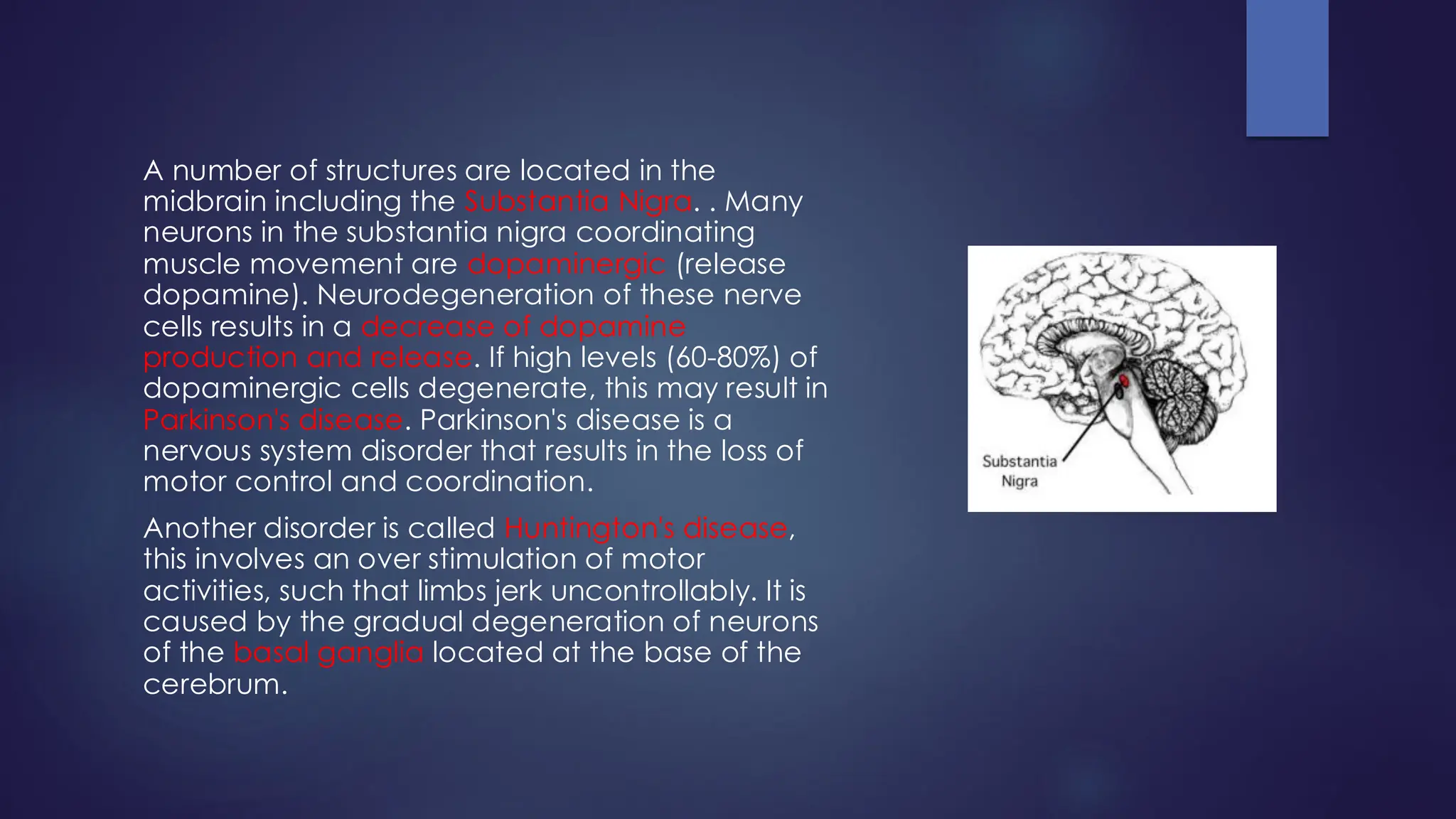
A number ofstructures are located in the
midbrain including the Substantia Nigra. . Many
neurons in the substantia nigra coordinating
muscle movement are dopaminergic (release
dopamine). Neurodegeneration of these nerve
cells results in a decrease of dopamine
production and release. If high levels (60-80%) of
dopaminergic cells degenerate, this may result in
Parkinson's disease. Parkinson's disease is a
nervous system disorder that results in the loss of
motor control and coordination.
Another disorder is called Huntington's disease,
this involves an over stimulation of motor
activities, such that limbs jerk uncontrollably. It is
caused by the gradual degeneration of neurons
of the basal ganglia located at the base of the
cerebrum.
18.
CEREBELLUM
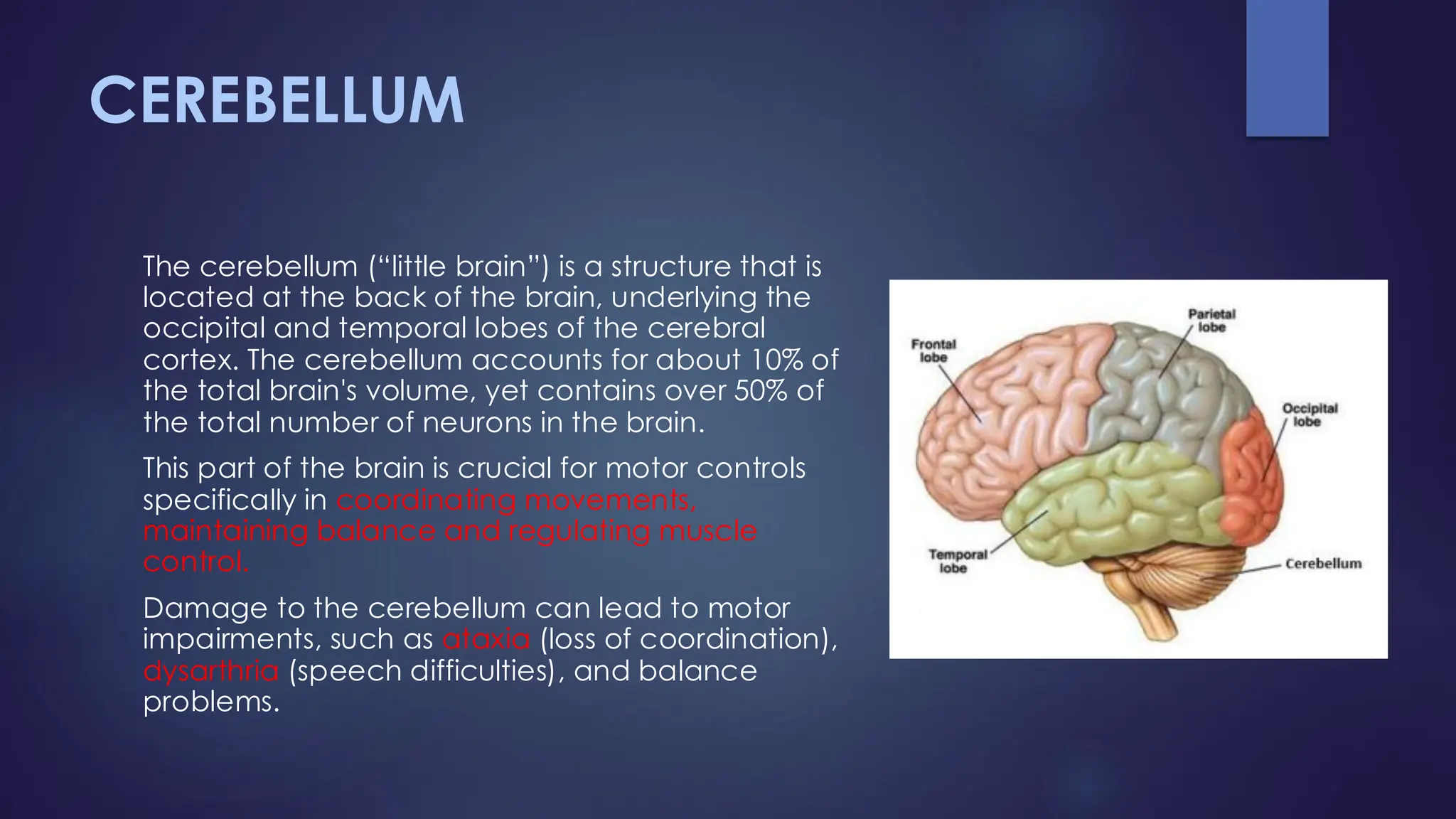
The cerebellum (“littlebrain”) is a structure that is
located at the back of the brain, underlying the
occipital and temporal lobes of the cerebral
cortex. The cerebellum accounts for about 10% of
the total brain's volume, yet contains over 50% of
the total number of neurons in the brain.
This part of the brain is crucial for motor controls
specifically in coordinating movements,
maintaining balance and regulating muscle
control.
Damage to the cerebellum can lead to motor
impairments, such as ataxia (loss of coordination),
dysarthria (speech difficulties), and balance
problems.
19.
PONS
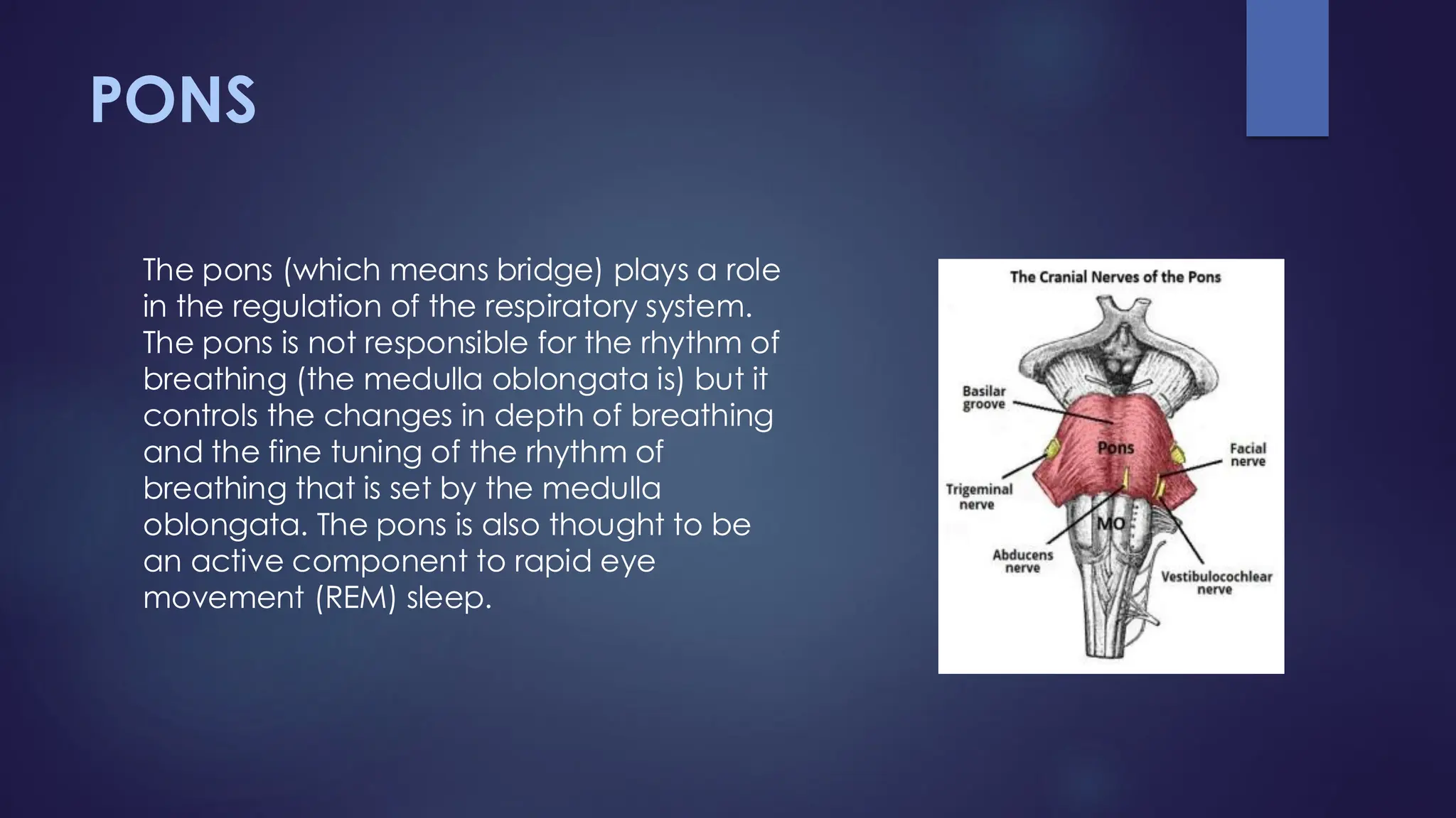
The pons (whichmeans bridge) plays a role
in the regulation of the respiratory system.
The pons is not responsible for the rhythm of
breathing (the medulla oblongata is) but it
controls the changes in depth of breathing
and the fine tuning of the rhythm of
breathing that is set by the medulla
oblongata. The pons is also thought to be
an active component to rapid eye
movement (REM) sleep.
20.
MEDULLA OBLONGATA
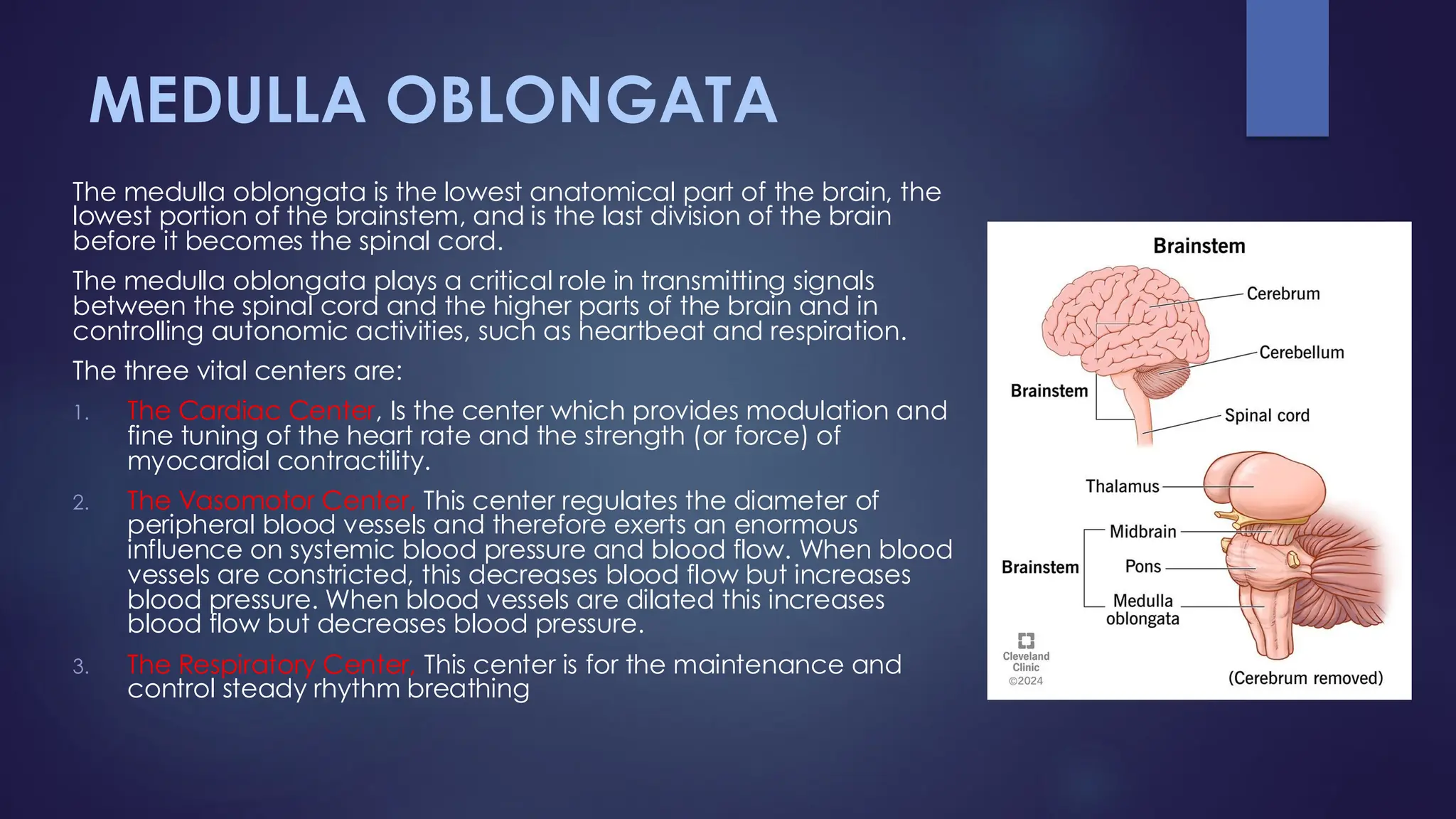
The medullaoblongata is the lowest anatomical part of the brain, the
lowest portion of the brainstem, and is the last division of the brain
before it becomes the spinal cord.
The medulla oblongata plays a critical role in transmitting signals
between the spinal cord and the higher parts of the brain and in
controlling autonomic activities, such as heartbeat and respiration.
The three vital centers are:
1. The Cardiac Center, Is the center which provides modulation and
fine tuning of the heart rate and the strength (or force) of
myocardial contractility.
2. The Vasomotor Center, This center regulates the diameter of
peripheral blood vessels and therefore exerts an enormous
influence on systemic blood pressure and blood flow. When blood
vessels are constricted, this decreases blood flow but increases
blood pressure. When blood vessels are dilated this increases
blood flow but decreases blood pressure.
3. The Respiratory Center, This center is for the maintenance and
control steady rhythm breathing
21.
THE CENTRAL NERVOUSSYSTEM –
THE SPINAL CORD
The basic structure of the spinal cord is that it is the downward
continuation of medulla oblongata. All spinal nerves are ‘mixed’
nerves meaning they all contain axons of both sensory (incoming) and
motor (outgoing) neurons, thus information is going in both directions.
Spinal cord is cylindrical in shape. Length of the spinal cord is about 45
cm in males and about 43 cm in females.
Cerebrospinal fluid (CSF) is a clear, colorless fluid that is found in the
brain and spinal cord. The Central Nervous System (CNS) gets its
oxygen and nutrients from CSF and not directly from blood.
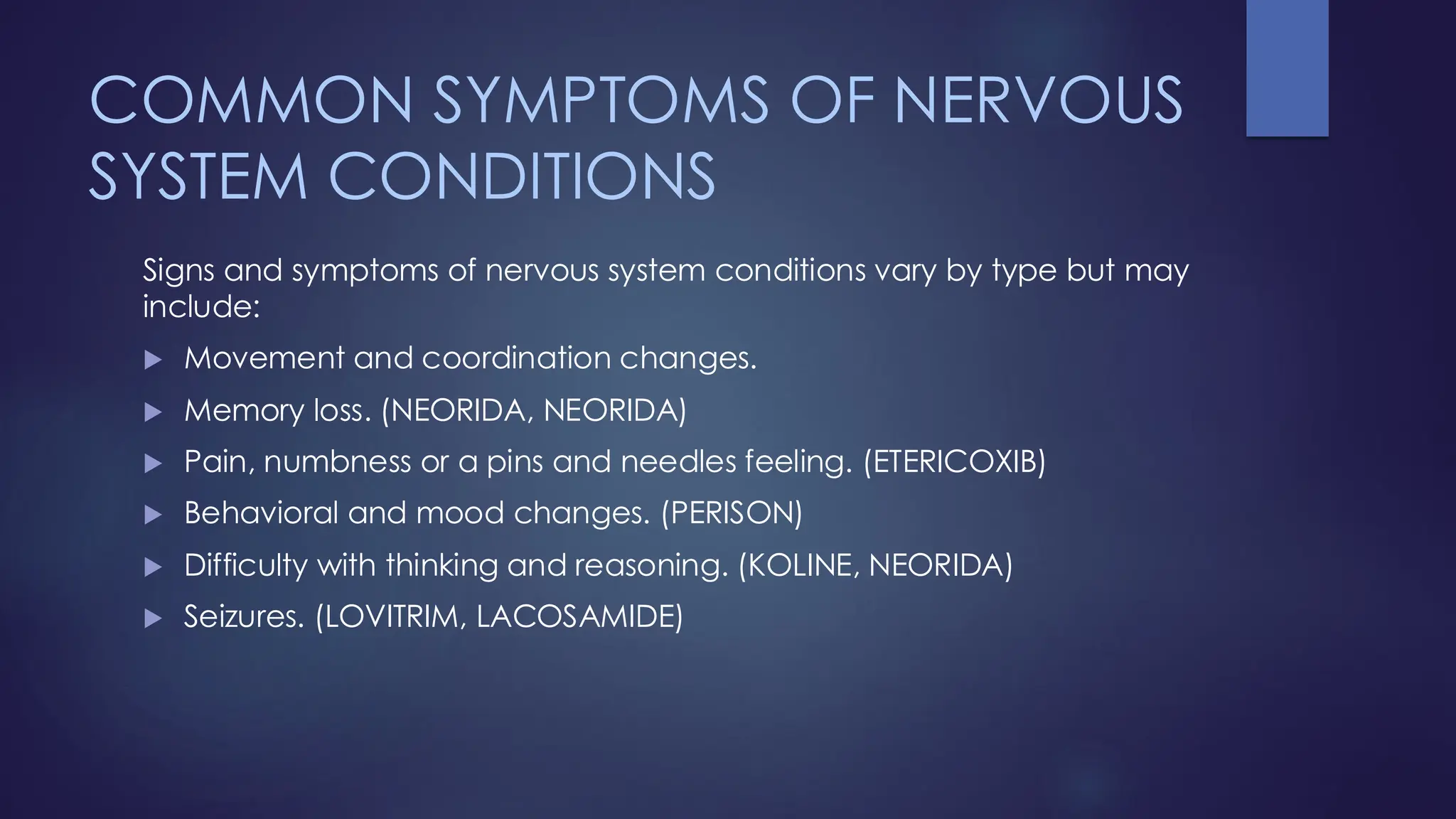
COMMON SYMPTOMS OFNERVOUS
SYSTEM CONDITIONS
Signs and symptoms of nervous system conditions vary by type but may
include:
Movement and coordination changes.
Memory loss. (NEORIDA, NEORIDA)
Pain, numbness or a pins and needles feeling. (ETERICOXIB)
Behavioral and mood changes. (PERISON)
Difficulty with thinking and reasoning. (KOLINE, NEORIDA)
Seizures. (LOVITRIM, LACOSAMIDE)