3. CLASSIFICATION OF WHITE FIBERS
A-ASSOCIATION FIBERS.
B-COMMISSURAL FIBERS.
C-PROJECTION FIBERS.
4. CLASSIFICATION OF WHITE MATTER
1.Association fibers
•Connect different parts of cerebral cortex of
same cerebral hemisphere.
•Short association fibers connect adjacent gyri
to each other.
•Long association fibers connect gyri located at
a distance from each other.
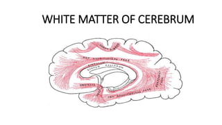
7. EXAMPLES OF LONG ASSOCIATION FIBERS
1. Cingulum:-
•lies in cingulate gyrus.
•Extends from paraterminal gyrus to uncus.
•Part of Papez circuit ( part of limbic system)
2. Uncinate fasciculus:-
•Connects inferior frontal gyrus and orbital gyri of frontal lobe to
the cortex.
•Connects sensory and motor speech areas.
8. 3.Superior longitudinal fasciculus:-
•Long bundle beginning in frontal lobe.
•Arches back to occipital lobe via parietal lobe, then
turning in temporal lobe.
•So, it connects occipital lobe to frontal eye field.
4.Inferior Longitudinal fasciculus:-
•Connects occipital lobe to temporal lobe.
11. EXAMPLES OF COMMISSURAL FIBERS
1.Anterior commissure
• Situated in anterior wall of third ventricle
• Connects right and left temporal lobes
• Crosses midline in the upper part of lamina terminalis, just anterior
to anterior columns of fornix and interventricular foramen.
• Fibers divide in anterior and posterior bundles.
(a): Anterior bundle :- smaller connects olfactory regions of two
sides.
(b): Posterior bundle:-larger, connects lower and anterior parts of
temporal lobes.
12. 2. Posterior and habenular commissure:-
• Part of epithalamus.
• Lie in the posterior part of roof of third ventricle.
• Posterior commissure crosses the midline
through inferior lamina of stalk of pineal gland.
• Habenular commissure crosses the midline
through superior lamina of stalk of pineal gland.
15. EXAMPLES OF PROJECTION FIBERS
1.CORONA RADIATA:-Mass of white matter composed of projection
fibers, converging from cerebral cortex to internal capsule and fan out
from internal capsule to cortex.
2. INTERNAL CAPSULE:-gives passage to descending & ascending fibers.
(a) Descending fibers (corticospinal, corticopontine and corticonuclear)
arise in the cortex and terminate in lower motor neurons (anterior horn
cells of spinal cord, pontine nuclei and cranial nerve nuclei).
(b) Ascending fibers arise in neurons of thalamus as thalamic radiations
(anterior, posterior, superior and inferior), terminate in cerebral cortex.
3. FORNIX :- projection fibers, part of limbic system, arising from
hippocampus (part of limbic system) and terminating in mamillary body
(part of hypothalamus).
19. CORPUS CALLOSUM
Located in the floor of longitudinal fissure.
•Shape of an arch.
PARTS:-
1.Genu :- anterior end.
2.Trunk :- central part.
3.Splenium:- posterior bulbous part.
4.Rostrum:- prolongation from genu to the upper end of lamina
terminalis.
23. RELATIONS
• Upper surface covered with thin layer of grey matter called indusium
griseum. It contains two bands of white matter termed lateral and
medial longitudinal stria.
1.SPLENIUM:- related above to falx cerebri and inferior sagittal sinus.
• Below related to great cerebral vein, pineal body and midbrain
tectum.
2.BODY:-lower surface of body gives attachment to septum
pellucidum anteriorly and is closely related to body of fornix posteriorly.
• On each side it forms roof of central part of lateral ventricle.
• Upper surface forms floor of longitudinal fissure .
24. 3.GENU :-
• Anteriorly related anterior cerebral vessels.
• Posteriorly gives attachment to septum pellucidum and
forms the anterior wall of the anterior horn of lateral
ventricle on each side.
4.ROSTRUM:-
• Upper surface gives attachment to septum pellucidum in
the midline and on each side forms the floor of anterior
horn of lateral ventricle.
26. CONNECTIONS
• Rostrum connects orbital surfaces of frontal lobes.
• Genu interconnect two frontal lobes and these fibers form a fork like
bundle called forceps minor.
• Body has wide cortical connections. Majority of fibers run
transversely and intersect with fibers of corona radiata.
-Some fibers of body and adjacent splenium do not intersect with
corona radiata and form a sheet called tapetum. Tapetum forms roof
and lateral walls of posterior and inferior horns of lateral ventricle.
• Splenial fibers interconnect occipital lobes. They curve backwards and
medially and form forceps major. Forceps major bulges in the medial
wall of posterior horn of lateral ventricle and gives rise to an elevation
called bulb of posterior horn
31. APPLIED
(a) CALLOSAL SYNDROME/SPLIT BRAIN SYNDROME:-
• Corpus callosum congenitally absent
• Two hemispheres are disconnected. So, non dominant hemisphere
(right) is dissociated from dominant hemisphere (left).
• Senses perceived by left hemisphere are unable to go to right
hemisphere.
SYMPTOMS
1. Hemialexia:- Patient unable to read matter in the left visual field.
2. Left ideomotor apraxia:- patient not able to carry out work with left
hand in response to verbal command.
3. Left agraphia:-not able to write with left hand.
4. Left tactile anomia:-not able to name an object held in left hand with
eyes closed.