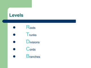
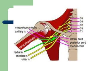
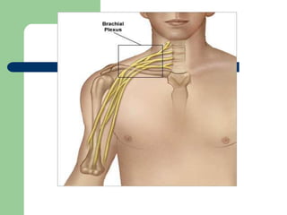
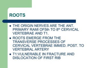
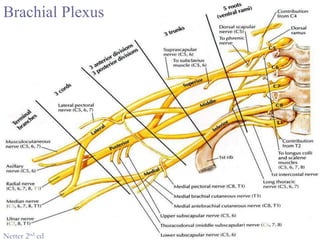
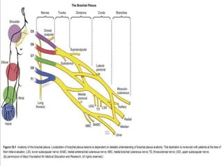
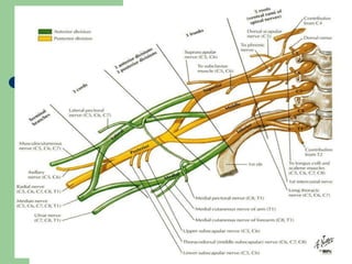
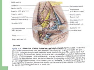
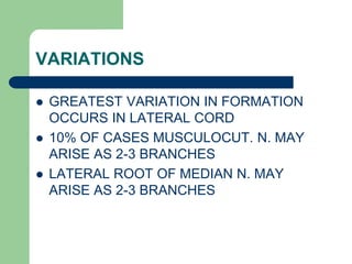
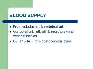
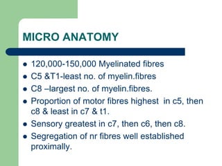
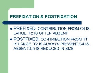
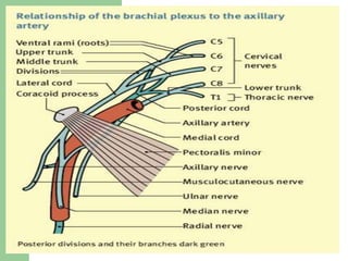
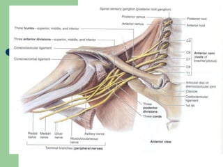
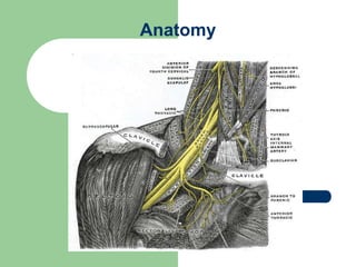
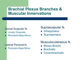
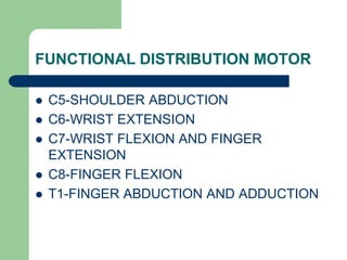
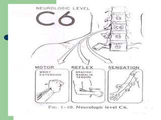
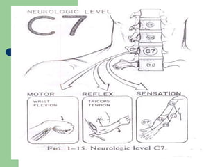
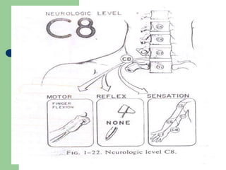
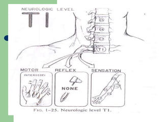
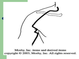
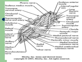
SURGICAL ANATOMY OF
BRACHIAL PLEXUS
Sudhir suggala
ANATOMY
 A network of nerves starting from 5 nerve
roots in upper spine & ending in nerves that
control movement & sensation in the upper
limb.
ANATOMY contd…
 Located where neck joins shoulder behind
clavicle & between spine & upper arm just
distal to axilla.
 5 roots ant.primary rami after giving
segmental supply to prevertebral muscles
&scalene muscles.
Levels
 Roots
 Trunks
 Divisions
 Cords
 Branches
ROOTS
 THE ORIGIN NERVES ARE THE ANT.
PRIMARY RAMI OF5th TO 8th CERVICAL
VERTEBRAE AND T1.
 ROOTS EMERGE FROM THE
TRANSVERSE PROCESSES OF
CERVICAL VERTEBRAE IMMED. POST. TO
VERTEBRAL ARTERY
 T1:VULNERABLE IN FRACTURE AND
DISLOCATION OF FIRST RIB
NERVES FROM ROOTS .
 DORSAL SCAPULAR N. C5
 ARISES FROM C5 JUST LAT. TO LONG
THORACIC
 TRAV. POST TO Plx
 INNERVATES LEV.SCAPULAE, RHOMBOIDES
Brachial Plexus
Netter 2nd ed
ROOTS contd…
 LONG THORACIC N. OF BELL
C567
 IMMED. AFTER THEY ORIG. FROM
INTERVERTEBRAL FORAMEN
 ALSO PIERCES S.MED. AS IT PASSES
POST TO Plx
TRUNKS
 IN THE POST. TRIANGLE OF NECK
 C56: UPPER/SUPERIOR TRUNK
 C7; MIDDLE TRUNK
 C8 T1: LOWER/INFERIOR TRUNK
 TRUNKS LIE IN FRONT OF ONE
ANOTHER
 RELATION b/w C5 AND PHRENIC NERVE
NERVES FROM TRUNKS
 SUPRASCAPULAR N. C5,6
 SENSORY TO SHOULDER JT.
 MOTOR TO SUPRASPINATUS AND
INFRASPINATUS
 N.TO SUBCLAVIUS C5,6
DIVISONS
 LIE DEEP TO CLAVICLE
 POSTERIOR DIV. OF UPPER AND MIDDLE
TRUNK LARGER
 10% OF CASES NO POST. DIVISON OF
LOWER TRUNK
CORDS
 NAMED LAT. ,POST. AND MEDIAL ACC.
TO RELATION WITH AXILLARY ARTERY
 LATERAL CORD: ANT. DIV OF UPPER &
MIDDLE TRUNK
 MEDIAL CORD: ANT. DIV. OF LOWER
TRUNK
 POSTERIOR CORD:POST./DORSAL
DIVISONS OF ALL 3 TRUNKS
FROM LAT.CORD
 Lateral pectoral nerve- c5,c6,c7
 Pierce clavipectoral fascia.
 Communicate with medial pectoral across front
of 1st part of axillary art.
 Br. To P.major & P.minor
 No cut. Br.
FROM LAT. CORD contd…
 Musculocutaneous nr.-c5,c6,c7
 To biceps & brachialis.
 Becomes lateral cut. Nr. Of forearm.
 Lateral root of median nr.-c5,c6,c7
 Continuation of lat.cord
FROM MEDIAL CORD
Medial pectoral nr.- behind 1st part of ax.art
Perforates p.minor
No cut.br.
Medial root of median nr-. Crosses ax. Art to join lat root.
Medial cut.nr. Of arm – smallest & most medial.
Medial cut. Nr. Of forearm –
Large nr.
Runs down bet ax.art & ax. Vein in front of ulnar nr.
Ulnar nr.- largest br. C7 C8 T1
To most of the hand muscles.
FROM POST CORD contd…
 Radial nr. – c5,c6,c7,c8,T1
 Cont.of post cord
 Largest in the whole plexus
 Gives post cut nr. Of forearm.
FROM POST.CORD
 Upper subscapular- c5,c6
 Small nr.
 Enters upper part of subscapularis.
 Thoracodorsal – c6,c7,c8
 nr.to lat. Dorsi.
 A large nerve.
 Runs down post.ax. Wall.
FROM POST CORD contd…
 Lower subscapular –c5,c6
 To lower part of subscapularis
 Ends in teres major.
 Axillary nr. – c5,c6
 Circumflex nr.
 A large terminal br.
 No supply in axilla.
 Br.to shoulder jt.
 Ant. & post div.
FROM POST CORD contd…
 Radial nr. – c5,c6,c7,c8,T1
 Cont.of post cord
 Largest in the whole plexus
 Gives post cut nr. Of forearm.
VARIATIONS
 GREATEST VARIATION IN FORMATION
OCCURS IN LATERAL CORD
 10% OF CASES MUSCULOCUT. N. MAY
ARISE AS 2-3 BRANCHES
 LATERAL ROOT OF MEDIAN N. MAY
ARISE AS 2-3 BRANCHES
BLOOD SUPPLY
 From subclavian & vertebral art.
 Vertebral art.- c5, c6. & more proximal
cervical nerves
 C8, T1,- br. From costocervical trunk.
MICRO ANATOMY
 120,000-150,000 Myelinated fibres
 C5 &T1-least no. of myelin.fibres
 C8 –largest no. of myelin.fibres.
 Proportion of motor fibres highest in c5, then
c8 & least in c7 & t1.
 Sensory greatest in c7, then c6, then c8.
 Segregation of nr fibres well established
proximally.
PREFIXATION & POSTFIXATION
 PREFIXED: CONTRIBUTION FROM C4 IS
LARGE ,T2 IS OFTEN ABSENT
 POSTFIXED: CONTRIBUTION FROM T1
IS LARGE, T2 IS ALWAYS PRESENT,C4 IS
ABSENT,C5 IS REDUCED IN SIZE
Anatomy
BRACHIAL PLEXUS SHEATH
 C.t. of prevertebral fascia
 Blends with fascia of biceps & brachialis.
 C.t. septae compartmentalize components of
the sheath.
 Controversies – septae may limit spread of
solutions during BP block.
Brachial Plexus Branches &
Muscular Innervations
Dorsal Scapular N.
 Levator Scapulae
 Rhomboid Major/Minor
Lateral Pectoral N.
 Pectoralis Major/Minor
Suprascapular N.
 Infraspinatus
 Supraspinatus
Musculocutaneous N.
 Biceps Brachii
 Brachialis
 Coracobrachialis
Brachial Plexus Branches &
Muscular Innervations
Axillary N.
 Deltoid
 Teres Minor
Upper Subscapular N.
 Subscapularis
Middle Subscapular or
Thoracodorsal N.
 Latissimus Dorsi
Lower Subscapular N.
 Subscapularis
 Teres Major
Brachial Plexus Branches &
Muscular Innervations
Median N.
 Abductor Pollicis
Brevis/Longus
 Flexor Carpi Radialis
 Flexor Digitorum Superficialis
 Flexor Digitorum Profundus
(Lat. 2)
 Flexor Pollicis Brevis (Lat.) &
Longus
 Lumbricales (Lat. 2)
 Opponens Pollicis
 Palmaris Longus
 Pronator Quadratus
 Pronator Teres *
Radial N.
 Abductor Pollicis Brevis
 Anconeus
 Brachioradialis
 Extensor Carpi Radialis
Brevis/Longus
 Extensor Carpi Ulnaris
 Extensor Digiti Minimi
 Extensor Digitorum Communis
 Extensor Indicis
 Extensor Pollicis
Brevis/Longus
 Supinator
 Triceps Brachii
Brachial Plexus Branches &
Muscular Innervations
Ulnar N.
 Abductor Digiti Minimi
 Adductor Pollicis
 Dorsal Interossei
 Flexor Carpi Ulnaris
 Flexor Digiti Minimi
 Flexor Digitorum Profundus
(Med. 2)
 Flexor Pollicis Brevis (Med.)
 Lumbricals (Med. 2)
 Opponens Digiti Minimi
 Palmar Interossei
Long Thoracic N.
 Serratus Anterior
Medial Pectoral N.
 Pectoralis Major
Medial Brachial Cutaneous N.
(sensory)
Medial Antebrachial
Cutaneous N. (sensory)
FUNCTIONAL DISTRIBUTION MOTOR
 C5-SHOULDER ABDUCTION
 C6-WRIST EXTENSION
 C7-WRIST FLEXION AND FINGER
EXTENSION
 C8-FINGER FLEXION
 T1-FINGER ABDUCTION AND ADDUCTION
SENSATION
 C5-LATERAL ARM
 C6-LAT.FOREARM ,THUMB,INDEX
FINGER
 C7-MIDDLE FINGER
 C8-MEDIAL FOREARM,RING,SMALL
FINGER
 T1-MEDIAL ARM
 T2-AXILLA
TRANSITIONAL ZONE
 BERTHOLD (1993):JUNC. b/w ROOTS AND
SPINAL CORD
 A CENTRAL-PERIPHERAL TRANSITIONAL
ZONE
 2 TYPES OF INTRADURAL INJURY-
RUPTURE AND AVULSION
RUPTURE & AVULSION
 INTRADURAL RUPTURE IS TEARING OF
ROOTLETS AT OR PERIPHERAL TO T.Z
 AVULSION IS ROOTLETS ARE TORN
FROM SPINAL CORD ITSELF AND INJURY
IS CENTRAL TO TRANSITIONAL ZONE .
 UPPER PLEXUS INJURY (ERB’S
PARALYSIS)
 LOWER PLEXUS INJURY(KLUMPKE’S
PARALYSIS)
 INJURY AT LEVEL OF ROOTS
CORDS INJURY
 LATERAL
 POSTERIOR
 MEDIAL
NEOPLASTIC RADIATION
THANK YOU

Brachial plexus injury

  • 1.
    SURGICAL ANATOMY OF BRACHIALPLEXUS Sudhir suggala
  • 2.
    ANATOMY  A networkof nerves starting from 5 nerve roots in upper spine & ending in nerves that control movement & sensation in the upper limb.
  • 3.
    ANATOMY contd…  Locatedwhere neck joins shoulder behind clavicle & between spine & upper arm just distal to axilla.  5 roots ant.primary rami after giving segmental supply to prevertebral muscles &scalene muscles.
  • 4.
    Levels  Roots  Trunks Divisions  Cords  Branches
  • 7.
    ROOTS  THE ORIGINNERVES ARE THE ANT. PRIMARY RAMI OF5th TO 8th CERVICAL VERTEBRAE AND T1.  ROOTS EMERGE FROM THE TRANSVERSE PROCESSES OF CERVICAL VERTEBRAE IMMED. POST. TO VERTEBRAL ARTERY  T1:VULNERABLE IN FRACTURE AND DISLOCATION OF FIRST RIB
  • 8.
    NERVES FROM ROOTS.  DORSAL SCAPULAR N. C5  ARISES FROM C5 JUST LAT. TO LONG THORACIC  TRAV. POST TO Plx  INNERVATES LEV.SCAPULAE, RHOMBOIDES
  • 9.
  • 11.
    ROOTS contd…  LONGTHORACIC N. OF BELL C567  IMMED. AFTER THEY ORIG. FROM INTERVERTEBRAL FORAMEN  ALSO PIERCES S.MED. AS IT PASSES POST TO Plx
  • 13.
    TRUNKS  IN THEPOST. TRIANGLE OF NECK  C56: UPPER/SUPERIOR TRUNK  C7; MIDDLE TRUNK  C8 T1: LOWER/INFERIOR TRUNK  TRUNKS LIE IN FRONT OF ONE ANOTHER  RELATION b/w C5 AND PHRENIC NERVE
  • 15.
    NERVES FROM TRUNKS SUPRASCAPULAR N. C5,6  SENSORY TO SHOULDER JT.  MOTOR TO SUPRASPINATUS AND INFRASPINATUS  N.TO SUBCLAVIUS C5,6
  • 16.
    DIVISONS  LIE DEEPTO CLAVICLE  POSTERIOR DIV. OF UPPER AND MIDDLE TRUNK LARGER  10% OF CASES NO POST. DIVISON OF LOWER TRUNK
  • 17.
    CORDS  NAMED LAT.,POST. AND MEDIAL ACC. TO RELATION WITH AXILLARY ARTERY  LATERAL CORD: ANT. DIV OF UPPER & MIDDLE TRUNK  MEDIAL CORD: ANT. DIV. OF LOWER TRUNK  POSTERIOR CORD:POST./DORSAL DIVISONS OF ALL 3 TRUNKS
  • 18.
    FROM LAT.CORD  Lateralpectoral nerve- c5,c6,c7  Pierce clavipectoral fascia.  Communicate with medial pectoral across front of 1st part of axillary art.  Br. To P.major & P.minor  No cut. Br.
  • 19.
    FROM LAT. CORDcontd…  Musculocutaneous nr.-c5,c6,c7  To biceps & brachialis.  Becomes lateral cut. Nr. Of forearm.  Lateral root of median nr.-c5,c6,c7  Continuation of lat.cord
  • 20.
    FROM MEDIAL CORD Medialpectoral nr.- behind 1st part of ax.art Perforates p.minor No cut.br. Medial root of median nr-. Crosses ax. Art to join lat root. Medial cut.nr. Of arm – smallest & most medial. Medial cut. Nr. Of forearm – Large nr. Runs down bet ax.art & ax. Vein in front of ulnar nr. Ulnar nr.- largest br. C7 C8 T1 To most of the hand muscles.
  • 21.
    FROM POST CORDcontd…  Radial nr. – c5,c6,c7,c8,T1  Cont.of post cord  Largest in the whole plexus  Gives post cut nr. Of forearm.
  • 22.
    FROM POST.CORD  Uppersubscapular- c5,c6  Small nr.  Enters upper part of subscapularis.  Thoracodorsal – c6,c7,c8  nr.to lat. Dorsi.  A large nerve.  Runs down post.ax. Wall.
  • 23.
    FROM POST CORDcontd…  Lower subscapular –c5,c6  To lower part of subscapularis  Ends in teres major.  Axillary nr. – c5,c6  Circumflex nr.  A large terminal br.  No supply in axilla.  Br.to shoulder jt.  Ant. & post div.
  • 24.
    FROM POST CORDcontd…  Radial nr. – c5,c6,c7,c8,T1  Cont.of post cord  Largest in the whole plexus  Gives post cut nr. Of forearm.
  • 25.
    VARIATIONS  GREATEST VARIATIONIN FORMATION OCCURS IN LATERAL CORD  10% OF CASES MUSCULOCUT. N. MAY ARISE AS 2-3 BRANCHES  LATERAL ROOT OF MEDIAN N. MAY ARISE AS 2-3 BRANCHES
  • 26.
    BLOOD SUPPLY  Fromsubclavian & vertebral art.  Vertebral art.- c5, c6. & more proximal cervical nerves  C8, T1,- br. From costocervical trunk.
  • 27.
    MICRO ANATOMY  120,000-150,000Myelinated fibres  C5 &T1-least no. of myelin.fibres  C8 –largest no. of myelin.fibres.  Proportion of motor fibres highest in c5, then c8 & least in c7 & t1.  Sensory greatest in c7, then c6, then c8.  Segregation of nr fibres well established proximally.
  • 28.
    PREFIXATION & POSTFIXATION PREFIXED: CONTRIBUTION FROM C4 IS LARGE ,T2 IS OFTEN ABSENT  POSTFIXED: CONTRIBUTION FROM T1 IS LARGE, T2 IS ALWAYS PRESENT,C4 IS ABSENT,C5 IS REDUCED IN SIZE
  • 31.
  • 32.
    BRACHIAL PLEXUS SHEATH C.t. of prevertebral fascia  Blends with fascia of biceps & brachialis.  C.t. septae compartmentalize components of the sheath.  Controversies – septae may limit spread of solutions during BP block.
  • 33.
    Brachial Plexus Branches& Muscular Innervations Dorsal Scapular N.  Levator Scapulae  Rhomboid Major/Minor Lateral Pectoral N.  Pectoralis Major/Minor Suprascapular N.  Infraspinatus  Supraspinatus Musculocutaneous N.  Biceps Brachii  Brachialis  Coracobrachialis
  • 34.
    Brachial Plexus Branches& Muscular Innervations Axillary N.  Deltoid  Teres Minor Upper Subscapular N.  Subscapularis Middle Subscapular or Thoracodorsal N.  Latissimus Dorsi Lower Subscapular N.  Subscapularis  Teres Major
  • 35.
    Brachial Plexus Branches& Muscular Innervations Median N.  Abductor Pollicis Brevis/Longus  Flexor Carpi Radialis  Flexor Digitorum Superficialis  Flexor Digitorum Profundus (Lat. 2)  Flexor Pollicis Brevis (Lat.) & Longus  Lumbricales (Lat. 2)  Opponens Pollicis  Palmaris Longus  Pronator Quadratus  Pronator Teres * Radial N.  Abductor Pollicis Brevis  Anconeus  Brachioradialis  Extensor Carpi Radialis Brevis/Longus  Extensor Carpi Ulnaris  Extensor Digiti Minimi  Extensor Digitorum Communis  Extensor Indicis  Extensor Pollicis Brevis/Longus  Supinator  Triceps Brachii
  • 36.
    Brachial Plexus Branches& Muscular Innervations Ulnar N.  Abductor Digiti Minimi  Adductor Pollicis  Dorsal Interossei  Flexor Carpi Ulnaris  Flexor Digiti Minimi  Flexor Digitorum Profundus (Med. 2)  Flexor Pollicis Brevis (Med.)  Lumbricals (Med. 2)  Opponens Digiti Minimi  Palmar Interossei Long Thoracic N.  Serratus Anterior Medial Pectoral N.  Pectoralis Major Medial Brachial Cutaneous N. (sensory) Medial Antebrachial Cutaneous N. (sensory)
  • 37.
    FUNCTIONAL DISTRIBUTION MOTOR C5-SHOULDER ABDUCTION  C6-WRIST EXTENSION  C7-WRIST FLEXION AND FINGER EXTENSION  C8-FINGER FLEXION  T1-FINGER ABDUCTION AND ADDUCTION
  • 39.
    SENSATION  C5-LATERAL ARM C6-LAT.FOREARM ,THUMB,INDEX FINGER  C7-MIDDLE FINGER  C8-MEDIAL FOREARM,RING,SMALL FINGER  T1-MEDIAL ARM  T2-AXILLA
  • 48.
    TRANSITIONAL ZONE  BERTHOLD(1993):JUNC. b/w ROOTS AND SPINAL CORD  A CENTRAL-PERIPHERAL TRANSITIONAL ZONE  2 TYPES OF INTRADURAL INJURY- RUPTURE AND AVULSION
  • 49.
    RUPTURE & AVULSION INTRADURAL RUPTURE IS TEARING OF ROOTLETS AT OR PERIPHERAL TO T.Z  AVULSION IS ROOTLETS ARE TORN FROM SPINAL CORD ITSELF AND INJURY IS CENTRAL TO TRANSITIONAL ZONE .
  • 59.
     UPPER PLEXUSINJURY (ERB’S PARALYSIS)  LOWER PLEXUS INJURY(KLUMPKE’S PARALYSIS)  INJURY AT LEVEL OF ROOTS
  • 60.
    CORDS INJURY  LATERAL POSTERIOR  MEDIAL
  • 61.
  • 62.