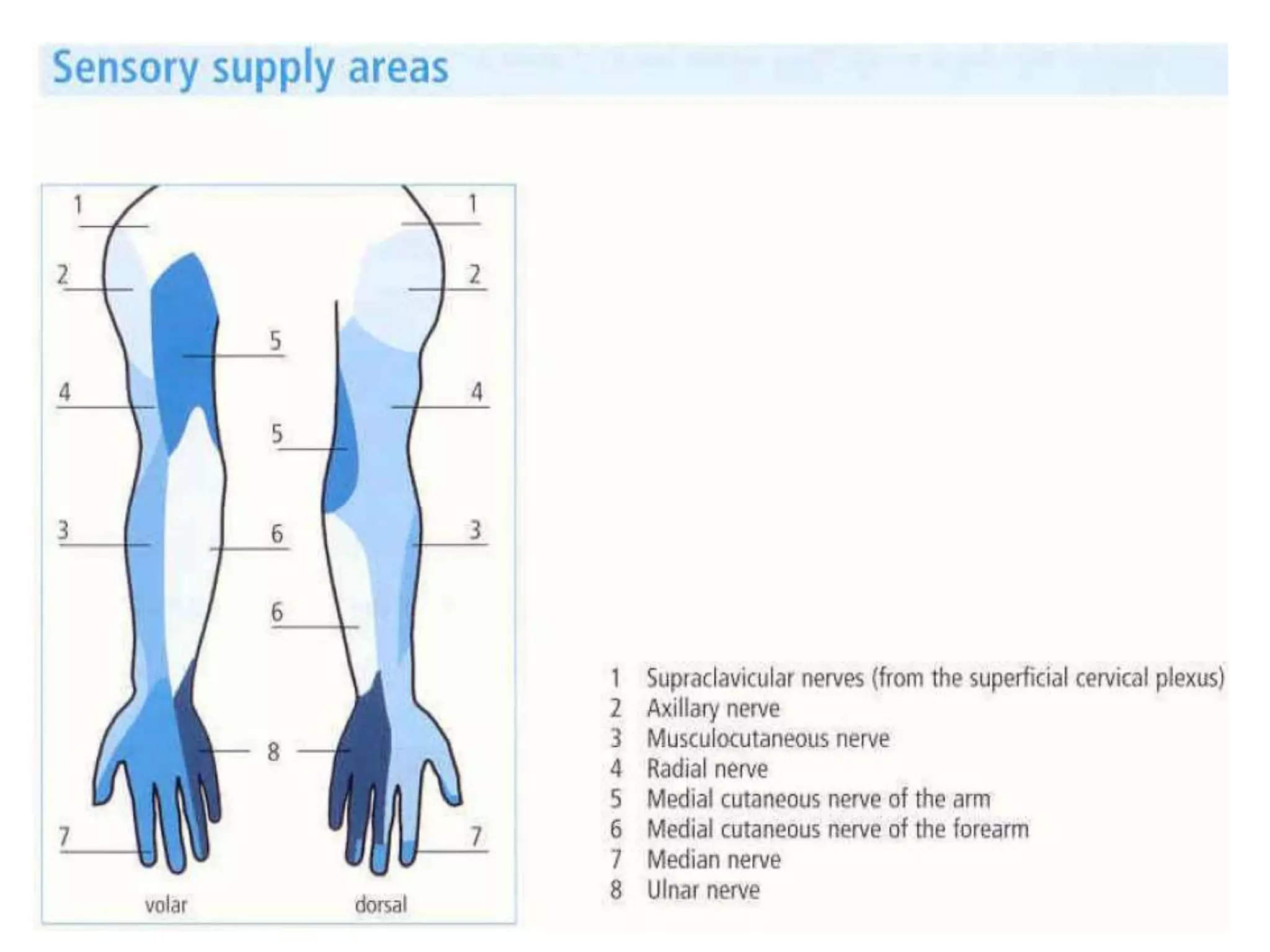
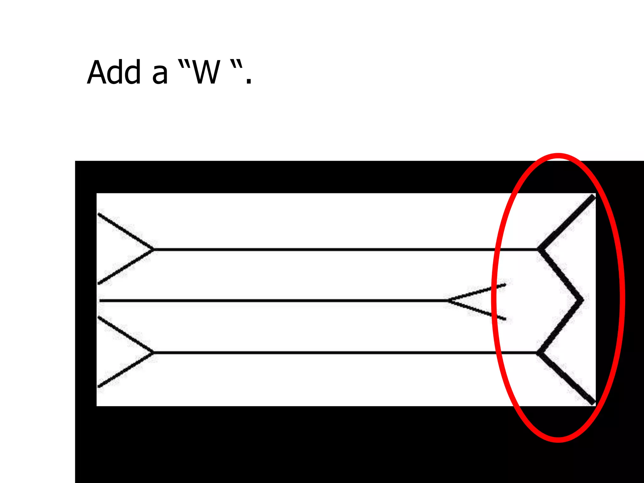
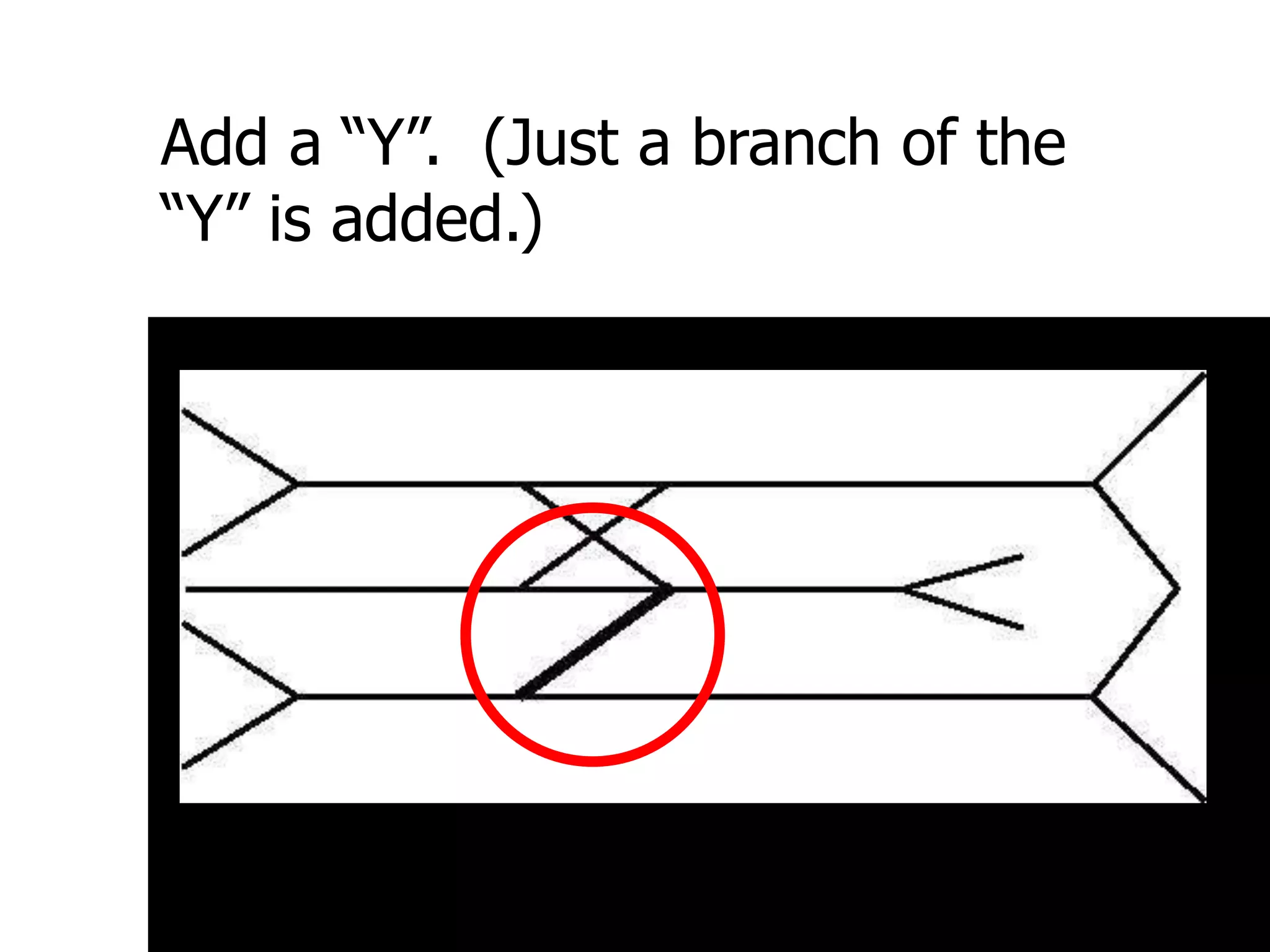
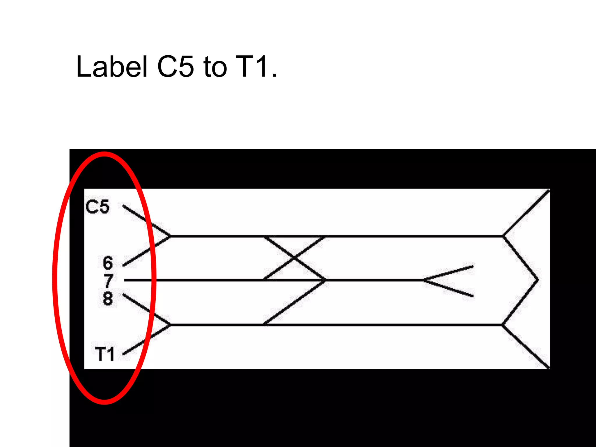
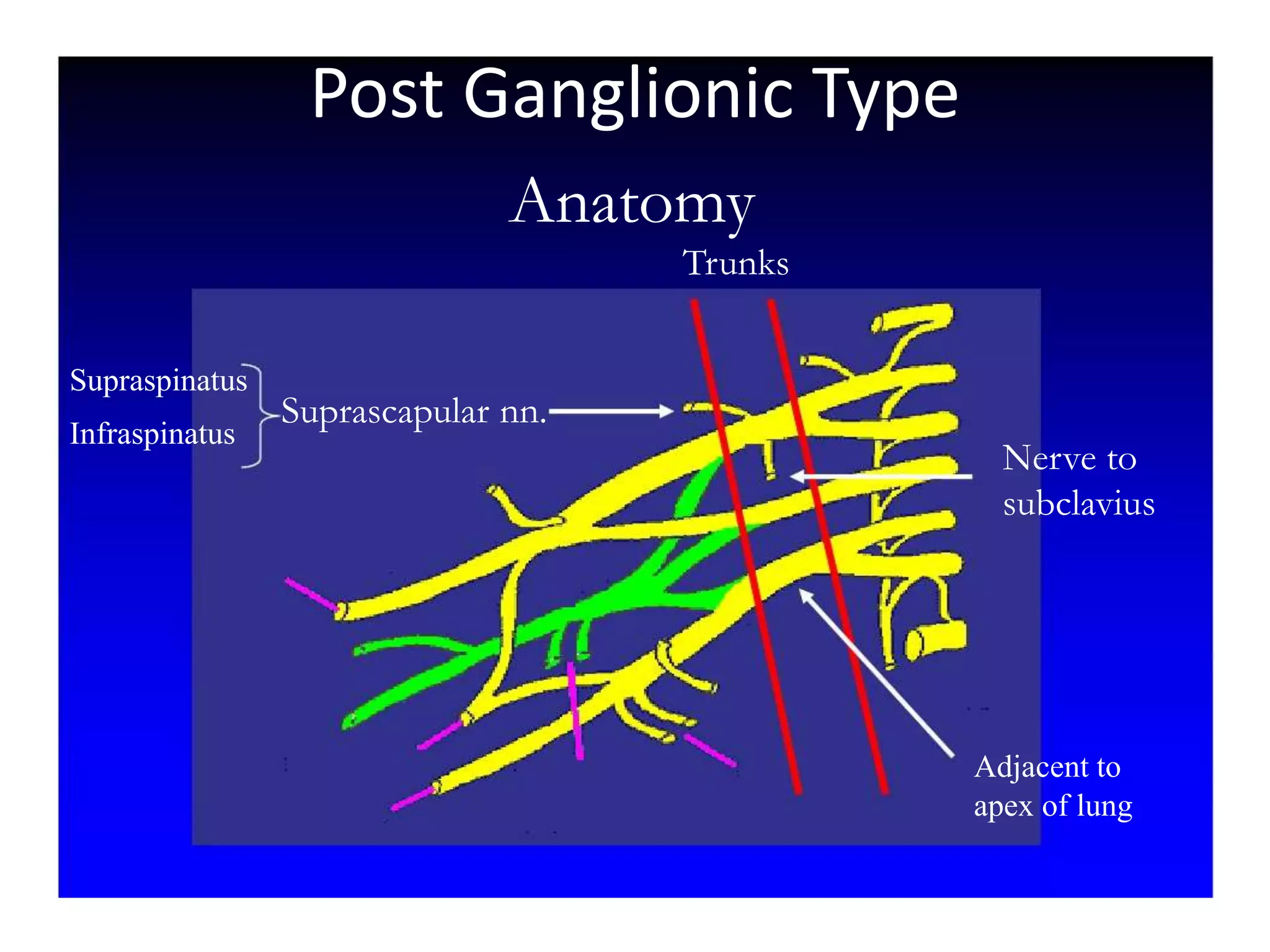
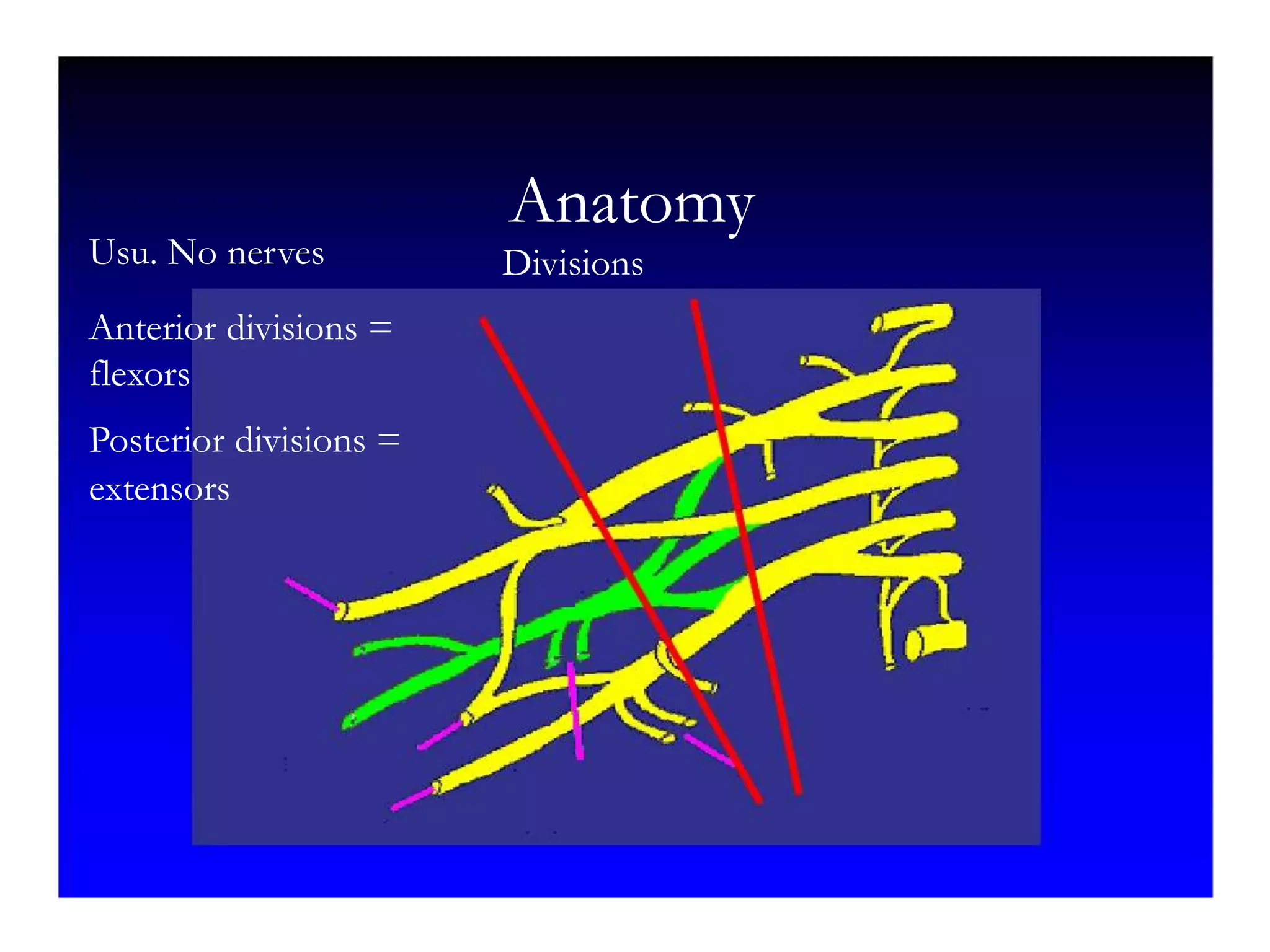
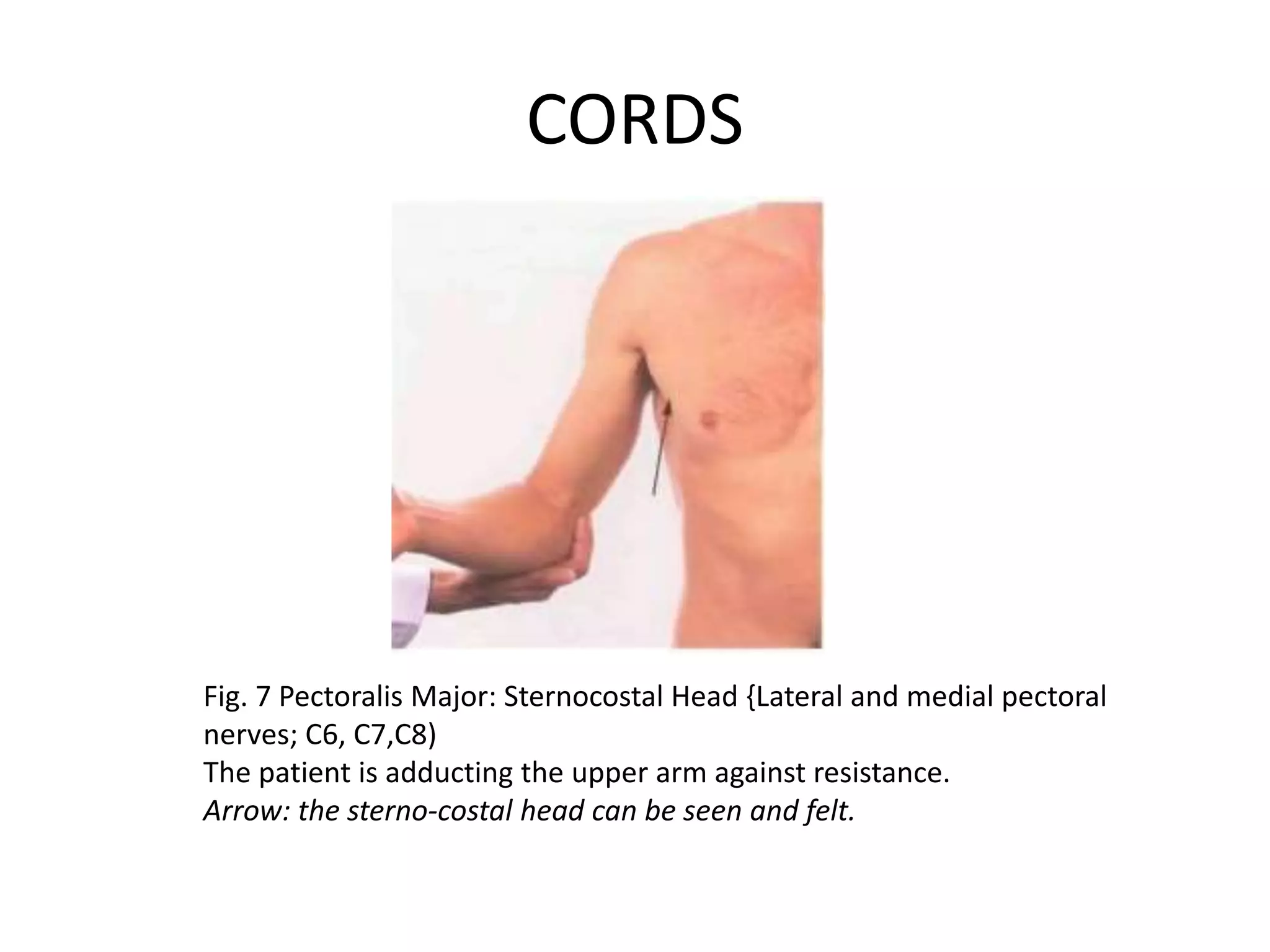
Brachial Plexus
Novra Yuditya Santoso
Branches
The brachial plexus is subdivided into roots, trunks,
divisions, cords, and branches
Composed of :
5 Roots
3 trunks – sup, med, inf
6 Divisions – ant, post
3 Cords – lat, post, med
5 Branches
Roots
- Ventral rami of spinal nerves C5-T1
- The roots emerge from the transverse
processes of the cervical vertebrae
immediately posterior to the vertebral artery
Trunks
- The 5 roots unite to form 3 trunks
- Pass between anterior and middle scalene muscles
- The ventral rami of C5 and C6 unite to form the upper
trunk
- The ventral ramus of C7 continues as the middle trunk
- The ventral rami of C8 and T1 unite to form the lower
trunk
Divisions
- Each trunks splits into an anterior division and
a posterior division
- The anterior divisions usually supply flexor
muscles
- The posterior divisions usually supply extensor
muscles.
Cords
- lateral, posterior and medial cords.
- The divisions of the upper and middle trunks
unite to form the lateral cord
- The division of the lower trunk forms the
medial cord
- The posterior divisions from each of the 3
trunks unite to form the posterior cord.
Branches
Musculocutaneous Nerve
• Roots: C5, C6, C7
• Motor Functions: Innervates the
brachialis, biceps brachii and
coracobrachialis muscles
• Sensory Functions: Gives off the
lateral cutaneous branch of the
forearm, which innervates the lateral
half of the anterior forearm, and a
small lateral portion of the posterior
forearm
Branches
Axillary Nerve
• Roots: C5 and C6.
• Motor Functions: Innervates
the teres minor and deltoid
muscles.
Branches
Axillary Nerve
• Roots: C5 and C6.
• Sensory Functions: Gives off the
superior lateral cutaneous nerve
of arm, which innervates the
inferior region of the deltoid
Branches
Median Nerve
• Roots: C6 – T1
• Motor Functions:
Innervates most of the
flexor muscles in the
forearm, the thenar
muscles.
Branches
- Sensory Functions: Gives off the
palmar cutaneous branch, which
innervates the lateral part of the
palm, and the digital cutaneous
branch, which innervates the
lateral three and a half fingers on
the anterior (palmar) surface of
the hand.
Branches
• Radial Nerve
• Roots: C5-C8 and T1
• Motor Functions: Innervates the
triceps brachii, and the extensor
muscles in the posterior
compartment of the forearm.
• Sensory Functions: Innervates the
posterior aspect of the arm and
forearm, and the posterior, lateral
aspect of the hand.
Branches
• Sensory Functions: Innervates the
posterior aspect of the arm and
forearm, and the posterior, lateral
aspect of the hand.
Branches
Ulnar Nerve
• Roots: C8 and T1.
• Motor Functions: Innervates the
muscles of the hand (apart from
the thenar muscles and two lateral
lumbricals), flexor carpi ulnaris
and medial half of flexor digitorum
profundus.
Branches
• Sensory Functions: Innervates the
anterior and posterior surfaces of
the medial one and half fingers,
and associated palm area.
Other branches of the brachial plexus
Nerve Roots Muscles Cutaneous
Roots Dorsal scapular nerve C5 Rhomboid
Levator scapulae
Roots Long thoracic nerve C5 C6 C7 Serratus anterior
Nerve Roots Muscles Cutaneous
Upper
Trunk
Nerve to subclavius C5 C6 Subclavious
Upper
Trunk
Suprascapular nerve C5 C6 Supraspinatous
Infraspinatous
Nerve Roots Muscles Cutaneous
Lateral
cord
Lateral pectoral nerve C5 C6 C7 Pectoralis Major By
communication
with Medial
Pectoral Nerve
Nerve Roots Muscles Cutaneous
Medial
cord
Medial pectoral Nr C8 T1 Pectoralis major
Pectoralis minor
Medial
cord
Medial cutaneous nerve
of arm
C8 T1 front and medial
skin of the arm
Medial
cord
Medial cutaneous nerve
of forearm
C8 T1 medial skin of the
forearm
Nerve Roots Muscles Cutaneous
Post
cord
Upper subscapular nerve C5 C6 Sub scapilaris
(upper part)
Post
cord
Thoracodorsal Nr
(Middle subscapular)
C6 C7 C8 Latismus Dorsi
Post cord Lower scapular Nr C5 C6 Subscapularis
(lower part)
Teres major
Draw two headless arrows to the
right.
Add a headless arrow to the left.
Add a “W “.
Add an “X”.
Add a “Y”. (Just a branch of the
“Y” is added.)
Label C5 to T1.
Label the major branches:
MC = musculocutaneous,
M = median U = ulnar
R = radial AX = axillary
This diagram includes the main
branches and main nerve roots with
the proper connections.
The first “3” is the branches to C5,
6, and 7 which form LTN = long
thoracic nerve.
Next, each of the headless arrows
has three nerves attached to it. To
the top headless arrow, add its “3.”
Label these “3”: DSN = dorsoscapular
nerve; SS = suprascapular nerve;
LP = lateral pectoral nerve.
Add the “3” to the middle
headless arrow.
Label the second headless arrow “3”:
SS = subscapular
TD = thoracodorsal nerve.
Add the final “3” on the bottom
headless arrow.
Label the last “3.” MP = medial pectoral,
MBC = medial brachial cutaneous, and MABC = medial
antebrachial cutaneous. Remember: the brachial cutaneous
goes to the brachium or arm, and the MABC goes to the antebrachium or
forearm. The nerve to the forearm starts distally.
Label roots, trunks, divisions, cords,
terminal branches.
The complete brachial plexus
diagram:
THE WAY OF THINKING IN
BRACHIAL PLEXUS INJURY
In case of Brachial Plexus Injury :
1. Determine whether Obstetric / Trauma
2. In case of trauma determine Pre or Post
Ganglion Type
3. In case of trauma determine Complete or
Incomplete lession
4. In Case of Post Ganglionic Type determine
Level of Injury (RTDCB)
Obstetric Case
1. Erb type (C5-C6)
2. Klumke type (C7-Th1)
Trauma Setting
• History taking , Mechanisme Of Injury and Physical
Examination are needed
• Don’t forget Live saving in acute setting (ATLS)
PHYSICAL EXAMINATION
• Always remember ATLS
• HEAD TO TOE
- eye : horner’s syndrome
- neck : C-spine evaluation
- chest : asymetri , other injury
- abdomen
- extremity
• Local status :
L : skin colour, bruising, soft tissue swelling,
scar, muscle wasting, deformity
F : warm/not, skin dry/wet, tenderness,
muscle tone, pulse
M : A/P ROM
SPINAL CORD WITH MOTOR AND SENSORY
CELL BODIES
Pre Ganglion Type
1. Horner Syndrome (APEM) only shown if Th1
is involved
2. Winging scapula (+)
3. Vascular Injury (+/-)
4. X ray Cervical Fracture
5. MRI
Muscle chart
Level Nerve Muscle
Root Dorsalscapular
Long Thoracic
Rhomboid
Serratus Anterior
Trunk Suprascapular Supraspinatus
Infraspinatus
Cord Thoracodorsal
Lateral/Medial Pectoral
Lattisimus Dorsi
Pectoralis
Branch Axilla
Musculocutaneous
Radial
Median
Ulnar
Deltoid
Biceps
Tricep,ECRL,ECRB,EDC, ECU,APL,EPB, EPL
FCR,FDS,FPL,Thenar
FCU, FDP 34 ,Adductor Policis, Interosseus
ROM
Remember “Do Passive ROM first in order to exclude
possibility of joint contracture”
Fig. 1 Trapezius (Spinal accessory nerve and
C3, C4)
The patient is elevating the shoulder against
resistance.
Arrow: the thick upper part of the muscle can
be seen and felt.
Anatomy
Roots
Rhomboids
Levator Scapulae
Prefixed C4
C5
C6
C7
C8
T1
Postfixed T2
Long Thoracic
Dorsal Scapular
Serratus Anterior
Post Ganglionic Type
ROOT
Fig. 2 Rhomboids (Dorsal scapular nerve; C4, C5)
The patient is pressing the palm of his hand backwards against
the examiner's hand.
Arrow: the muscle bellies can be felt and sometimes seen.
ROOT
Fig. 3 Serratus anterior (Long thoracic nerve; C5, C6, C7)
The patient is pushing against a wall. The left serratus anterior is
paralysed and there is winging of the scapula
Anatomy
Trunks
Suprascapular nn.
Supraspinatus
Infraspinatus
Nerve to
subclavius
Adjacent to
apex of lung
Post Ganglionic Type
TRUNK
Fig. 4 Supraspinatus (Suprascapular nerve; C5, C6)
The patient is abducting the upper arm against resistance.
Arrow: the muscle belly can be felt and sometimes seen
TRUNK
Fig. 5 Infraspinatus {Suprascapular nerve; C5, C6)
Anatomy
Divisions
Usu. No nerves
Anterior divisions =
flexors
Posterior divisions =
extensors
Anatomy
Cords
Lateral pectoral n.
Pect major
Medial pectoral n.
Medial brachial cutaneous
Medial antebrach. Cutan.
Upper & lower subscapular n
Thoracodorsal n Lat dorsi
Teres major
subscapularis
CORDS
Fig. 6 Pectoralis Major; Clavicular Head (Lateral pectoral nerve; C5, C6)
The upper arm is above the horizontal and the patient is pushing forward
against the examiner's hand,
Arrow, the clavicular head of pectoralis major can be seen and felt.
CORDS
Fig. 7 Pectoralis Major: Sternocostal Head {Lateral and medial pectoral
nerves; C6, C7,C8)
The patient is adducting the upper arm against resistance.
Arrow: the sterno-costal head can be seen and felt.
CORDS
Fig. 8 Latissimus Dorsi (Thoracodorsal nerve; C6, C7, C8)
The upper arm is horizontal and the patient is adducting it against
resistance. Lower
arrow: the muscle belly can be seen and felt. The upper arrow points to
teres major.
Anatomy
Branches
Biceps
Brachialis (lat cut n of forearm)
coracobrachialis
musculocutaneous
Deltoid, teres minor
Axillary
radial
median
Ulnar
BRANCHES
Fig. 9 Biceps (Muscuiocutaneous nerve; C5, C6)
The patient is flexing the supinated forearm
against resistance.
Arrow: the muscle belly can be seen and fett
BRANCHES
Fig. 10 Deltoid (Axillary nerve; C5, C6)
The patient is abducting the upper arm against resistance.
Arrow: the anterior and middle fibres of the muscle can be
seen and felt.
BRANCHES
Fig. 11 Triceps (Radial nerve; C6, C7. C8)
The patient is extending the forearm at the elbow against
resistance.
Arrows: the long and lateral heads of the muscle can be seen
and felt.
BRANCHES
Fig. 12 First Dorsal Interosseous Muscle (Ulnar nerve; C8,
T1)
The patient is abducting the index finger against
resistance.
Arrow: the muscle belly can be felt and usually seen
BRANCHES
Fig. 13 Flexor Digitorium Superficialis (Median
nerve; C7, C8, T1)
THANK YOU
Treatment
Depend on time of presentation :
1. Emergency Treatment
2. Immediate Treatment (3 weeks – 6 months)
3. Late Treatment ( more than 6 months)
Type of treatment
Soft Tissue
1. Emergency presentation Nerve repair
2. Immediate presentation nerve procedure
3. Late Presentation muscle or bony procedure
Nerve, Muscle or Bony Procedure ?
• Nerve Procedure if 3 weeks – 6 months of
presentation
• Muscle or Bony Procedure if more than 6
months of presentation
Priority of Treatment
1. Elbow Flexion Reconstruction
- Soft tissue procedure : Nerve (ex. Nerve
transfer of N Intercostal or N Accesorius spinalis
into Musculocutaneous or Oberlin procedure)
and muscle procedure (ex. Steindler flexorplasty
or Free gracilis muscle transfer wich is innervated
by N Intercostal)
- Bony procedure
OBERLIN
PROCEDURE
Free gracilis muscle transfer, yang
diinervasi oleh N Intercostal
2. Shoulder Reconstruction
Goal : shoulder abduksi, external rotation and shoulder
stability
- Soft tissue : nerve transfer (ex. transfer distal part of
N Accesorius Spinalis into N Suprascapularis or triceps
branch of N Radialis transferred into N Axillaris, nerve
transfer by using N Phrenicus ) and Muscle transfer (Saha
Procedure)
- Bony Procedure (Arthrodesis Glenohumeral Joint)
3. Wrist and Hand Function
- Soft tissue procedure : muscle procedure(ex.Jones
Procedure and gracilis free functioning muscle transfer )
actually hand function is difficult to be achieved by nerve
transfer.
- Bony Procedure for wrist joint
Notes
• Steindler Flexorplasty (by re-route position of common
flexor muscle origin into anterior distal humerus)
• Saha Procedure (Transfer of Trapezius muscle into
Greater Tuberosity of Humerus)
• Jones procedure consist of :
PL into APL (Extention thumb)
PT into ECRL (Extention wrist joint)
FCU / FCR into EDC (Extention MCP)

Brachial Plexus NO (1).ppt

  • 1.
  • 2.
    Branches The brachial plexusis subdivided into roots, trunks, divisions, cords, and branches Composed of : 5 Roots 3 trunks – sup, med, inf 6 Divisions – ant, post 3 Cords – lat, post, med 5 Branches
  • 3.
    Roots - Ventral ramiof spinal nerves C5-T1 - The roots emerge from the transverse processes of the cervical vertebrae immediately posterior to the vertebral artery
  • 4.
    Trunks - The 5roots unite to form 3 trunks - Pass between anterior and middle scalene muscles - The ventral rami of C5 and C6 unite to form the upper trunk - The ventral ramus of C7 continues as the middle trunk - The ventral rami of C8 and T1 unite to form the lower trunk
  • 5.
    Divisions - Each trunkssplits into an anterior division and a posterior division - The anterior divisions usually supply flexor muscles - The posterior divisions usually supply extensor muscles.
  • 6.
    Cords - lateral, posteriorand medial cords. - The divisions of the upper and middle trunks unite to form the lateral cord
  • 7.
    - The divisionof the lower trunk forms the medial cord - The posterior divisions from each of the 3 trunks unite to form the posterior cord.
  • 8.
    Branches Musculocutaneous Nerve • Roots:C5, C6, C7 • Motor Functions: Innervates the brachialis, biceps brachii and coracobrachialis muscles • Sensory Functions: Gives off the lateral cutaneous branch of the forearm, which innervates the lateral half of the anterior forearm, and a small lateral portion of the posterior forearm
  • 9.
    Branches Axillary Nerve • Roots:C5 and C6. • Motor Functions: Innervates the teres minor and deltoid muscles.
  • 10.
    Branches Axillary Nerve • Roots:C5 and C6. • Sensory Functions: Gives off the superior lateral cutaneous nerve of arm, which innervates the inferior region of the deltoid
  • 11.
    Branches Median Nerve • Roots:C6 – T1 • Motor Functions: Innervates most of the flexor muscles in the forearm, the thenar muscles.
  • 12.
    Branches - Sensory Functions:Gives off the palmar cutaneous branch, which innervates the lateral part of the palm, and the digital cutaneous branch, which innervates the lateral three and a half fingers on the anterior (palmar) surface of the hand.
  • 13.
    Branches • Radial Nerve •Roots: C5-C8 and T1 • Motor Functions: Innervates the triceps brachii, and the extensor muscles in the posterior compartment of the forearm. • Sensory Functions: Innervates the posterior aspect of the arm and forearm, and the posterior, lateral aspect of the hand.
  • 14.
    Branches • Sensory Functions:Innervates the posterior aspect of the arm and forearm, and the posterior, lateral aspect of the hand.
  • 15.
    Branches Ulnar Nerve • Roots:C8 and T1. • Motor Functions: Innervates the muscles of the hand (apart from the thenar muscles and two lateral lumbricals), flexor carpi ulnaris and medial half of flexor digitorum profundus.
  • 16.
    Branches • Sensory Functions:Innervates the anterior and posterior surfaces of the medial one and half fingers, and associated palm area.
  • 17.
    Other branches ofthe brachial plexus Nerve Roots Muscles Cutaneous Roots Dorsal scapular nerve C5 Rhomboid Levator scapulae Roots Long thoracic nerve C5 C6 C7 Serratus anterior
  • 18.
    Nerve Roots MusclesCutaneous Upper Trunk Nerve to subclavius C5 C6 Subclavious Upper Trunk Suprascapular nerve C5 C6 Supraspinatous Infraspinatous
  • 19.
    Nerve Roots MusclesCutaneous Lateral cord Lateral pectoral nerve C5 C6 C7 Pectoralis Major By communication with Medial Pectoral Nerve
  • 20.
    Nerve Roots MusclesCutaneous Medial cord Medial pectoral Nr C8 T1 Pectoralis major Pectoralis minor Medial cord Medial cutaneous nerve of arm C8 T1 front and medial skin of the arm Medial cord Medial cutaneous nerve of forearm C8 T1 medial skin of the forearm
  • 21.
    Nerve Roots MusclesCutaneous Post cord Upper subscapular nerve C5 C6 Sub scapilaris (upper part) Post cord Thoracodorsal Nr (Middle subscapular) C6 C7 C8 Latismus Dorsi Post cord Lower scapular Nr C5 C6 Subscapularis (lower part) Teres major
  • 23.
    Draw two headlessarrows to the right.
  • 24.
    Add a headlessarrow to the left.
  • 25.
  • 26.
  • 27.
    Add a “Y”.(Just a branch of the “Y” is added.)
  • 28.
  • 29.
    Label the majorbranches: MC = musculocutaneous, M = median U = ulnar R = radial AX = axillary
  • 30.
    This diagram includesthe main branches and main nerve roots with the proper connections.
  • 31.
    The first “3”is the branches to C5, 6, and 7 which form LTN = long thoracic nerve.
  • 32.
    Next, each ofthe headless arrows has three nerves attached to it. To the top headless arrow, add its “3.”
  • 33.
    Label these “3”:DSN = dorsoscapular nerve; SS = suprascapular nerve; LP = lateral pectoral nerve.
  • 34.
    Add the “3”to the middle headless arrow.
  • 35.
    Label the secondheadless arrow “3”: SS = subscapular TD = thoracodorsal nerve.
  • 36.
    Add the final“3” on the bottom headless arrow.
  • 37.
    Label the last“3.” MP = medial pectoral, MBC = medial brachial cutaneous, and MABC = medial antebrachial cutaneous. Remember: the brachial cutaneous goes to the brachium or arm, and the MABC goes to the antebrachium or forearm. The nerve to the forearm starts distally.
  • 38.
    Label roots, trunks,divisions, cords, terminal branches.
  • 40.
    The complete brachialplexus diagram:
  • 41.
    THE WAY OFTHINKING IN BRACHIAL PLEXUS INJURY
  • 42.
    In case ofBrachial Plexus Injury : 1. Determine whether Obstetric / Trauma 2. In case of trauma determine Pre or Post Ganglion Type 3. In case of trauma determine Complete or Incomplete lession 4. In Case of Post Ganglionic Type determine Level of Injury (RTDCB)
  • 43.
    Obstetric Case 1. Erbtype (C5-C6) 2. Klumke type (C7-Th1)
  • 44.
    Trauma Setting • Historytaking , Mechanisme Of Injury and Physical Examination are needed • Don’t forget Live saving in acute setting (ATLS)
  • 45.
    PHYSICAL EXAMINATION • Alwaysremember ATLS • HEAD TO TOE - eye : horner’s syndrome - neck : C-spine evaluation - chest : asymetri , other injury - abdomen - extremity
  • 46.
    • Local status: L : skin colour, bruising, soft tissue swelling, scar, muscle wasting, deformity F : warm/not, skin dry/wet, tenderness, muscle tone, pulse M : A/P ROM
  • 47.
    SPINAL CORD WITHMOTOR AND SENSORY CELL BODIES
  • 48.
    Pre Ganglion Type 1.Horner Syndrome (APEM) only shown if Th1 is involved 2. Winging scapula (+) 3. Vascular Injury (+/-) 4. X ray Cervical Fracture 5. MRI
  • 50.
    Muscle chart Level NerveMuscle Root Dorsalscapular Long Thoracic Rhomboid Serratus Anterior Trunk Suprascapular Supraspinatus Infraspinatus Cord Thoracodorsal Lateral/Medial Pectoral Lattisimus Dorsi Pectoralis Branch Axilla Musculocutaneous Radial Median Ulnar Deltoid Biceps Tricep,ECRL,ECRB,EDC, ECU,APL,EPB, EPL FCR,FDS,FPL,Thenar FCU, FDP 34 ,Adductor Policis, Interosseus
  • 51.
    ROM Remember “Do PassiveROM first in order to exclude possibility of joint contracture” Fig. 1 Trapezius (Spinal accessory nerve and C3, C4) The patient is elevating the shoulder against resistance. Arrow: the thick upper part of the muscle can be seen and felt.
  • 52.
    Anatomy Roots Rhomboids Levator Scapulae Prefixed C4 C5 C6 C7 C8 T1 PostfixedT2 Long Thoracic Dorsal Scapular Serratus Anterior Post Ganglionic Type
  • 53.
    ROOT Fig. 2 Rhomboids(Dorsal scapular nerve; C4, C5) The patient is pressing the palm of his hand backwards against the examiner's hand. Arrow: the muscle bellies can be felt and sometimes seen.
  • 54.
    ROOT Fig. 3 Serratusanterior (Long thoracic nerve; C5, C6, C7) The patient is pushing against a wall. The left serratus anterior is paralysed and there is winging of the scapula
  • 56.
  • 57.
    TRUNK Fig. 4 Supraspinatus(Suprascapular nerve; C5, C6) The patient is abducting the upper arm against resistance. Arrow: the muscle belly can be felt and sometimes seen
  • 58.
    TRUNK Fig. 5 Infraspinatus{Suprascapular nerve; C5, C6)
  • 59.
    Anatomy Divisions Usu. No nerves Anteriordivisions = flexors Posterior divisions = extensors
  • 61.
    Anatomy Cords Lateral pectoral n. Pectmajor Medial pectoral n. Medial brachial cutaneous Medial antebrach. Cutan. Upper & lower subscapular n Thoracodorsal n Lat dorsi Teres major subscapularis
  • 62.
    CORDS Fig. 6 PectoralisMajor; Clavicular Head (Lateral pectoral nerve; C5, C6) The upper arm is above the horizontal and the patient is pushing forward against the examiner's hand, Arrow, the clavicular head of pectoralis major can be seen and felt.
  • 63.
    CORDS Fig. 7 PectoralisMajor: Sternocostal Head {Lateral and medial pectoral nerves; C6, C7,C8) The patient is adducting the upper arm against resistance. Arrow: the sterno-costal head can be seen and felt.
  • 64.
    CORDS Fig. 8 LatissimusDorsi (Thoracodorsal nerve; C6, C7, C8) The upper arm is horizontal and the patient is adducting it against resistance. Lower arrow: the muscle belly can be seen and felt. The upper arrow points to teres major.
  • 65.
    Anatomy Branches Biceps Brachialis (lat cutn of forearm) coracobrachialis musculocutaneous Deltoid, teres minor Axillary radial median Ulnar
  • 66.
    BRANCHES Fig. 9 Biceps(Muscuiocutaneous nerve; C5, C6) The patient is flexing the supinated forearm against resistance. Arrow: the muscle belly can be seen and fett
  • 67.
    BRANCHES Fig. 10 Deltoid(Axillary nerve; C5, C6) The patient is abducting the upper arm against resistance. Arrow: the anterior and middle fibres of the muscle can be seen and felt.
  • 68.
    BRANCHES Fig. 11 Triceps(Radial nerve; C6, C7. C8) The patient is extending the forearm at the elbow against resistance. Arrows: the long and lateral heads of the muscle can be seen and felt.
  • 69.
    BRANCHES Fig. 12 FirstDorsal Interosseous Muscle (Ulnar nerve; C8, T1) The patient is abducting the index finger against resistance. Arrow: the muscle belly can be felt and usually seen
  • 70.
    BRANCHES Fig. 13 FlexorDigitorium Superficialis (Median nerve; C7, C8, T1)
  • 71.
  • 80.
    Treatment Depend on timeof presentation : 1. Emergency Treatment 2. Immediate Treatment (3 weeks – 6 months) 3. Late Treatment ( more than 6 months)
  • 81.
    Type of treatment SoftTissue 1. Emergency presentation Nerve repair 2. Immediate presentation nerve procedure 3. Late Presentation muscle or bony procedure
  • 82.
    Nerve, Muscle orBony Procedure ? • Nerve Procedure if 3 weeks – 6 months of presentation • Muscle or Bony Procedure if more than 6 months of presentation
  • 83.
    Priority of Treatment 1.Elbow Flexion Reconstruction - Soft tissue procedure : Nerve (ex. Nerve transfer of N Intercostal or N Accesorius spinalis into Musculocutaneous or Oberlin procedure) and muscle procedure (ex. Steindler flexorplasty or Free gracilis muscle transfer wich is innervated by N Intercostal) - Bony procedure
  • 85.
  • 86.
    Free gracilis muscletransfer, yang diinervasi oleh N Intercostal
  • 87.
    2. Shoulder Reconstruction Goal: shoulder abduksi, external rotation and shoulder stability - Soft tissue : nerve transfer (ex. transfer distal part of N Accesorius Spinalis into N Suprascapularis or triceps branch of N Radialis transferred into N Axillaris, nerve transfer by using N Phrenicus ) and Muscle transfer (Saha Procedure) - Bony Procedure (Arthrodesis Glenohumeral Joint)
  • 88.
    3. Wrist andHand Function - Soft tissue procedure : muscle procedure(ex.Jones Procedure and gracilis free functioning muscle transfer ) actually hand function is difficult to be achieved by nerve transfer. - Bony Procedure for wrist joint
  • 89.
    Notes • Steindler Flexorplasty(by re-route position of common flexor muscle origin into anterior distal humerus) • Saha Procedure (Transfer of Trapezius muscle into Greater Tuberosity of Humerus) • Jones procedure consist of : PL into APL (Extention thumb) PT into ECRL (Extention wrist joint) FCU / FCR into EDC (Extention MCP)

Editor's Notes

  • #3 Netter;s concise orthopaedic anatomy 2nd edition
  • #4 Netter;s concise orthopaedic anatomy 2nd edition
  • #5 Netter;s concise orthopaedic anatomy 2nd edition http://emedicine.medscape.com/article/1877731-overview#showall
  • #6 Netter;s concise orthopaedic anatomy 2nd edition http://emedicine.medscape.com/article/1877731-overview#showall
  • #7 Netter;s concise orthopaedic anatomy 2nd edition http://emedicine.medscape.com/article/1877731-overview#showall
  • #8 http://emedicine.medscape.com/article/1877731-overview#showall
  • #9 http://teachmeanatomy.info/upper-limb/nerves/brachial-plexus/
  • #10 http://teachmeanatomy.info/upper-limb/nerves/brachial-plexus/
  • #11 http://teachmeanatomy.info/upper-limb/nerves/brachial-plexus/
  • #12 http://teachmeanatomy.info/upper-limb/nerves/brachial-plexus/
  • #13 http://teachmeanatomy.info/upper-limb/nerves/brachial-plexus/
  • #14 http://teachmeanatomy.info/upper-limb/nerves/brachial-plexus/
  • #15 http://teachmeanatomy.info/upper-limb/nerves/brachial-plexus/
  • #16 http://teachmeanatomy.info/upper-limb/nerves/brachial-plexus/
  • #17 http://teachmeanatomy.info/upper-limb/nerves/brachial-plexus/
  • #18 Netter;s concise orthopaedic anatomy 2nd edition
  • #19 Netter;s concise orthopaedic anatomy 2nd edition
  • #20 Netter;s concise orthopaedic anatomy 2nd edition
  • #21 Netter;s concise orthopaedic anatomy 2nd edition
  • #22 Netter;s concise orthopaedic anatomy 2nd edition
  • #23 http://emedicine.medscape.com/article/1877731-overview#showall