Brachial Plexus Injury
•Download as PPTX, PDF•
13 likes•1,445 views

The brachial plexus is formed from nerve roots exiting the cervical and thoracic spinal cord. It can be injured through trauma, tumors, or birth injuries. A brachial plexus injury causes weakness, numbness, pain and deformities in the arm and hand. Physical examination tests specific muscles innervated by different nerve roots to localize the level of injury. Imaging studies and electrodiagnostic tests help evaluate the severity and location of injury to guide treatment.

Recommended
More Related Content
What's hot
What's hot (20)
Similar to Brachial Plexus Injury
Similar to Brachial Plexus Injury (20)
Recently uploaded
Recently uploaded (20)
Brachial Plexus Injury
- 1. Brachial Plexus Injury Dr Sandip Biswas PG Resident Sharda Hospital
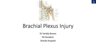
- 2. Anatomy • The dorsal and ventral rootlets exit the spinal cord and merge to form the spinal nerves which leaves the inter-vertebral foramina and divide into dorsal and ventral rami • Ventral Rami of C5 through C8 and T1 spinal nerves gives origin to Brachial Plexus
- 3. The plexus consists of roots, trunks, divisions, cords and branches. • Roots : Lower 4 cervical (C5-8) and the 1st thoracic. Situated between the Scalenus Anterior and Medius muscle deep to sternocleidomastoid muscle. The origin of the plexus may shift one segment either upward or downward resulting in a PRE FIXED PLEXUS or POST FIXED PLEXUS respectively. In a prefixed plexus, the contribution by C4 is large and in that from T2 is often absent. In a post fixed plexus, the contribution by T1 is large, T2 is always present, C4 is absent, and C5 is reduced in size.
- 4. Trunks : Derived from roots Located in the antero-inferior portion of post triangle of neck C5-6 > ant primary rami unite > upper trunk. C8-T1 > ant primary rami unite > lower trunk. C7 > ant primary rami continues as middle trunk. Each trunk ends by splitting into 1) Anterior 2) Posterior divisions.
- 5. CORDS: it forms 3 cords The Posterior Cord is formed from the three posterior divisions of the trunks (C5-C8,T1) The Lateral Cord is the anterior divisions from the upper and middle trunks (C5-C7) The Medial Cord is simply a continuation of the anterior division of the lower trunk (C8,T1)
- 6. BRANCHES: • Branches of the brachial plexus may be described as supraclavicular and infraclavicular. Supraclavicular branches • Supraclavicular branches arise from roots or from trunks as follows: From roots 1. Dorsal scapular nerve C5 2. Long thoracic nerve C5, 6 ,7 From trunks 1. Nerve to subclavius C5, 6 2. Suprascapular nerve C5, 6
- 7. Infraclavicular branches • branches come from the cords Lateral cord • Lateral pectoral C5, 6, 7 • Musculocutaneous C5, 6 7 • Lateral root of median C(5), 6, 7 Medial cord • Medial pectoral C8, T1 • Medial cutaneous of forearm C8, T1 • Medial cutaneous of arm C8, T1 • Ulnar C(7), 8, T1 Posterior cord • Upper subscapular C5, 6 • Thoracodorsal C6, 7,8 • Lower subscapular C5, 6 • Axillary C5, 6 • Radial C5, 6, 7, 8, (T1)
- 8. Etiology • Trauma • Non-penetrating (traction injury- high velocity trauma in RTA or fall of heavy object over shoulder) • Penetrating (knife, gunshot wound) • Nerve Entrapment • Thoracic Outlet Syndrome • During Birth –Shoulder Dystocia, Breech Presentation when the child's shoulder is pulled down • Infection –viral • Tumours • Schwannomas • Iatrogenic • Axillary or Scalene Anaesthesia • Surgical Biopsy
- 9. Mechanisms of Injury to the Brachial Plexus Traction: direct blow to the shoulder with the neck laterally flexed toward the unaffected shoulder (e.g. gymnast falls on beam) Direct trauma: direct blow to the supraclavicular fossa over Erb’s point Compression: Occurs when the neck is flexed laterally toward the patient’s affected shoulder, compressing / irritating the nerves, resulting in point tenderness over involved vertebrae of affected nerve(s)
- 10. Preganglionic Lesion: • avulsion of nerve roots from spinal cord • disruption proximal to dorsal root ganglion • usually from high speed injuries • no proximal stump, no neuroma formation (negative Tinel’s) • Pseudomeningocele, denervation of neck muscles are common • Horner's sign (ptosis, miosis, anhidrosis, enophthalmos) • Associated spine fractures TYPES OF BRACHIAL PLEXUS INJURIES
- 11. • Severe burning sensation in upper limb with insensate limb • Normal Histamine test - intact triple response (redness, wheal, flare) • Palsy of serratus anterior(long thoracic nerve) – medial winging of scapula and rhomboids (dorsal scapular nerve) – lateral winging of scapula • elevated hemidiaphragm (phrenic nerve) • Upper motor neuron signs in lower limb • Nerve conduction velocity detects normal sensory action potential due to intact dorsal root ganglion • Electromyography – denervation potential in cervical paraspinal muscles • Cannot recover and surgically irreparable, so poor prognosis
- 12. Postganglionic: • roots remain intact • usually from traction injuries • proximal stump present and neuroma formation (positive Tinel’s) • deep dorsal neck muscles are intact, and pseudomeningocele will not develop • Abnormal Histamine Test (only redness and wheal, but no flare) • Surgically reparable , better prognosis
- 13. • Based on Anatomical Location • Supraclavicular lesions ( 65%) - affects roots, trunks, and divisions • Typically occurs in motorcycle accidents as the rider collides with the ground or another vehicle, his neck and shoulder are wrenched apart • Infraclavicular lesions (25%)- Affects cords, branches • Usually associated with fractures or dislocations of shoulder joint and axillary artery injury • Based on the level of injury • Upper plexus palsy (Erb’s palsy) involves C5-C6+/- C7roots. • Lower plexus palsy (Klumpke’s palsy) involves C8-T1 roots (and sometimes also C7) • Total plexus lesions involve all nerve roots C5-T1
- 14. ERB'S PARALYSIS: • Site of injury: The region of the upper trunk of the brachial plexus is called Erb's point(where 6 nerves meet namely c5, c6 nerve roots, anterior and posterior divisions, suprascapular nerve, and nerve to subclavius • Causes of injury: Undue separation of the head from the shoulder, which is commonly encountered in 1)birth injury 2) fall on shoulder, and 3)during Anaesthesia • Nerve roots involved: Mainly C5 and partly C6. • Muscles paralysed: Mainly biceps, deltoid, brachialis and brachioradialis. Partly supraspinatus, infraspinatus and supinator
- 15. Deformity • Arm: Hangs by the side, it is adducted and medially rotated • Forearm: Extended and pronated • Abduction impossible because of paralysis of deltoid & supraspinatus m/s. • ER impossible because of paralysis of infraspinatus & teres minor m/s. Active flexion impossible because of paralysis biceps, brachialis & brachioradialis. • Paralysis of supinator m/s causes pronation deformity of forearm. • The deformity is known as "Policeman's tip hand"
- 16. KLUMPKE’S PALSY: • Site of injury: Lower trunk of the brachial plexus. • Cause of injury: Undue abduction of the arm, as in clutching something with the hand after a fall from a height, or sometimes in birth injury. • Nerve roots involved: Mainly T1 and partly C8. • Muscles paralysed: • Intrinsic muscles of the hand (T1) • flexors of the wrist and fingers (C8) – flexor carpi ulnaris and ulnar half of flexor digitorum profundus • Deformity: (position of the hand): claw hand due to the unopposed action of the long flexors and extensors of the fingers. In a claw hand there is hyperextension at the metacarpophalangeal joints and flexion at the interphalangeal joints.
- 17. • Disability: Claw hand • Cutaneous anaesthesia and analgesia in a narrow zone along the ulnar border of the forearm and hand. • Horner's syndrome: ptosis, miosis, anhidrosis, enophthalmos and loss of ciliospinal reflex- may be associated- injury to sympathetic fibres to the head and neck that leave the spinal cord through nerve T1. • Vasomotor changes: The skin areas with sensory loss is warmer due to arteriolar dilation. skin is dry due to the absence of sweating as there is loss of sympathetic activity. • Tropic changes: Long standing case of paralysis leads to dry and scaly skin. Nails crack easily with atrophy of the pulp of fingers.
- 18. Clinical features : History : • The mechanism of injury should be considered. • Birth injury : Usually 5th and 6th root. • Motor cycle accidents. • Stab and bullet wounds. • Symptoms vary depending upon the type and location of the injury to the brachial plexus. • The most common symptoms of BPI include: • Weakness or numbness • Loss of sensation • Loss of movement (paralysis) • Pain • drooping of the left eyelid • pupillary constriction • anhidrosis
- 19. • The pain from brachial plexus injuries results from injury to the spinal cord where the nerve rootlets are avulsed from the cord. • Pain is neuropathic in nature. The pain can last for a very long time. • Brachial plexus injuries that occur at the level of the spinal cord often produce greater pain than injuries more distant from the spinal cord. • In addition, injuries nearer the spinal cord may cause a burning numbness, paresthesias or dysesthesias.
- 20. Physical examination : • Examination of all nerve groups controlled by the brachial plexus to identify the specific location of the nerve injury and its severity. • In addition, some patients display specific signs that help determine the location of the nerve injury: Narrowing of the eye pupils, drooping of the eyelid, and lack of ability for the face to sweat (Horner's syndrome) is a sign that the injury is close to the spinal cord. • A shooting nerve-like pain on taping along the affected nerves (Tinel sign) suggests an injury farther from the spinal cord. Over time, if the location of the Tinel sign moves down the arm toward the hand, it is a sign that the injury is repairing itself. • During the physical examination, assess the arm and shoulder for stability and range of motion • Important muscles to test – serratus anterior (long thoracic nerve) and rhomboids (dorsal scapular nerve) if they are functioning then it is more likely the C5 injury is postganglionic Pulses • Check radial, ulnar and brachial pulses arterial injuries common with complete BPIs
- 21. Is it a pre-ganglionic or post-ganglionic lesion? The following are clues to a pre-ganglionic injury- • Horner's syndrome The T1 root lies close to the T1 sympathetic ganglion. Evidence of injury to the T1 sympathetic chain as evidenced by a Horner’s syndrome would infer that the T1 root has probably been injured. • If rhomboids( dorsal scapular nerve) or serratus anterior (long thoracic nerve) are weak then a pre-ganglionic injury should be suspected. • Chest X-ray, look for elevated (paralysed) hemi-diaphragm (phrenic nerve palsy C3,4,5). • Fractures of the transverse processes of the cervical vertebrae or a fractured first rib indicate a high-energy injury with likely intradural injury of the lower two roots. • Scapulothoracic dissociation is often associated with root avulsion and major vascular injury.
- 22. Sequence of Clinical Examination • Inspection start with the patient stood with both arms and torso exposed. Look at the face for Horner's syndrome Look for surgical scars Comment on muscle wasting – shoulder girdle, arm, forearm or hand • Exclude fixed contractures by gentle passive movements. • Motor testing If a muscle is weak, repeat testing in the horizontal plane in order to eliminate gravity e.g. abducting the shoulder to test elbow flexion/extension power. Muscle testing is an active process involving Look (for contraction and movement of the limb) Feel (for contracted muscle/tendon) Move (to test resistance) Start proximally and work distally
- 23. Standing from the back • Trapezius (spinal accessory - XI, C3,4) Can you shrug your shoulders • Rhomboids (dorsal scapular nerve – C4,5) Push your shoulder blades together • Serratus anterior (long thoracic nerve - C5,6,7) The classic test is wall-press test. In BPI, the patient may be unable to lift the arm. The arm should be supported by the examiner with one hand and the patient asked to push forward as if trying to open a door. At the same time the examiner should hold the lower pole of the scapula with another hand.
- 24. • Latissimus dorsi (thoracodorsal nerve – C6,7,8) While the arm is supported in a flexed position, ask the patient to push down (while the examiner palpates for musle contraction). • Deltoids (axillary nerve – C5,6) Extend, abduct and flex the shoulder to test the posterior, middle and anterior parts respectively (unless the muscle is clearly wasted). Abduction internal rotation Actively and maximally abduct the shoulder in internal rotation with the elbow flexed. Abduction lag relative to the normal side indicates a positive sign.
- 25. Standing from the front Pectoralis major (lateral and medial pectoral nerves) • Clavicular head (C5,6) Atrophy would imply lateral cord injury. Ask the patient to touch their contralateral shoulder (and the examiner palpates for evidence of contraction). • Sternocostal head (C7,8,T1) Atrophy would imply medial cord injury. Ask the patient to push against the hip (and the examiner palpates the axillary fold). Rotator cuffs • Supraspinatus (suprascapular nerve - C5,6) Test shoulder abduction in the scapular plane with the thumb pointing downwards. • Infraspinatus (suprascapular nerve - C5,6) Test external rotation with the shoulder in adduction and the elbow flexed. • Teres minor (axillary nerve – C5,6) Test external rotation with the shoulder in abduction and the elbow flexed. • Subscapularis (upper and lower subscapular nerves – C5,6,7) Belly-press sign. Ask the patient to bring the elbows forward while pressing the belly. A flexed wrist relative to the normal side indicates a positive sign.
- 26. Next, proceed with the following composite testing to demonstrate the myotomes (levels)- • Elbow flexion (C5,6) • • Elbow extension (C7,8) • • Forearm supination (C6) • • Forearm pronation (C7,8) • • Wrist flexion/extension (C6,7) • • MCPJ flexion/extension (C7,8) • • Grip (C8) • • Fingers abduction (T1)
- 27. Sensory testing • Establish normal sensation in an uninjured area (such as forehead or sternum). • First, assess the dermatomes (C5-lateral elbow; C6-thumb tip; C7-middle finger tip; C8-little finger tip; T1-medial elbow) and then if necessary such as in infraclavicular BPI, examine according to the terminal branch distribution. Check for Tinel's signs (and take note of the dermatomal distribution). Palpate for the radial pulse and check the reflexes.
- 28. INVESTIGATIONS • Imaging Studies • Electromyogram • Nerve Conduction Velocity • Intraoperative Nerve Action Potential • Myelography • CT scan for any tumours • MRI
- 29. Imaging studies : X-ray of cervical spine : • Fracture of cervical vertebrae are strongly associated with pre-ganglionic injuries. Chest x-ray : • May show 1st and 2nd rib fracture or an elevated hemidiaphragm, which denotes phrenic nerve paralysis and proximal injury to upper plexus. Fractures of scapula and clavicle and humerus may indicate infraclavicular plexus injuries.
- 30. EMG : • Most important use of EMG studies is for serial evaluation of injury to search for signs of re- innervation. • A decreased in number of fibrillation potentials and positive sharp potentials >typically seen in dennervated muscles > regenerating axons have reached the motor end plates. • The appearance of prolonged, polyphasic and low-amplitude indicate > re-innervation. • Seen several weeks before the onset of voluntary muscle contraction and signify that a further period of observation is in order.
- 31. Intra operative nerve action potential (NAP) : • This study is performed during surgical exploration of the plexus, which is usually done 3-4 months after injury. • If a nerve action potential can be recorded. Substantial number of regenerating axons have traversed the lesion site. • Conversely if an action potential cannot be elicited The abnormal segment is resected because spontaneous recovery is likely to be poor. • NAP is best for evaluating a neuroma in continuity. If an NAP can be transmitted across the area of injury, the patient has 93% chance that useful motor function will develop in the muscles supplied by that nerve.
- 33. CT Myelography : • If plexus injury is strongly suspected a myelogram and subsequent CT scan should be obtained 2-3 months after injury. • It may be inaccurate early after the injury because clotted blood may occlude the opening of the pseudomeningocele. • A delay of 6-12 weeks is recommended before myelogram is advised. • Advantages: -detect partial root avulsion -excellent visualization of bony structures -no CSF flow artifacts -multiplanar reconstruction. • Disadvantages: - high radiation dose -poor visualization of lower brachial plexus due to bony artifacts.
- 34. MRI • MRI provides additional anatomic and physiologic information on injuries. 1. Signal intensity changes in the spinal cord 2. Enhancement of nerve roots 3. Enhancement of paraspinal muscles
- 35. 1. Signal intensity changes in the spinal cord • Hyperintense areas on T2-weighted images suggest oedema in the acute phase and myelomalacia in the chronic phase. • Hypointense lesions on T2-weighted images reflect hemosiderin deposition on due to hemorrhage . 2. Enhancement of nerve roots • Enhancement of intradural nerve roots and root stumps suggests functional impairment of nerve roots despite morphologic continuity • Breakdown of the blood-nerve barrier and dilatation of radicular veins are postulated as the mechanisms of intradural nerve root enhancement.
- 36. 3. Enhancement of paraspinal muscles • Abnormal enhancement of paraspinal muscles is an accurate indirect sign of root avulsion injury. • Denervated muscles show enhancement as early as 24 hours after a nerve is injured. • The presumed mechanisms for muscle enhancement are 1. dilatation of the vascular bed and 2. enlargement of the extracellular space. Axial T2-weighted (A) and coronal MIP 3D STIR SPACE (B) images show the avulsed left T1 nerve root (large arrows) and C8 nerve root (small arrow) with pseudomeningocele formation
- 37. MANAGEMENT • Closed Brachial Plexus Injury • Open Brachial Plexus Injury
- 38. Closed injury • In the case of closed BPI wounds and when there are no other emergent injuries, surgical exploration and recovery may not take place immediately. Recommendations include • managing pain, and • starting rehabilitation. • Upper & Lower Plexuses injuries caused by traction can be divided into four groups 1)Injuries at C5 & C6 2)Injuries at C5,C6 & C7 3)Degenerative lesions of entire plexus 4)Injuries at C7,C8 & T1 (rare)
- 39. • spontaneous recovery in group 1 & 2 cases • But in case of Degenerative entire plexus injuries there is partial recovery. • EMG should be done at 3 to 4 wks. • At 6 to 8wks additional studies like myelography can be done if return of functions not seen. • Exploration is justified at 3 to 6 months after injury if function has not returned.
- 40. CONSERVATIVE TREATMENT • to maintain the range of motion of the extremity • to strengthen the remaining functional muscles • to protect the denervated dermatomes and • to manage pain. Significant pain is observed in complete palsy especially in root avulsions. Pain is excruciating and exhausting for the patient NSAIDs and opioid drugs useful during the first stages but do not appear to help with neuropathic pain, which requires careful use of antiepileptic drugs (gabapentin and carbamazepine) or antidepressants such as amitriptyline. About 30% of patients report significant pain relief with this type of treatment.
- 41. OPEN INJURIES • Open wounds in BPI are uncommon and vary from small penetrating injuries to high energy injuries. • INDICATIONS OF SURGERY: Injuries caused by sharp objects or missiles. When patient seen soon after injury & pt's general condition permits exploration & primary repair can be done When patient not seen soon after injury but only after initial management, It is best to wait for wound healing & stabilization of any other injuries. During this period locate neurological deficit for level of injury. EMG to be performed 3 to 4 wks. after injury.
- 42. Exploration of plexus & neurorrhaphy, autogenous interfascicular nerve grafting or neurolysis is indicated 3 to 6 wks after injury. Motor function recover to a grade of 3 or better in half of pts. Best results obtained in upper trunk , lateral cord & posterior cord injuries. Poor prognosis can be expected in lower trunk injuries.
- 43. SURGICAL GOALS In order of priority as follows: 1)Restoration of elbow flexion 2)Restoration of shoulder abduction 3)Restoration of sensation of medial border of forearm & hand. • Depending on extent of injury various surgical techniques may be required: Primary neurorrhaphy - rarely possible due to traction and usually only possible for acute and sharp penetration injuries Neurolysis (freeing up the nerve from scar tissue) Nerve grafting Neurotization (nerve transfer)
- 44. NEUROLYSIS • When the nerve lesion is in continuity, neurolysis may help. It is of great importance to maintain the interfascicular structure and the nerve sheath. • Because of the risk of vascular damage, interfascicular neurolysis is not done. Instead an anterior epineurectomy is performed, excising the fibrous tissue. • Use of direct nerve stimulation before and after neurolysis helps us demonstrate the improvement in nerve conductance.
- 45. NERVE GRAFTING • Nerve grafting is the predominant technique for clear cut injuries with a healthy proximal stump and with no axial damage. • The outcome is influenced by - 1. the length of the nerve graft 2. the presence of scar tissue at the wound site 3. the number of grafts used 4. the presence of a healthy proximal stump available for grafting 5. the nerve gap to be covered. • The sural nerve, the sensory branch of ulnar nerve, and the medial cutaneous nerve of the forearm are the usual donor nerves. • Generally, use of nerve grafts shorter than 10 cm results in better functional and clinical outcomes compared with longer grafts
- 46. NEUROTIZATION • This type of procedure is used for preganglionic root injury • The nerve transfer may be extraplexus or intraplexus. • Intraplexus transfer options include intact nerve roots. Other choices - medial thoracic nerve and inferior medial cord/ulnar nerve, phrenic nerve , pectoral nerve Oberlin et al. described nerve transfer to the biceps muscle using part of the ulnar nerve for C5-C6 avulsion of the brachial plexus • Extraplexus transfer options include the use of intercostal, spinal accessory nerves (CN XI) , hypoglossal nerve (CN XII)
- 47. • In ROOT avulsion of upper plexus in which no proximal neural stump is available for nerve grafting, neurotization between intercostal nerves or FCU motor fascicles of ulnar nerve & musculocutaneous nerve to restore the ELBOW FLEXION may be considered. • NEUROTIZATION of the suprascapular nerve using the spinal accessory nerve and neurotization of the axillary nerve with fascicles of radial nerve innervating the lateral, medial, or long head of triceps can be used to restore SHOULDER ABDUCTION AND EXTERNAL ROTATION • After Brachial plexus repair 12 to 18 mths are required to determine extent of neural regeneration. • If recovery inadequate> Peripheral reconstruction considered
- 48. Secondary Operations • In the absence of spontaneous recovery or when the first surgical procedure does not provide satisfactory outcomes then a second operation may be required. • In such cases there should be specific signs of neurological denervation or no possibility of neurological recovery, or sufficient time should have passed with no functional improvement. • Secondary options include 1. Arthrodesis 2. Tendon transfer and 3. Functional free muscle transplantation
- 49. ARTHRODESIS • INDICATIONS • In complete brachial plexus traumatic injuries • unstable and painful shoulders • When planning shoulder arthrodesis certain parameters should be taken into consideration. 1. good thoracic-shoulder functionality . 2. the mobility of the peripheral hand is important as shoulder arthrodesis has no clinical effect on a paralytic hand. 3. The acromioclavicular joint, sterno-clavicular joint, and scapulothoracic joint should be intact. Any dysfunction may affect the success of arthrodesis. • NOTE: The shoulder should be fused with only 30 degrees of abduction, 30 degrees flexion, and 30 degrees of internal rotation to allow the patient to be independent in his daily life with a mean range of 60 degrees abduction and flexion through the scapulothoracic joint.
- 50. TENDON TRANSFERS • Tendon transfers are useful in restoring upper extremity function after BPI. • An absolute indication for tendon transfer is upper or lower brachial plexus traumatic injury with only partial paralysis. • Many tendon transfer techniques have been described for treating partial shoulder paralysis. • the most common procedures are the following: 1.Trapezius to deltoid transfer to restore abduction of the shoulder 2.Latissimus dorsi transfer to improve external rotation. 3. Anterior transfer of the posterior branch of the deltoid muscle to restore non-functional anterior segment.
- 51. Restoration of elbow flexion • The surgical goal is to restore good muscle strength through a range of elbow motion (30 to 130 degrees). • The most commonly used procedures are as follows: i)transfer of the common origin of the flexor forearm muscles to a proximal section . May lead to disappointing outcomes such as elbow stiffness or over pronation; (ii)transfer of latissimus dorsi muscle to the tendon of the biceps brachialis provides great muscle strength, but this muscle is often denervated (iii)transfer of pectoralis major brachial branch tendon to brachial biceps (Clark technique). A fused shoulder is required for the best postoperative result; (iv)transfer of triceps tendon to biceps provides good results
- 52. RECOVERY AND REHABILITATION • Because nerve regeneration occurs slowly at a rate of approximately 1 mm/day, recovery from a brachial plexus injury takes time, and patients may not experience results for several months. • A positive mindset and the support of family, friends, and healthcare professionals are important to recovery and rehabilitation. • During this recovery process, occupational therapists teach patients how to use the unaffected arm to perform daily activities like eating and personal hygiene. • Physical therapy of the shoulder, elbow, wrist, and fingers involves specific exercises to prevent stiffness, contractures, or muscle atrophy.
- 53. • A physical therapist can also recommend assistive devices, such as splinting or supportive bracing to help support a limp arm and joints. • Compression gloves and sleeves may be used to prevent swelling in the affected arm, which can lead to pain and joint contractures. • Pain may be managed with medications, therapy, and assistive devices. • In addition, patients will require healthy coping skills in order to make adjustments in their lives.