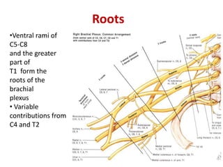
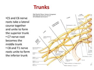
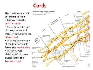
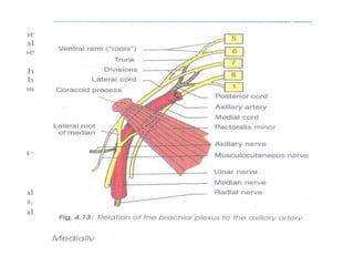
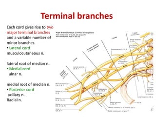
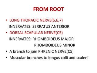
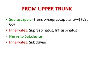
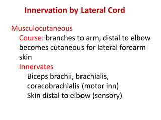
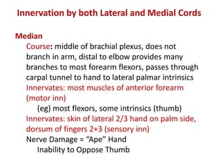
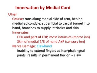
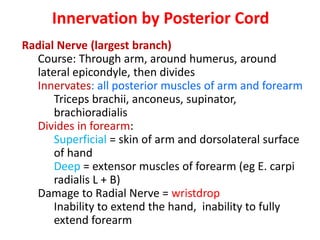
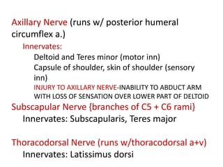
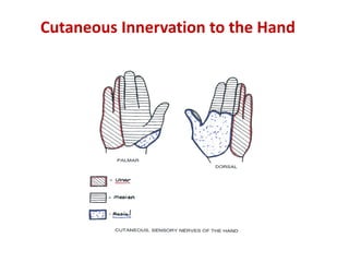
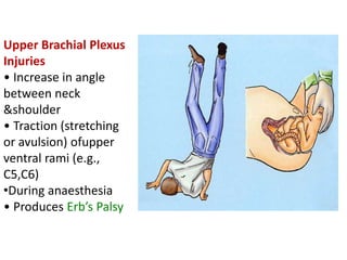
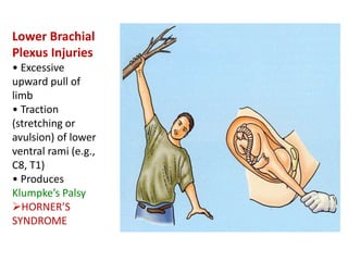
•Roots
• Trunks
• Divisions
• Cords
• Terminal
branches
Anatomy
Roots
•Ventral rami of
C5-C8
and the greater
part of
T1 form the
roots of the
brachial
plexus
• Variable
contributions from
C4 and T2
Trunks
•C5 and C6 nerve
roots take a lateral
course together
and unite to form
the superior trunk
• C7 nerve root
becomes the
middle trunk
• C8 and T1 nerve
roots unite to form
the inferior trunk
•At the lateral border of the first rib and behind the
middle third of the clavicle, the trunks divide into
anterior and posterior divisions.
• Anterior division -supply flexors
•Posterior division - supply extensors
• The divisions continue the journey down into the
apex of the axilla where they further reorganize into
cords
Divisions
Cords
The cords are named
according to their
relationship to the
axillary artery
• The anterior divisions
of the superior and
middle trunks form the
lateral cord
• The anterior division
of the inferior trunk
forms the medial cord
• The posterior
divisions of all three
trunks forms the
Posterior cord
Cords
The cords complete the journey at the lateral
border of the pectoralis minor where they give
rise to the terminal branches of the brachial
plexus
Terminal branches
Each cord gives rise to two
major terminal branches
and a variable number of
minor branches.
• Lateral cord
musculocutaneous n.
lateral root of median n.
• Medial cord
ulnar n.
medial root of median n.
• Posterior cord
axillary n.
Radial n.
FROM ROOT
• LONG THORACIC NERVE(5,6,7)
INNERVATES: SERRATUS ANTERIOR
• DORSAL SCAPULAR NERVE(C5)
INNERVATES: RHOMBOIDEUS MAJOR
RHOMBOIDEUS MINOR
• A branch to join PHRENIC NERVE(C5)
• Muscular branches to longus colli and scaleni
FROM UPPER TRUNK
• Suprascapular (runs w/suprascapular a+v) {C5,
C6}
• Innervates: Supraspinatus, Infraspinatus
• Nerve to Subclavius
• Innervates: Subclavius
Musculocutaneous
Course: branches to arm, distal to elbow
becomes cutaneous for lateral forearm
skin
Innervates
Biceps brachii, brachialis,
coracobrachialis (motor inn)
Skin distal to elbow (sensory)
Innervation by Lateral Cord
Median
Course: middle of brachial plexus, does not
branch in arm, distal to elbow provides many
branches to most forearm flexors, passes through
carpal tunnel to hand to lateral palmar intrinsics
Innervates: most muscles of anterior forearm
(motor inn)
(eg) most flexors, some intrinsics (thumb)
Innervates: skin of lateral 2/3 hand on palm side,
dorsum of fingers 2+3 (sensory inn)
Nerve Damage = “Ape” Hand
Inability to Oppose Thumb
Innervation by both Lateral and Medial Cords
Ulnar
Course: runs along medial side of arm, behind
medial epicondyle, superficial to carpal tunnel into
hand, branches to supply intrinsics and skin
Innervates:
FCU and part of FDP, most intrinsics (motor inn)
Skin of medial 2/3 of hand A+P (sensory inn)
Nerve Damage: Clawhand
Inability to extend fingers at interphalangeal
joints, results in permanent flexion = claw
Innervation by Medial Cord
Radial Nerve (largest branch)
Course: Through arm, around humerus, around
lateral epicondyle, then divides
Innervates: all posterior muscles of arm and forearm
Triceps brachii, anconeus, supinator,
brachioradialis
Divides in forearm:
Superficial = skin of arm and dorsolateral surface
of hand
Deep = extensor muscles of forearm (eg E. carpi
radialis L + B)
Damage to Radial Nerve = wristdrop
Inability to extend the hand, inability to fully
extend forearm
Innervation by Posterior Cord
Axillary Nerve (runs w/ posterior humeral
circumflex a.)
Innervates:
Deltoid and Teres minor (motor inn)
Capsule of shoulder, skin of shoulder (sensory
inn)
INJURY TO AXILLARY NERVE-INABILITY TO ABDUCT ARM
WITH LOSS OF SENSATION OVER LOWER PART OF DELTOID
Subscapular Nerve {branches of C5 + C6 rami}
Innervates: Subscapularis, Teres major
Thoracodorsal Nerve (runs w/thoracodorsal a+v)
Innervates: Latissimus dorsi
Cutaneous Innervation to the Hand
Upper Brachial Plexus
Injuries
• Increase in angle
between neck
&shoulder
• Traction (stretching
or avulsion) ofupper
ventral rami (e.g.,
C5,C6)
•During anaesthesia
• Produces Erb’s Palsy
Lower Brachial
Plexus Injuries
• Excessive
upward pull of
limb
• Traction
(stretching or
avulsion) of lower
ventral rami (e.g.,
C8, T1)
• Produces
Klumpke’s Palsy
HORNER’S
SYNDROME
“Obstetrical” or “Birth palsy”
• Becoming increasingly rare
• Categorized on basis of damage
• Upper (C5,6), Erb’s: 60-90%
• All (C5-T1), both palsies: 10%
• Lower (C8, T1), Klumpke’s Palsy1-5%
• INJURY TO LONG THORACIC NERVE-(N. OF
BELL) WINGING OF SCAPULA
• INJURY TO LATERAL CORD- DISLOCATION OF
HUMERUS-
• DEFORMITY-MIDPRONE FOREARM
• LOSS OF FLEXION AT WRIST & FOREARM
• SENSORY LOSS ON RADIAL SIDE OF
FOREARM
• VASOMOTOR AND TROPHIC CHANGES
• INJURY TO MEDIAL CORD
• CAUSE-SUBCORACOID DISLOCATION
• DEFORMITY -CLAW HAND
• SENSORY LOSS ON ULNAR SIDE OF FOREARM
• VASOMOTOR AND TROPHIC CHANGES

Brachial plexuses

  • 1.
    •Roots • Trunks • Divisions •Cords • Terminal branches Anatomy
  • 2.
    Roots •Ventral rami of C5-C8 andthe greater part of T1 form the roots of the brachial plexus • Variable contributions from C4 and T2
  • 3.
    Trunks •C5 and C6nerve roots take a lateral course together and unite to form the superior trunk • C7 nerve root becomes the middle trunk • C8 and T1 nerve roots unite to form the inferior trunk
  • 4.
    •At the lateralborder of the first rib and behind the middle third of the clavicle, the trunks divide into anterior and posterior divisions. • Anterior division -supply flexors •Posterior division - supply extensors • The divisions continue the journey down into the apex of the axilla where they further reorganize into cords Divisions
  • 5.
    Cords The cords arenamed according to their relationship to the axillary artery • The anterior divisions of the superior and middle trunks form the lateral cord • The anterior division of the inferior trunk forms the medial cord • The posterior divisions of all three trunks forms the Posterior cord
  • 7.
    Cords The cords completethe journey at the lateral border of the pectoralis minor where they give rise to the terminal branches of the brachial plexus
  • 8.
    Terminal branches Each cordgives rise to two major terminal branches and a variable number of minor branches. • Lateral cord musculocutaneous n. lateral root of median n. • Medial cord ulnar n. medial root of median n. • Posterior cord axillary n. Radial n.
  • 9.
    FROM ROOT • LONGTHORACIC NERVE(5,6,7) INNERVATES: SERRATUS ANTERIOR • DORSAL SCAPULAR NERVE(C5) INNERVATES: RHOMBOIDEUS MAJOR RHOMBOIDEUS MINOR • A branch to join PHRENIC NERVE(C5) • Muscular branches to longus colli and scaleni
  • 10.
    FROM UPPER TRUNK •Suprascapular (runs w/suprascapular a+v) {C5, C6} • Innervates: Supraspinatus, Infraspinatus • Nerve to Subclavius • Innervates: Subclavius
  • 11.
    Musculocutaneous Course: branches toarm, distal to elbow becomes cutaneous for lateral forearm skin Innervates Biceps brachii, brachialis, coracobrachialis (motor inn) Skin distal to elbow (sensory) Innervation by Lateral Cord
  • 12.
    Median Course: middle ofbrachial plexus, does not branch in arm, distal to elbow provides many branches to most forearm flexors, passes through carpal tunnel to hand to lateral palmar intrinsics Innervates: most muscles of anterior forearm (motor inn) (eg) most flexors, some intrinsics (thumb) Innervates: skin of lateral 2/3 hand on palm side, dorsum of fingers 2+3 (sensory inn) Nerve Damage = “Ape” Hand Inability to Oppose Thumb Innervation by both Lateral and Medial Cords
  • 13.
    Ulnar Course: runs alongmedial side of arm, behind medial epicondyle, superficial to carpal tunnel into hand, branches to supply intrinsics and skin Innervates: FCU and part of FDP, most intrinsics (motor inn) Skin of medial 2/3 of hand A+P (sensory inn) Nerve Damage: Clawhand Inability to extend fingers at interphalangeal joints, results in permanent flexion = claw Innervation by Medial Cord
  • 14.
    Radial Nerve (largestbranch) Course: Through arm, around humerus, around lateral epicondyle, then divides Innervates: all posterior muscles of arm and forearm Triceps brachii, anconeus, supinator, brachioradialis Divides in forearm: Superficial = skin of arm and dorsolateral surface of hand Deep = extensor muscles of forearm (eg E. carpi radialis L + B) Damage to Radial Nerve = wristdrop Inability to extend the hand, inability to fully extend forearm Innervation by Posterior Cord
  • 15.
    Axillary Nerve (runsw/ posterior humeral circumflex a.) Innervates: Deltoid and Teres minor (motor inn) Capsule of shoulder, skin of shoulder (sensory inn) INJURY TO AXILLARY NERVE-INABILITY TO ABDUCT ARM WITH LOSS OF SENSATION OVER LOWER PART OF DELTOID Subscapular Nerve {branches of C5 + C6 rami} Innervates: Subscapularis, Teres major Thoracodorsal Nerve (runs w/thoracodorsal a+v) Innervates: Latissimus dorsi
  • 16.
  • 17.
    Upper Brachial Plexus Injuries •Increase in angle between neck &shoulder • Traction (stretching or avulsion) ofupper ventral rami (e.g., C5,C6) •During anaesthesia • Produces Erb’s Palsy
  • 18.
    Lower Brachial Plexus Injuries •Excessive upward pull of limb • Traction (stretching or avulsion) of lower ventral rami (e.g., C8, T1) • Produces Klumpke’s Palsy HORNER’S SYNDROME
  • 19.
    “Obstetrical” or “Birthpalsy” • Becoming increasingly rare • Categorized on basis of damage • Upper (C5,6), Erb’s: 60-90% • All (C5-T1), both palsies: 10% • Lower (C8, T1), Klumpke’s Palsy1-5%
  • 20.
    • INJURY TOLONG THORACIC NERVE-(N. OF BELL) WINGING OF SCAPULA • INJURY TO LATERAL CORD- DISLOCATION OF HUMERUS- • DEFORMITY-MIDPRONE FOREARM • LOSS OF FLEXION AT WRIST & FOREARM • SENSORY LOSS ON RADIAL SIDE OF FOREARM • VASOMOTOR AND TROPHIC CHANGES
  • 21.
    • INJURY TOMEDIAL CORD • CAUSE-SUBCORACOID DISLOCATION • DEFORMITY -CLAW HAND • SENSORY LOSS ON ULNAR SIDE OF FOREARM • VASOMOTOR AND TROPHIC CHANGES