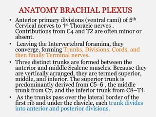
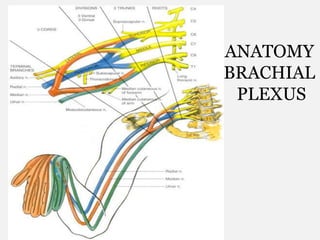
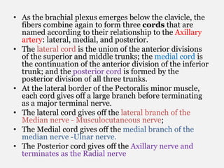
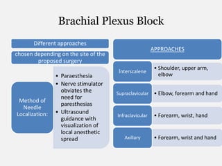
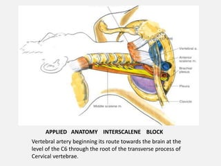
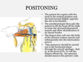
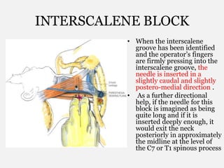
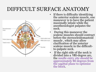
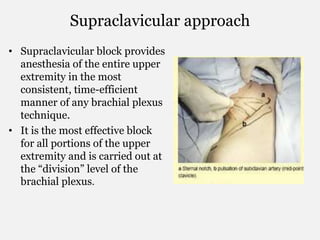
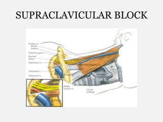
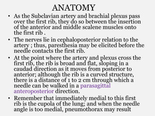
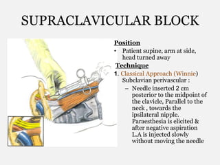
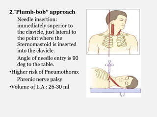
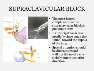
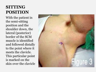
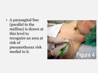
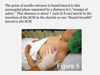
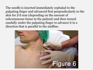
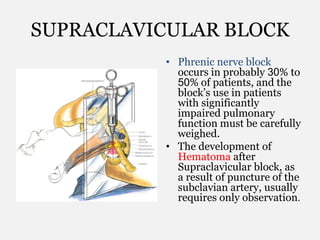
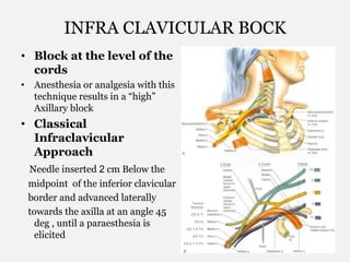
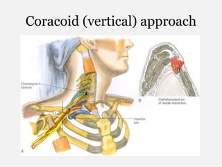
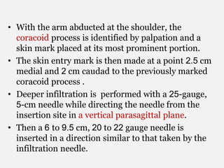
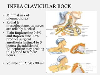
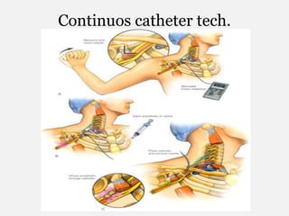
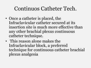
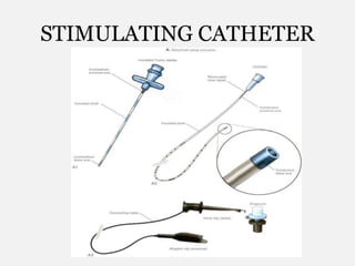
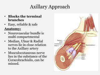
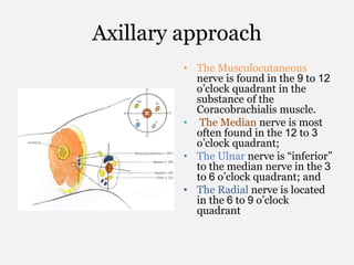
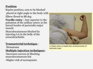
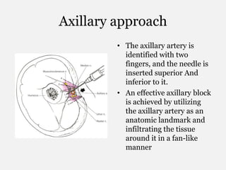
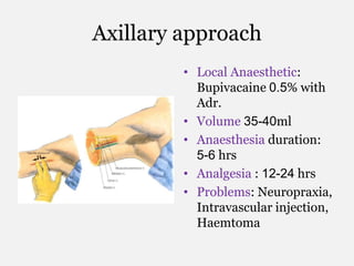
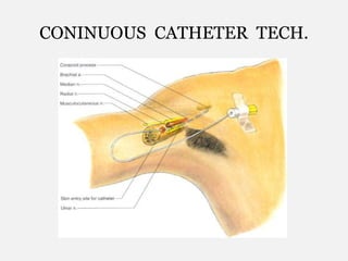
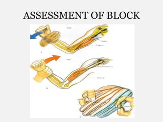
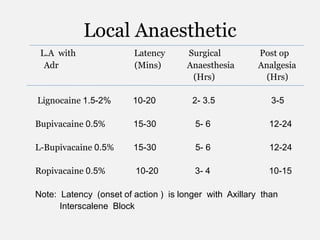
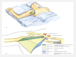
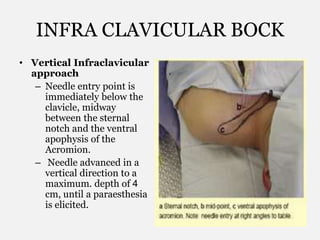
2. ANATOMY BRACHIAL PLEXUS
⢠Anterior primary divisions (ventral rami) of 5th
Cervical nerves to 1st Thoracic nerves .
Contributions from C4 and T2 are often minor or
absent.
⢠Leaving the Intervertebral foramina, they
converge, forming Trunks, Divisions, Cords, and
then finally Terminal nerves.
⢠Three distinct trunks are formed between the
anterior and middle Scalene muscles. Because they
are vertically arranged, they are termed superior,
middle, and inferior. The superior trunk is
predominantly derived from C5â6 , the middle
trunk from C7, and the inferior trunk from C8âT1.
⢠As the trunks pass over the lateral border of the
first rib and under the clavicle, each trunk divides
into anterior and posterior divisions.
4. ⢠As the brachial plexus emerges below the clavicle, the
fibers combine again to form three cords that are
named according to their relationship to the Axillary
artery: lateral, medial, and posterior.
⢠The lateral cord is the union of the anterior divisions
of the superior and middle trunks; the medial cord is
the continuation of the anterior division of the inferior
trunk; and the posterior cord is formed by the
posterior division of all three trunks.
⢠At the lateral border of the Pectoralis minor muscle,
each cord gives off a large branch before terminating
as a major terminal nerve.
⢠The lateral cord gives off the lateral branch of the
Median nerve - Musculocutaneous nerve;
⢠The Medial cord gives off the medial branch of the
median nerve -Ulnar nerve.
⢠The Posterior cord gives off the Axillary nerve and
terminates as the Radial nerve
6. Brachial Plexus Block
Different approaches
chosen depending on the site of the
proposed surgery
⢠Paraesthesia
⢠Nerve stimulator
obviates the
need for
paresthesias
⢠Ultrasound
guidance with
visualization of
local anesthetic
spread
Method of
Needle
Localization:
APPROACHES
⢠Shoulder, upper arm,
elbow
Interscalene
⢠Elbow, forearm and handSupraclavicular
⢠Forearm, wrist, handInfraclavicular
⢠Forearm, wrist and handAxillary
7. ELICITATION OF PARAESTHESIA
⢠When a needle makes direct contact with a sensory
nerve, a paresthesia is elicited in its area of sensory
distribution.
⢠Needle should contact with the nerve rather than
penetrating it, and that the injection is in
proximity to the nerve (perineural) rather than
within its substance (intraneural).
⢠The high pressures generated by a direct
intraneural injection can cause hydrostatic
(ischemic) injury to nerve fibers.
⢠A Perineural injection may produce a brief
accentuation of the paresthesia, whereas an
Intraneural injection produces an intense, searing
pain that serves as a warning to immediately
terminate the injection and reposition the needle.
8. NERVE STIMULATION
⢠One lead of a low-output nerve stimulator is attached to a
needle and the other lead is grounded elsewhere on the
patient.
⢠The special needles that are used are insulated and permit
current flow only at the tip for precise localization of nerves,
whereas the nerve stimulators used deliver a linear, constant
current output of 0.1â6.0 mA.
⢠Muscle contractions occur and increase in intensity as the
needle approaches the nerve and diminish when the needle
moves away.
⢠Moreover, the evoked contractions require much less current
as the needle approaches the nerve.
⢠Optimal positioning produces evoked contractions with 0.5
mA or less, but successful blocks can often be obtained with
needle positions that produce contractions with as much as
1 mA.
⢠Characteristically, the evoked response rapidly diminishes
(fades) after injection of 1â2 mL of local anesthetic
9. INTERSCALENE BLOCK
⢠Interscalene block (classic anterior approach) is
especially effective for surgery of the Shoulder or
Upper arm.
⢠The roots of the brachial plexus are most easily
blocked with this technique.
⢠This block is ideal for reduction of a dislocated
shoulder and often can be achieved with as little as
10 to 15 mL of local anesthetic.
⢠The block also can be performed with the arm in
almost any position and thus can be useful when
brachial plexus block needs to be repeated during
a prolonged upper extremity procedure
10. Surface Anatomy
⢠Surface anatomy of importance to
anesthesiologists includes that of the larynx,
sternocleidomastoid muscle, and external jugular
vein.
⢠Interscalene block is most often performed at the
level of the C6 vertebral body, which is at the level
of the cricoid cartilage.
⢠Thus, by projecting a line laterally from the cricoid
cartilage, the level at which one should roll the
fingers off the sternocleidomastoid muscle onto
the belly of the anterior scalene and then into the
interscalene groove can be identified.
⢠With firm pressure, it is possible to feel the
transverse process of C6 in most individuals, and
in some people it is possible to elicit a paresthesia
by deep palpation.
12. ANATOMY
⢠It is always important to visualize what lies
under the palpating fingers, and again the key to
carrying out successful Interscalene block is
identifying the Interscalene groove.
⢠We should make out from the surface anatomy,
how closely the lateral border of the anterior
scalene muscle deviates from the border of the
Sternocleidomastoid. .
⢠The anterior scalene muscle and the Interscalene
groove are oriented at an oblique angle to the
long axis of the Sternocleidomastoid muscle.
13.
14. APPLIED ANATOMY INTERSCALENE BLOCK
Vertebral artery beginning its route towards the brain at the
level of the C6 through the root of the transverse process of
Cervical vertebrae.
15. POSITONING
⢠The patient lies supine with the
neck in the neutral position and
the head turned slightly opposite
the site to be blocked.
⢠The anesthesiologist then asks the
patient to lift the head off the table
to tense the sternocleidomastoid
muscle and allow identification of
its lateral border.
⢠The fingers then roll onto the belly
of the anterior scalene muscle and
subsequently into the interscalene
groove.
⢠This maneuver should be carried
out in the horizontal plane
through the cricoid cartilageâthat
is, at the level of C6. To roll the
fingers effectively , the operator
should stand at the patientâs side.
16. INTERSCALENE BLOCK
⢠When the interscalene
groove has been identified
and the operatorâs fingers
are firmly pressing into the
interscalene groove, the
needle is inserted in a
slightly caudal and slightly
postero-medial direction .
⢠As a further directional
help, if the needle for this
block is imagined as being
quite long and if it is
inserted deeply enough, it
would exit the neck
posteriorly in approximately
the midline at the level of
the C7 or T1 spinous process
17. DIFFICULT SURFACE ANATOMY
⢠If there is difficulty identifying
the anterior scalene muscle, one
maneuver is to have the patient
maximally inhale while the
anesthesiologist palpates the
neck.
⢠During this maneuver the
scalene muscles should contract
before the sternocleidomastoid
muscle , which may allow
clarification of the anterior
scalene muscle in the difficult-
to-palpate neck.
⢠If the right side of the neck is
divided into a 180-degree arc,
the needle entry site should be
approximately 60 degrees from
the sagittal plane to optimize
the block.
18. Injection
CONFIRMATON: 1. By Eliciting paraesthesia in
the arm.
2. If using a nerve stimulater, activity of the Phrenic
nerve indicates needle is too anterior, whereas
stimulation of Trapezius â needle is too posterior.
Motor activity of the arm, wrist or hand should be
sought.
After confirmation & negative aspiraton inject the
L.A. slowly in a free flowing manner. If there is
any resistance to the flow, u might be injecting in
the nerve bundle.
19. INTERSCALENE BLOCK
⢠Most of the injection difficulties that result in complications of the
block can be avoided if one remembers that it should be an
extremely âsuperficialâ block; if the palpating fingers apply
Sufficient pressure, no more than 1 to1.5cm of the needle should be
necessary to reach the plexus.
⢠Local Anaesthetic : Bupivacaine 0.5%, Ropivacaine 0.5% with
Adr (1 : 200000).
Volume 35-40 ml .
⢠Duration: 12-18 Hrs
⢠Potential Problems: Common - Phrenic nerve palsy (dyspnoea),
Honer syndome, Recurrent LN Block (Hoarseness)
Rare - Vertebral artery injection ( Siezures) , Pneumothorax,
Inadvertant spninal & epiduralBlock.
⢠Contraindications: Pt. with Sigificant Lung Ds.
20. Supraclavicular approach
⢠Supraclavicular block provides
anesthesia of the entire upper
extremity in the most
consistent, time-efficient
manner of any brachial plexus
technique.
⢠It is the most effective block
for all portions of the upper
extremity and is carried out at
the âdivisionâ level of the
brachial plexus.
22. ANATOMY
⢠As the Subclavian artery and brachial plexus pass
over the first rib, they do so between the insertion
of the anterior and middle scalene muscles onto
the first rib .
⢠The nerves lie in cephaloposterior relation to the
artery ; thus, paresthesia may be elicited before the
needle contacts the first rib.
⢠At the point where the artery and plexus cross the
first rib, the rib is broad and flat, sloping in a
caudad direction as it moves from posterior to
anterior; although the rib is a curved structure,
there is a distance of 1 to 2 cm through which a
needle can be walked in a parasagittal
anteroposterior direction.
⢠Remember that immediately medial to this first
rib is the cupola of the lung; and when the needle
angle is too medial, pneumothorax may result
23. SUPRACLAVICULAR BLOCK
Position
⢠Patient supine, arm at side,
head turned away
Technique
1. Classical Approach (Winnie)
Subclavian perivascular :
â Needle inserted 2 cm
posterior to the midpoint of
the clavicle, Parallel to the
neck , towards the
ipsilateral nipple.
Paraesthesia is elicited &
after negative aspiration
L.A is injected slowly
without moving the needle
24. 2.âPlumb-bobâ approach
Needle insertion:
immediately superior to
the clavicle, just lateral to
the point where the
Sternomastoid is inserted
into the clavicle.
Angle of needle entry is 90
deg to the table.
â˘Higher risk of Pneumothorax
Phrenic nerve palsy
â˘Volume of L.A : 25-30 ml
25. SUPRACLAVICULAR BLOCK
⢠The most feared
complication of the
supraclavicular block is
pneumothorax.
⢠Its principal cause is a
needle/syringe angle that
âaimsâ toward the cupola
of the lung.
⢠Special attention should
be directed toward
walking the needle in a
strictly anteroposterior
direction
26. SITTING
POSITION
With the patient in
the semi-sitting
position and the
shoulder down, the
lateral (posterior)
border of the SCM
muscle is identified
and followed distally
to the point where it
meets the clavicle.
This particular point
is marked on the
skin over the clavicle
27. ⢠A parasagital line
(parallel to the
midline) is drawn at
this level to
recognize an area at
risk of
pneumothorax risk
medial to it.
28. The point of needle entrance is found lateral to this
parasagital plane separated by a distance k/a âmargin of
safetyâ. This distance is about 1 inch (2.5 cm) lateral to the
insertion of the SCM in the clavicle or one âthumb breadthâ
lateral to the SCM
29. The needle is inserted immediately cephalad to the
palpating finger and advanced first perpendicularly to the
skin for 2-5 mm (depending on the amount of
subcutaneous tissue in the patient) and then turned
caudally under the palpating finger to advance it in a
direction that is parallel to the midline.
30. SUPRACLAVICULAR BLOCK
⢠Phrenic nerve block
occurs in probably 30% to
50% of patients, and the
blockâs use in patients
with significantly
impaired pulmonary
function must be carefully
weighed.
⢠The development of
Hematoma after
Supraclavicular block, as
a result of puncture of the
subclavian artery, usually
requires only observation.
31. INFRA CLAVICULAR BOCK
⢠Block at the level of the
cords
⢠Anesthesia or analgesia with this
technique results in a âhighâ
Axillary block
⢠Classical
Infraclavicular
Approach
Needle inserted 2 cm Below the
midpoint of the inferior clavicular
border and advanced laterally
towards the axilla at an angle 45
deg , until a paraesthesia is
elicited
33. ⢠With the arm abducted at the shoulder, the
coracoid process is identified by palpation and a
skin mark placed at its most prominent portion.
⢠The skin entry mark is then made at a point 2.5 cm
medial and 2 cm caudad to the previously marked
coracoid process .
⢠Deeper infiltration is performed with a 25-gauge,
5-cm needle while directing the needle from the
insertion site in a vertical parasagittal plane.
⢠Then a 6 to 9.5 cm, 20 to 22 gauge needle is
inserted in a direction similar to that taken by the
infiltration needle.
34. ⢠If a Paresthesia technique is used, a distal
upper extremity paresthesia is sought.
⢠If a nerve stimulator technique is used, a distal
upper extremity motor response is sought.
⢠If needle redirection is needed , should be
redirected in a Cephalocaudad arc .
⢠The depth of contact with the brachial plexus
depends on body habitus and needle
angulation;
⢠It ranges from 2.5 to 3 cm in slender patients,
4-5 cm in larger pts.
35. INFRA CLAVICULAR BOCK
⢠Minimal risk of
pneumothorax
⢠Radial &
musculocutaneous nerves
are reliably blocked
⢠Plain Bupivacaine 0.5%
and Ropivacaine 0.5%
produce surgical
anesthesia lasting 4 to 6
hours; the addition of
Epinephrine may prolong
this period to 8 to 12
hours.
⢠Volume of LA: 20 - 30 ml
37. Continuos Catheter Tech.
⢠Once a catheter is placed, the
Infraclavicular catheter secured at its
insertion site is much more effective than
any other brachial plexus continuous
catheter technique.
⢠This reason alone makes the
Infraclavicular block, a preferred
technique for continuous catheter brachial
plexus analgesia
39. Axillary Approach
⢠Blocks the terminal
branches
⢠Easy, reliable & safe
Anatomy:
⢠Neurovascular bundle is
multi compartmental
⢠Median, Ulnar & Radial
nerves lie in close relation
to the Axillary artery
⢠Musculocutaneous nerve
lies in the substance of the
Coracobrachialis, can be
missed.
40. Axillary approach
⢠The Musculocutaneous
nerve is found in the 9 to 12
oâclock quadrant in the
substance of the
Coracobrachialis muscle.
⢠The Median nerve is most
often found in the 12 to 3
oâclock quadrant;
⢠The Ulnar nerve is âinferiorâ
to the median nerve in the 3
to 6 oâclock quadrant; and
⢠The Radial nerve is located
in the 6 to 9 oâclock
quadrant
41. Position
Supine position, arm to be blocked
placed at right angle to the body with
Elbow flexed to 90 deg
Needle entry : Just superior to the
pulsation of the axillary artery at the
lateral border of pectoralis major
muscle
Musculocutaneous blocked by
injecting LA in the belly of the
coracobrachialis
Transarterial technique :
Hematoma
Multiple injection techniques:
â˘Increases success in blocking
musculocutaneous but
â˘Higher risk of neuropraxia
42. Axillary approach
⢠The axillary artery is
identified with two
fingers, and the needle is
inserted superior And
inferior to it.
⢠An effective axillary block
is achieved by utilizing
the axillary artery as an
anatomic landmark and
infiltrating the tissue
around it in a fan-like
manner
46. QUICK ASSESSMENT OF BLOCK
⢠âpush, pull, pinch, pinchâ To check the FOUR peripheral nerves
of interest during a brachial plexus block.
⢠Ask the patient to resist the anesthesiologistâs pulling the
forearm away from the upper arm, motor innervation to the
Biceps muscle can be assessed. If this muscle has been
weakened, one can be certain that the local anesthetic has
reached the Musculocutaneous nerve.
⢠Likewise, by asking the patient to attempt to extend the forearm
by contracting the Triceps muscle, one can assess the Radial
nerve.
⢠Finally, pinching the fingers in the distribution of the Ulnar or
Median nerveâthat is, at the base of the fifth or second digit,
respectivelyâcan help to assess the adequacy of the block of
both the ulnar and median nerves.
⢠Typically, if these maneuvers are performed shortly after a
Brachial plexus block, motor weakness is evident before the
sensory block.
47. Local Anaesthetic
L.A with Latency Surgical Post op
Adr (Mins) Anaesthesia Analgesia
(Hrs) (Hrs)
Lignocaine 1.5-2% 10-20 2- 3.5 3-5
Bupivacaine 0.5% 15-30 5- 6 12-24
L-Bupivacaine 0.5% 15-30 5- 6 12-24
Ropivacaine 0.5% 10-20 3- 4 10-15
Note: Latency (onset of action ) is longer with Axillary than
Interscalene Block
52. INFRA CLAVICULAR BOCK
⢠Vertical Infraclavicular
approach
â Needle entry point is
immediately below the
clavicle, midway
between the sternal
notch and the ventral
apophysis of the
Acromion.
â Needle advanced in a
vertical direction to a
maximum. depth of 4
cm, until a paraesthesia
is elicited.