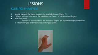
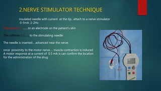
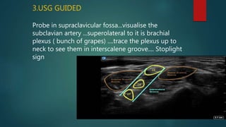
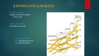
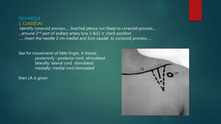
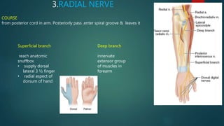
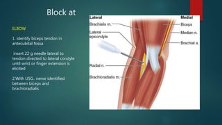
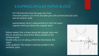
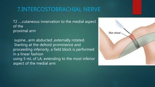
BRACHIAL PLEXUS
& UPPER LIMB BLOCKS
Dr VEENA MOHAN
JR1 ANAESTHESIA
Network of nerves ..formed by ventral rami of C5-T1 innervate the
upper limb
COMPONENT PARTS
• ROOTS - Five ventral rami (C5 -T1 )
• TRUNKS – Upper, Middle, and Lower
• DIVISIONS – Anterior & Posterior from each trunk,
• CORDS – Lateral, Medial and Posterior
• BRANCHES
LOCATION
ROOT– leave the intervertebral foramina, converge to form trunks
TRUNKS –formed b/w scalene anterior & medius muscle, passed over
lateral border of 1st rib
under the clavicle ,it divides to form
DIVISIONS - behind clavicle
CORDS- formed below clavicle ,, arranged around 2nd part of Axillary
artery
• From the Roots
1.Dorsal Scapular nerve
Rhomboideus major and minor muscles
2.Long Thoracic nerve
serratus anterior
BRANCHES
the Upper Trunk
1. Nerve to subclavius muscle
2. Suprascapular nerve
BRACHIAL PLEXUS: NERVES FROM CORDS
Lateral cord(C5,C6,C7)
• Lateral pectoral: pectoralis major
• Lateral root of median
• Musculocutaneous : coracobrachalis, brachialis, biceps brachii
Medial cord(C8,T1)
• Medial pectoral : pectoral major & minor
• Medial root of median
• Medial cutaneous N of forearm
• Medial cutaneous N of arm
• Ulnar : intrinsic hand muscles except thenar & lateral 2 lumbricals
Posterior cord(C5,C6)
• Upper subscapular :subscapularis
• Thoracodorsal :latissimus dorsi
• Lower suscapular :subscapularis
• Axillary: deltoid, teres minor
• Radial: triceps, supinator, anconeus, extensor muscles of forearm & brachioradialis
LESIONS
KLUMPKE PARALYSIS
 partial palsy of the lower roots of the brachial plexus. C8 and T1
 Affects intrinsic muscles of the hand and the flexors of the wrist and fingers.
 the“claw hand”
forearm is supinated and the wrist and fingers are hyperextended with flexion
at interphalangeal and metacarpo phalangeal joints.
injury to the upper trunk C5-C6.
• loss of sensation in the arm
• paralysis & atrophy of the deltoid, biceps, and brachialis
• the arm hangs by the side and is rotated medially;
• the forearm is extended and pronated
• "waiter's tip hand."
ERB’S PALSY
ANESTHETIC
INTERVENTION
nerve block involving the injection of local anesthetic as close to
the nerve as possible for pain relief.
• Short surgical procedure
• Postoperative analgesia
ADVANTAGES
• avoids side effects nausea and vomiting,
• avoiding complications of general & central neuraxial
anesthesia
• In hemodynamically unstable patient
PERIPHERAL NERVE BLOCK
HOW TO LOCALISE THE NERVE
1.PARESTHESIA TECHNIQUE
Knowing anatomic relationship and surface landmarks
…..
block needles inserted
….
advanced towards the target nerve
……
When comes in contact
….
the paresthesia develops
2.NERVE STIMULATOR TECHNIQUE
insulated needle with current at the tip.. attach to a nerve stimulator
0-5mA ,1-2Hz
The anode (+) …….to an electrode on the patient’s skin
The cathode (−)…….to the stimulating needle.
The needle is inserted… advanced near the nerve.
once proximity to the motor nerve…. muscle contraction is induced
A motor response at a current of 0.5 mA is can confirm the location
for the administration of the drug
3. USG GUIDED
High frequency(1-20MHz) sound waves emitted from piezoelectric crystals…. pass through tissue…. return
signal to transducer….. create as electrical voltage…. converted to 2D image
HYPOECHOIC
• appear black
• Allow
Soundwave to
pass
TRANSDUCER
HIGH FREQUENCY
• high resolution
• clear image
• less penetration
• for superficial
structures
• Linear probe
LOW FREQUENCY
• poor image quality
• better tissue
penetration
• for deeper structures
• Curvilinear probe
HYPERECHOIC
appears white/brighter
reflects more
IMAGE
IN PLANE
parallel to the ultrasound waves
OUT OF PLANE
not parallel to ultrasound waves
TECHNIQUE
BRACHIAL PLEXUS BLOCK
a fascial sleeve enclose brachial plexus extending from IV foramen to upper arm
Inject LA into this sheath—block the brachial plexus
• Interscalene block
• Supraclavicular block
• Infraclavicular block
• Axillary block
1.INTERSCALENE BLOCK
INDICATIONS
Surgery in shoulder, upperarm & forearm.
• Shoulder
• Distal clavicle
• Proximal Humerus
• Elbow
Postoperative analgesia for total shoulder arthroplasty
BLOCK: upper & middle trunk ....at interscalene groove.
PROCEDURE
1. Classic Technique (Winnie)
Supine position...
head to opp side.
..posterior border of SCM palpated..
..roll the finger posterolateraly from this border ...over
anterior scalenus muscle .
.. Reach interscalene groove....
Corresponds to C6..&..cricoid cartilage.
22G short bevel needle inserted perpendicular to skin at the level of C6,,, in the groove
after a motor response is obtained in
• Pectoralis muscle
• Deltoid muscle
• Triceps muscle
• Biceps muscle
• Any twitch of the hand or forearm
LA is given
• movement of the diaphragm: needle is inserted too anteriorly
• Twitching of trapezius/ scapula…..more posteriorly
OTHER APPROACHES
2. BORGEAT MODIFIED LATERAL APPROACH
places the needle 0.5 cm more caudaL. The angulation of the needle is more
acute and thus away from the midline
facilitates Continuous perineural catheter placement
CLASSICAL WINNIE MODIFIED LATERAL
3.USG GUIDED
Probe in supraclavicular fossa...visualise the
subclavian artery ...superolateral to it is brachial
plexus ( bunch of grapes) ....trace the plexus up to
neck to see them in interscalene groove.... Stoplight
sign
•COMPLICATIONS
• Ipsilateral diaphragmatic paresis
• Severe hypotension and bradycardia
(i.e., the Bezold- Jarisch reflex
• Inadvertent epidural or spinal block
• Nerve damage or neuritis
• intravascular injection with Seizure
 Horner’s syndrome.
• Puncture of the pleura -Pneumothorax.
• Hemothorax.
• HematomaandInfection.
CONTRAINDICATIONS
• Local infection
• Severe coagulopathy
• Local anesthetic allergy
• Patient refusal
2.SUPRACLAVICULAR BLOCK
INDICATIONS
Surgery of elbow, forearm, hand
BLOCK
Distal trunk & proximal division
PROCEDURE
1.Plumbob technique
Patient position supine head away from the block site
asked to raise the head to identify lateral border of
sternocleidomastoid
Needle entered just above clavicle -- posterior border of the
muscle & direct towards the floor
2. SUBCLAVIAN PERIVASCULAR APPROACH
Patient supine…, head turned only 30° to the opposite
side…..identify the interscalene groove….Follow down to the root
of the neck…..The subclavian artery is palpable……..
•Insert the needle at the lowest point of the interscalene groove
in the posterior part of the groove& posterior to the subclavian
artery if palpable ….Direct the needle parallel to the floor and
directly twitch should preferably be elicited in muscles below the
elbow i.e. flexors / extensors of wrist or fingers.
3.USG GUIDED
patient’s head turned 30 degree to contralateral side
………linear high frequency probe in supraclavicular fossa &
angled to thorax….identify subclavian artery …….Brachial
plexus seen as hypoechoic ,superficial and lateral to
subclavian artery as honeycomb appearance
COMPLICATIONS
1. Pneumothorax
2. Phrenic nerve palsy
3. Horners syndrome
4. Neuropathy
3 INFRACLAVICULAR BLOCK
INDICATION
surgery at distal to elbow
hand ,wrist
LEVEL OF BLOCK
at the level of cords
COMPLICATIONS
1. Vascular puncture
2. pneumothorax
TECHNIQUE
1 .CLASSICAL
identify coracoid process..... brachial plexus run Deep to coracoid process....
…around 2nd part of axillary artery b/w 3 &11 o’ clock position
….. insert the needle 2 cm medial and 2cm caudal to corocoid process…..
See for movememt of little finger, it moves
posteriorly : posterior cord stimulated
laterally: lateral cord stimulated
medially: medial cord stimulated
then LA is given
2. USG guided
Use curvilinear transducer …. place it over 2 cm medial and 2 cm caudal to
coracoid process… Axillary Artery & vein seen… Cords seen around the artery
medial cord : b/w axillary artery & vein
lateral Cord : lateral to artery)
posterior cord : posteriolateral to the artery
4 AXILLARY BLOCK
INDICATION
surgery to elbow & distal extremity
PROCEDURE
Patient is supine arm abducted 90 degree at shoulder,, flexed 90 degree at elbow ….
Arm lying across head
1.At Lateral border off pectoralis muscle cord forms terminal branches….. Palpate
axillary artery
Median nerve :superior ( wrist flexion ,thumb opposition, forearm pronation)
Ulnar nerve: inferior (wrist flexion,thumb adduction, 4th & 5th digit flexion)
Radial nerve: inferior posterior (digit wrist elbow–Extension, forearm supination
APPROCHES
1. TRANSARTERIAL: l identify axillary artery… give 30 to 40 ml LA in divided
doses posterior and anterior to the artery.
2. PERIVASCULAR: fanning small amount of LA inferior and superior to
axillary artery through constantly moving needle.
3. Parasthesia technique
4. Peripheral nerve stimulation technique
5. Usg guided
Use high frequency linear transducer in proximal axilla….
…visualise axillary artery and nerves are identified around the artery
Contraindication
• local infection
• Neuropathy
• bleeding risk
• systemic toxicity
disadvantages
• requires the arm be abducted 90
degrees at the shoulder,
• musculocutaneous nerve has usually
split away from the
Others… requires a separate
block
MUSCULOCUTANEOUS NERVE BLOCK
Nerve is deep within coracobrachialis , cannot block in axillary block
2 ways
1. After axillary block
redirect needle more
superiorly and Pierce
coracobrachialis
muscle
2. At elbow
Identify insertion of biceps .
Nerve is superficial at intercondylar line.
Needle inserted 1-2 cm lateral to it.
1.MEDIAN NERVE
COURSE
Enter arm…. run medial to brachial
artery ….enter anteriorcubital space
….give off motor branch to wrist &
finger flexors… reach wrist…lie behind
palmaris longus tendon in carpal
tunnel.
SELECTIVE NERVE BLOCK
Can block it
WRIST
identify palmers longus
( ask the patient to flex
against resistance)
Adjacent to it is median
nerve
ELBOW
Identify brachial artery medial
to it is median nerve
COURSE
Continuation of medial cord
In upper arm, medial to axillary and brachial artery
At elbow, pass under arcuate ligament
palpate proximal to medial epicondyle
At wrist,, lateral to FCU …medial to ulnar artery
2.ULNAR NERVE
TO BLOCK
ELBOW
22 g needle inserted to 1
fingerbreadth proximal to
arcuate ligament
elicit 4th or 5th digit
flexion or thumb
adduction
WRIST
insert needle
medial to ulnar
artery
3.RADIAL NERVE
COURSE
from posterior cord in arm. Posteriorly pass .enter spiral groove & leaves it
Superficial branch
reach anatomic
snuffbox
• supply dorsal
lateral 3 ½ finger
• radial aspect of
dorsum of hand
Deep branch
innervate
extensor group
of muscles in
forearm
Block at
ELBOW
1. identify biceps tendon in
antecubital fossa
insert 22 g needle lateral to
tendon directed to lateral condyle
until wrist or finger extension is
elicited
2.With USG.. nerve identified
between biceps and
brachioradialis
Block at WRIST
Superficial branch lateral to artery
4.DIGITAL NERVE BLOCK
• for minor operations on the fingers
• to supplement incomplete brachial plexus and terminal nerve blocks
Sensory innervation by 4 small digital nerves
that enter each digit at its base in each corner
At base of selected digit ,
medial & lateral aspect inject 2 -3ml LA injected
5.SUPRACLAVICULAR NERVE BLOCK
C3- C4 ….
sensory innervation to cape of shoulder…
area from neck to deltoid & from 2nd rib anteriorly to top
of scapula posteriorly
Superficial cervical plexus block
5-10 ml LA s/c along posterior border of SCM
I
6.SUPRASCAPULAR NERVE BLOCK
(C5–C6) branches from the supe-rior trunk
innervate posterior 2/3 of the shoulder joint, the acromioclavicular joint,
and the anterior axilla
suprascapular nerve is approached as it exits the supra-
scapular notch just above the scapular spine
Patient seated, line is drawn along the scapular spine and
then bi-sected by a second line drawn parallel to the
vertebral spine.
Needle entry is 2 cm along a third line that bisects the
upper
outer quadrant. The needle is directed parallel to the
vertebral .spine
7.INTERCOSTOBRACHIAL NERVE
T2 ….cutaneous innervation to the medial aspect
of the
proximal arm
supine…arm abducted ,externally rotated.
Starting at the deltoid prominence and
proceeding inferiorly, a field block is performed
in a linear fashion
using 5 mL of LA, extending to the most inferior
aspect of the medial arm
VEENA (1).pptx
VEENA (1).pptx

VEENA (1).pptx

  • 1.
    BRACHIAL PLEXUS & UPPERLIMB BLOCKS Dr VEENA MOHAN JR1 ANAESTHESIA
  • 2.
    Network of nerves..formed by ventral rami of C5-T1 innervate the upper limb COMPONENT PARTS • ROOTS - Five ventral rami (C5 -T1 ) • TRUNKS – Upper, Middle, and Lower • DIVISIONS – Anterior & Posterior from each trunk, • CORDS – Lateral, Medial and Posterior • BRANCHES
  • 4.
    LOCATION ROOT– leave theintervertebral foramina, converge to form trunks TRUNKS –formed b/w scalene anterior & medius muscle, passed over lateral border of 1st rib under the clavicle ,it divides to form DIVISIONS - behind clavicle CORDS- formed below clavicle ,, arranged around 2nd part of Axillary artery
  • 5.
    • From theRoots 1.Dorsal Scapular nerve Rhomboideus major and minor muscles 2.Long Thoracic nerve serratus anterior BRANCHES the Upper Trunk 1. Nerve to subclavius muscle 2. Suprascapular nerve
  • 6.
    BRACHIAL PLEXUS: NERVESFROM CORDS Lateral cord(C5,C6,C7) • Lateral pectoral: pectoralis major • Lateral root of median • Musculocutaneous : coracobrachalis, brachialis, biceps brachii Medial cord(C8,T1) • Medial pectoral : pectoral major & minor • Medial root of median • Medial cutaneous N of forearm • Medial cutaneous N of arm • Ulnar : intrinsic hand muscles except thenar & lateral 2 lumbricals Posterior cord(C5,C6) • Upper subscapular :subscapularis • Thoracodorsal :latissimus dorsi • Lower suscapular :subscapularis • Axillary: deltoid, teres minor • Radial: triceps, supinator, anconeus, extensor muscles of forearm & brachioradialis
  • 9.
    LESIONS KLUMPKE PARALYSIS  partialpalsy of the lower roots of the brachial plexus. C8 and T1  Affects intrinsic muscles of the hand and the flexors of the wrist and fingers.  the“claw hand” forearm is supinated and the wrist and fingers are hyperextended with flexion at interphalangeal and metacarpo phalangeal joints.
  • 10.
    injury to theupper trunk C5-C6. • loss of sensation in the arm • paralysis & atrophy of the deltoid, biceps, and brachialis • the arm hangs by the side and is rotated medially; • the forearm is extended and pronated • "waiter's tip hand." ERB’S PALSY
  • 11.
  • 12.
    nerve block involvingthe injection of local anesthetic as close to the nerve as possible for pain relief. • Short surgical procedure • Postoperative analgesia ADVANTAGES • avoids side effects nausea and vomiting, • avoiding complications of general & central neuraxial anesthesia • In hemodynamically unstable patient PERIPHERAL NERVE BLOCK
  • 13.
    HOW TO LOCALISETHE NERVE 1.PARESTHESIA TECHNIQUE Knowing anatomic relationship and surface landmarks ….. block needles inserted …. advanced towards the target nerve …… When comes in contact …. the paresthesia develops
  • 14.
    2.NERVE STIMULATOR TECHNIQUE insulatedneedle with current at the tip.. attach to a nerve stimulator 0-5mA ,1-2Hz The anode (+) …….to an electrode on the patient’s skin The cathode (−)…….to the stimulating needle. The needle is inserted… advanced near the nerve. once proximity to the motor nerve…. muscle contraction is induced A motor response at a current of 0.5 mA is can confirm the location for the administration of the drug
  • 15.
    3. USG GUIDED Highfrequency(1-20MHz) sound waves emitted from piezoelectric crystals…. pass through tissue…. return signal to transducer….. create as electrical voltage…. converted to 2D image HYPOECHOIC • appear black • Allow Soundwave to pass TRANSDUCER HIGH FREQUENCY • high resolution • clear image • less penetration • for superficial structures • Linear probe LOW FREQUENCY • poor image quality • better tissue penetration • for deeper structures • Curvilinear probe HYPERECHOIC appears white/brighter reflects more IMAGE
  • 16.
    IN PLANE parallel tothe ultrasound waves OUT OF PLANE not parallel to ultrasound waves TECHNIQUE
  • 17.
    BRACHIAL PLEXUS BLOCK afascial sleeve enclose brachial plexus extending from IV foramen to upper arm Inject LA into this sheath—block the brachial plexus • Interscalene block • Supraclavicular block • Infraclavicular block • Axillary block
  • 18.
    1.INTERSCALENE BLOCK INDICATIONS Surgery inshoulder, upperarm & forearm. • Shoulder • Distal clavicle • Proximal Humerus • Elbow Postoperative analgesia for total shoulder arthroplasty BLOCK: upper & middle trunk ....at interscalene groove.
  • 19.
    PROCEDURE 1. Classic Technique(Winnie) Supine position... head to opp side. ..posterior border of SCM palpated.. ..roll the finger posterolateraly from this border ...over anterior scalenus muscle . .. Reach interscalene groove.... Corresponds to C6..&..cricoid cartilage.
  • 20.
    22G short bevelneedle inserted perpendicular to skin at the level of C6,,, in the groove after a motor response is obtained in • Pectoralis muscle • Deltoid muscle • Triceps muscle • Biceps muscle • Any twitch of the hand or forearm LA is given • movement of the diaphragm: needle is inserted too anteriorly • Twitching of trapezius/ scapula…..more posteriorly
  • 21.
    OTHER APPROACHES 2. BORGEATMODIFIED LATERAL APPROACH places the needle 0.5 cm more caudaL. The angulation of the needle is more acute and thus away from the midline facilitates Continuous perineural catheter placement CLASSICAL WINNIE MODIFIED LATERAL
  • 22.
    3.USG GUIDED Probe insupraclavicular fossa...visualise the subclavian artery ...superolateral to it is brachial plexus ( bunch of grapes) ....trace the plexus up to neck to see them in interscalene groove.... Stoplight sign
  • 23.
    •COMPLICATIONS • Ipsilateral diaphragmaticparesis • Severe hypotension and bradycardia (i.e., the Bezold- Jarisch reflex • Inadvertent epidural or spinal block • Nerve damage or neuritis • intravascular injection with Seizure  Horner’s syndrome. • Puncture of the pleura -Pneumothorax. • Hemothorax. • HematomaandInfection. CONTRAINDICATIONS • Local infection • Severe coagulopathy • Local anesthetic allergy • Patient refusal
  • 24.
    2.SUPRACLAVICULAR BLOCK INDICATIONS Surgery ofelbow, forearm, hand BLOCK Distal trunk & proximal division PROCEDURE 1.Plumbob technique Patient position supine head away from the block site asked to raise the head to identify lateral border of sternocleidomastoid Needle entered just above clavicle -- posterior border of the muscle & direct towards the floor
  • 25.
    2. SUBCLAVIAN PERIVASCULARAPPROACH Patient supine…, head turned only 30° to the opposite side…..identify the interscalene groove….Follow down to the root of the neck…..The subclavian artery is palpable…….. •Insert the needle at the lowest point of the interscalene groove in the posterior part of the groove& posterior to the subclavian artery if palpable ….Direct the needle parallel to the floor and directly twitch should preferably be elicited in muscles below the elbow i.e. flexors / extensors of wrist or fingers.
  • 26.
    3.USG GUIDED patient’s headturned 30 degree to contralateral side ………linear high frequency probe in supraclavicular fossa & angled to thorax….identify subclavian artery …….Brachial plexus seen as hypoechoic ,superficial and lateral to subclavian artery as honeycomb appearance COMPLICATIONS 1. Pneumothorax 2. Phrenic nerve palsy 3. Horners syndrome 4. Neuropathy
  • 27.
    3 INFRACLAVICULAR BLOCK INDICATION surgeryat distal to elbow hand ,wrist LEVEL OF BLOCK at the level of cords COMPLICATIONS 1. Vascular puncture 2. pneumothorax
  • 28.
    TECHNIQUE 1 .CLASSICAL identify coracoidprocess..... brachial plexus run Deep to coracoid process.... …around 2nd part of axillary artery b/w 3 &11 o’ clock position ….. insert the needle 2 cm medial and 2cm caudal to corocoid process….. See for movememt of little finger, it moves posteriorly : posterior cord stimulated laterally: lateral cord stimulated medially: medial cord stimulated then LA is given
  • 29.
    2. USG guided Usecurvilinear transducer …. place it over 2 cm medial and 2 cm caudal to coracoid process… Axillary Artery & vein seen… Cords seen around the artery medial cord : b/w axillary artery & vein lateral Cord : lateral to artery) posterior cord : posteriolateral to the artery
  • 30.
    4 AXILLARY BLOCK INDICATION surgeryto elbow & distal extremity PROCEDURE Patient is supine arm abducted 90 degree at shoulder,, flexed 90 degree at elbow …. Arm lying across head 1.At Lateral border off pectoralis muscle cord forms terminal branches….. Palpate axillary artery Median nerve :superior ( wrist flexion ,thumb opposition, forearm pronation) Ulnar nerve: inferior (wrist flexion,thumb adduction, 4th & 5th digit flexion) Radial nerve: inferior posterior (digit wrist elbow–Extension, forearm supination
  • 31.
    APPROCHES 1. TRANSARTERIAL: lidentify axillary artery… give 30 to 40 ml LA in divided doses posterior and anterior to the artery. 2. PERIVASCULAR: fanning small amount of LA inferior and superior to axillary artery through constantly moving needle. 3. Parasthesia technique 4. Peripheral nerve stimulation technique
  • 32.
    5. Usg guided Usehigh frequency linear transducer in proximal axilla…. …visualise axillary artery and nerves are identified around the artery Contraindication • local infection • Neuropathy • bleeding risk • systemic toxicity disadvantages • requires the arm be abducted 90 degrees at the shoulder, • musculocutaneous nerve has usually split away from the Others… requires a separate block
  • 33.
    MUSCULOCUTANEOUS NERVE BLOCK Nerveis deep within coracobrachialis , cannot block in axillary block 2 ways 1. After axillary block redirect needle more superiorly and Pierce coracobrachialis muscle 2. At elbow Identify insertion of biceps . Nerve is superficial at intercondylar line. Needle inserted 1-2 cm lateral to it.
  • 34.
    1.MEDIAN NERVE COURSE Enter arm….run medial to brachial artery ….enter anteriorcubital space ….give off motor branch to wrist & finger flexors… reach wrist…lie behind palmaris longus tendon in carpal tunnel. SELECTIVE NERVE BLOCK
  • 35.
    Can block it WRIST identifypalmers longus ( ask the patient to flex against resistance) Adjacent to it is median nerve ELBOW Identify brachial artery medial to it is median nerve
  • 36.
    COURSE Continuation of medialcord In upper arm, medial to axillary and brachial artery At elbow, pass under arcuate ligament palpate proximal to medial epicondyle At wrist,, lateral to FCU …medial to ulnar artery 2.ULNAR NERVE
  • 37.
    TO BLOCK ELBOW 22 gneedle inserted to 1 fingerbreadth proximal to arcuate ligament elicit 4th or 5th digit flexion or thumb adduction WRIST insert needle medial to ulnar artery
  • 38.
    3.RADIAL NERVE COURSE from posteriorcord in arm. Posteriorly pass .enter spiral groove & leaves it Superficial branch reach anatomic snuffbox • supply dorsal lateral 3 ½ finger • radial aspect of dorsum of hand Deep branch innervate extensor group of muscles in forearm
  • 39.
    Block at ELBOW 1. identifybiceps tendon in antecubital fossa insert 22 g needle lateral to tendon directed to lateral condyle until wrist or finger extension is elicited 2.With USG.. nerve identified between biceps and brachioradialis
  • 40.
    Block at WRIST Superficialbranch lateral to artery
  • 41.
    4.DIGITAL NERVE BLOCK •for minor operations on the fingers • to supplement incomplete brachial plexus and terminal nerve blocks Sensory innervation by 4 small digital nerves that enter each digit at its base in each corner At base of selected digit , medial & lateral aspect inject 2 -3ml LA injected
  • 42.
    5.SUPRACLAVICULAR NERVE BLOCK C3-C4 …. sensory innervation to cape of shoulder… area from neck to deltoid & from 2nd rib anteriorly to top of scapula posteriorly Superficial cervical plexus block 5-10 ml LA s/c along posterior border of SCM I
  • 43.
    6.SUPRASCAPULAR NERVE BLOCK (C5–C6)branches from the supe-rior trunk innervate posterior 2/3 of the shoulder joint, the acromioclavicular joint, and the anterior axilla suprascapular nerve is approached as it exits the supra- scapular notch just above the scapular spine Patient seated, line is drawn along the scapular spine and then bi-sected by a second line drawn parallel to the vertebral spine. Needle entry is 2 cm along a third line that bisects the upper outer quadrant. The needle is directed parallel to the vertebral .spine
  • 44.
    7.INTERCOSTOBRACHIAL NERVE T2 ….cutaneousinnervation to the medial aspect of the proximal arm supine…arm abducted ,externally rotated. Starting at the deltoid prominence and proceeding inferiorly, a field block is performed in a linear fashion using 5 mL of LA, extending to the most inferior aspect of the medial arm