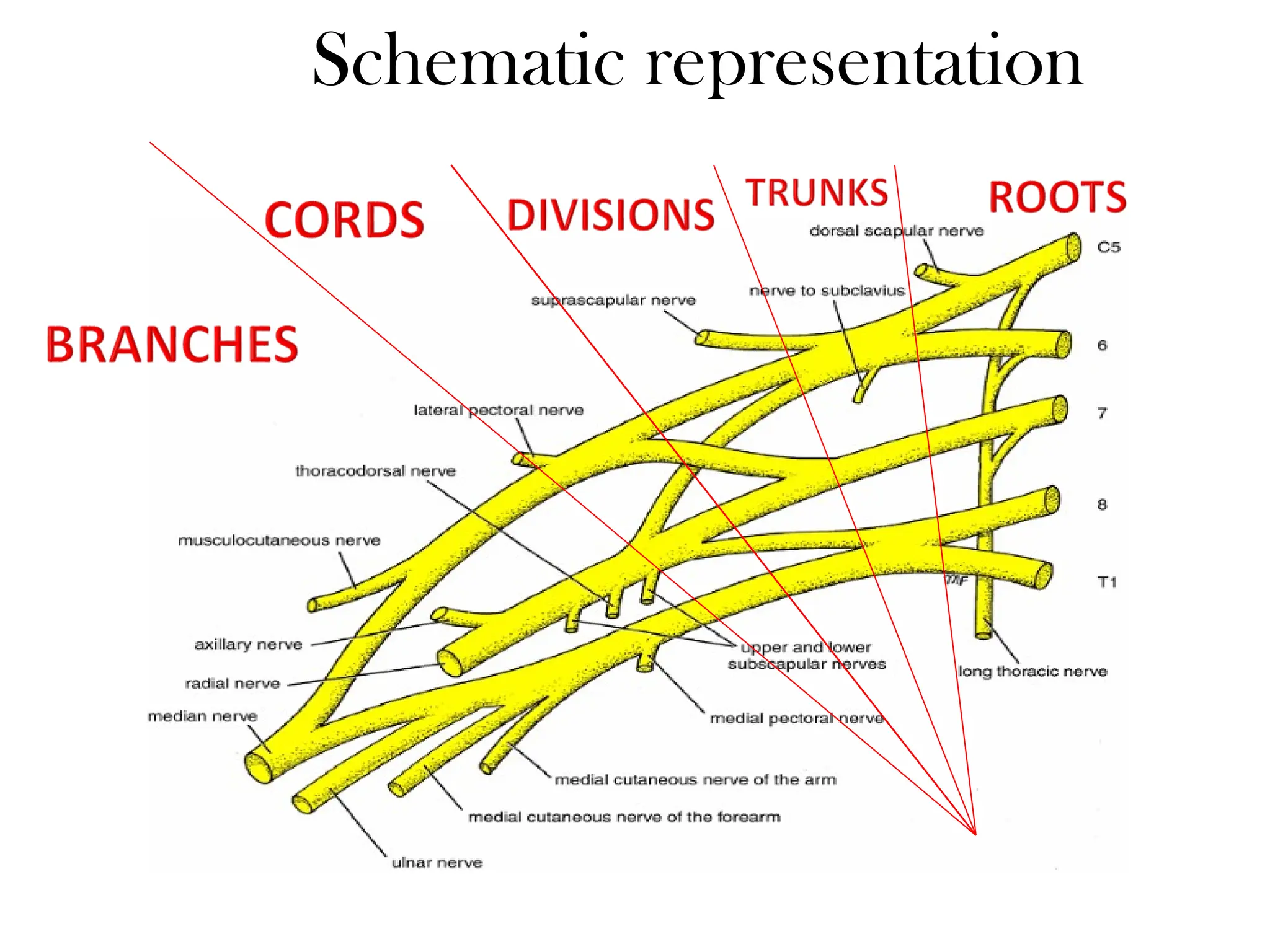
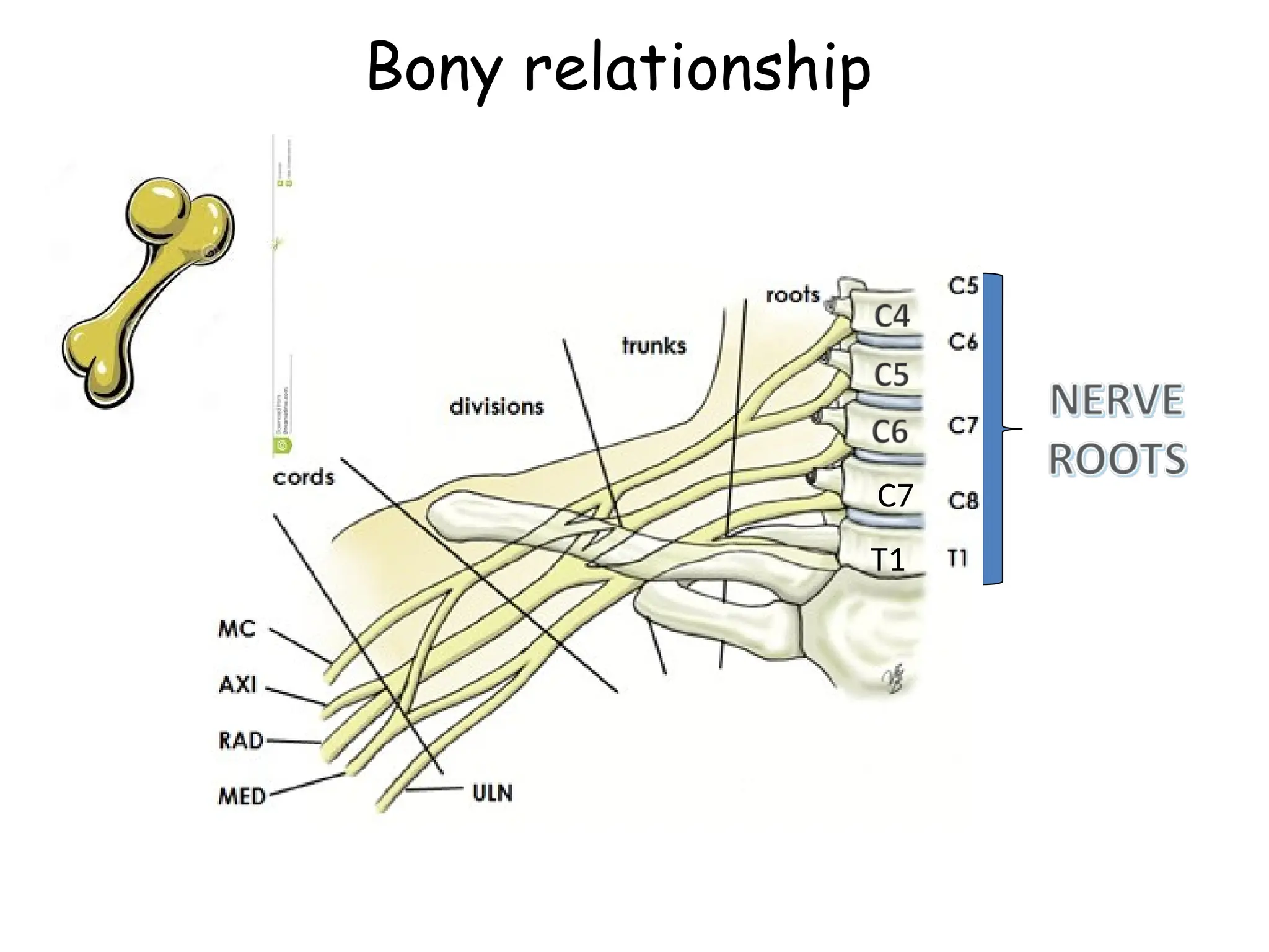
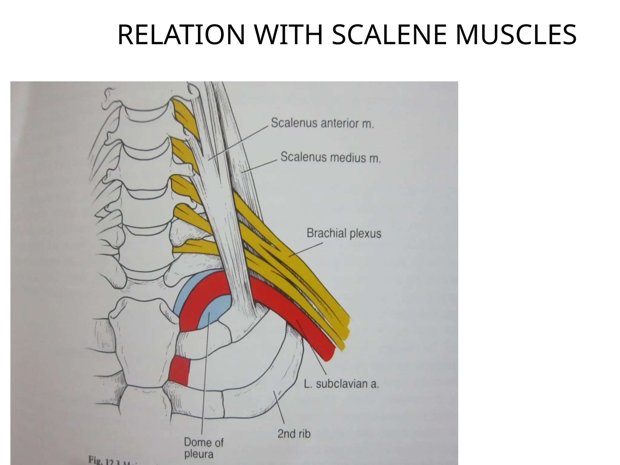
INTERSCALENE
BLOCK
• “Doctors
without
anatomy are
like moles.They
work in the dark
and the work of
their hands are
mounds”.
Anatomy
• The brachial plexus consists of trunks,divisons
and cords derived from the ventral roots of
C5to C8 & T1 nerves.
• Variable contributions may also from C4&T2
• The trunks are located b/w ant and middle
scalene muscles.
• At the lateral edge of the 1st
rib,the three
trunks separate into ant and post divisons.
Schematic representation
• The brachial plexus passes into the axilla,surrounded
by a fascial sheath descended from tha prevertebral
fascia separating the anterior & middle scalene.
• Within the axillary sheath ,thin septa divide the
brachial plexus into separate compartments,thus
limiting circumferential spread of injected drugs.
• That’s why multiple injection axillary block give
better results,and relative failure of single-injection
techniques.
Bony relationship
T1
C7
Relation with artery
RELATION WITH SCALENE MUSCLES
Interscalene Anatomy
SCM
Middle Scalene
Anterior Scalene
Omohyoid
• Anterior scalene-arises from the anterior
tubercles of the transverse processes of the
3,4,5,6th
cervical vertebrae.
• Inserted-into the sclene tubercle on the inner
border of the 1st
rib.
• Medial scalene-arises from posterior tubercles
of the six lowest cervical vertebrae & inserted
into upper surface of the 1st
rib.
• Clinical Applications
• The principal indication for an interscalene block is surgery on
or manipulation of the shoulder.
• Surgery in shoulder ,upper arm and forearm.
• Post operative analgesia for total shoulder arthroplasty
• Blockade occurs at the level of
the upper and middle trunks.
• Although this approach can also be
used for forearm and hand surgery, blockade of the inferior trunk
(C8 through T1) is often incomplete and requires supplementation
at the ulnar nerve for adequate surgical anesthesia in that
distribution.
Technique
• Five approaches;-
• Anterior approach
-winnie approach
-meier approach
-modified lateral approach of Borgeat.
• Posterior approach
-kappis /pippa approach
-boezaart approach.
• The patient is supine with the head flexed, not rotated, to the contralateral
side.
• A towel is placed under the head and the patient is asked to hold the
ipsilateral shoulder down as though reaching for the knee.
• The external jugular often crosses the interscalene groove at the level of
the cricoid cartilage.
• The SCM is palpated first.
• With the patient raising his head in a sniffing position the scalene muscle
stiffens.
• The interscalene groove between the anterior and medial scalene is found
by rolling the fingers off the lateral edge of the SCM.
• The groove can be felt at the peak of deep inspiration as the scalenes are
accessory muscles of ventilation.
• The groove is palpated as high as possible.
• The patient can then relax and the level at the cricoid is marked.
• The transverse process of C6 can usually be palpated and is directly
opposite to the entry point.
• Under sterile precautions and development of a skin wheal, a 22- to 25-
gauge, 4-cm needle is inserted perpendicular to the skin at a 45-degree
caudad and slightly posterior angle.
• needle is introduced perpendicular to the skin (not perpendicular to the
spinal axis) until a distinct pop is felt indicating penetration of the fascia.
• The needle is advanced until paresthesia is elicited.
• If bone is encountered within 2 cm of the skin, it is likely to be a
transverse process, and the needle may be “walked” across this structure
to locate the nerve.
• The plexus is very superficial (0.7-1.5cm),in obese pt no more than 2.5
cm.
• On PNS muscles below the shoulder is acceptable
(biceps or triceps).
• contraction of the diaphragm indicates phrenic nerve
stimulation and anterior needle placement; the needle
should be redirected posteriorly to locate the brachial
plexus.
• Ant to Post.-diaphragm(phrenic),rhomboids(dorsal
scapular),levator scapulae,trapezius(accessory).
• After the appropriate paresthesia or motor response is
obtained, the needle is stabilized.
• The use of flexible extension tubing facilitates
maintenance of the needle’s position while aspiration and
injection are taking place.
• After negative aspiration, 10to 40 mL of solution is injected
incrementally, depending on the desired extent of
blockade.
• Digital pressure above the injection site and downward
massage along with a 45-degree head-up position may
facilitate caudad spread and blockade of the lower trunk.
• Directing needles medially may cause inadvertent inj into
the vertebral artery,epidural space or cerebrospinal fluid .
Block assessment
• Successful neuraxial blockade relies upon spread of the
local anesthetic, therefore adequate “soak time” of 20-30
minutes after block administration should be allowed to
achieve adequate surgical anesthesia.
• Care must be taken when assessing block as injuries to
the arm or body can occur if the arm is not supported
and allowed to fall onto the patient or down to their side.
• Motor blockade occurs before sensory block.
• Motor and sensory block will occur proximally in the
shoulder 1st
and spreads distally.
• Axillary nerve motor block can be assessed by asking the patient
to abduct his upper arm against resistance.
• Axillary nerve sensory block can be assessed by checking for
anesthesia of the lateral aspect of the upper arm.
• Musculocutaneous nerve motor block is assessed by asking the
patient to flex at the elbow. Musculocutaneous nerve sensory
block is assessed by checking for anesthesia of the medial aspect
of the forearm.
• Radial nerve motor block can be assessed by asking the patient
to extend the forearm against resistance. Radial nerve sensory
block can be assessed by checking for anesthesia of the
webspace between the thumb and first finger.
• Median nerve motor block can be assessed by asking the
patient to make a fist while holding extension pressure on
the 1st
and 2nd
fingers.
• Median nerve sensory block can be assessed by checking
for anesthesia of the palmar surface of the thumb and 1st
two and half fingers.
• Ulnar nerve blockade is frequently missed by this block.
Ulnar nerve motor block can be assessed by asking the
patient to abduct his “pinky” finger against pressure.
Ulnar nerve sensory block can be assessed by checking for
anesthesia of the ring and “pinky” fingers.
Cutaneous distribution
Typical Areas of Block
• Winnie’s approach was subsequently modified by
Meier.
• In this modification the puncture point is at the
posterior edge of the scm bt located 2-3 cm more
cranially,at the level of the superior thyroid notch.
• The puncture is directed caudal at 30degree to
the skin and slightly lateral,aiming for the mid to
lateral point of the clavicle.
• Recommended for interscalene catheter insertion.
• The modified lateral approach of Borgeat uses
the same point as winnie,but directs the
needle along the interscalene space,towards
the post.part of the superior or middle trunk
in order to elicit triceps contractions.
Cervical approach
• The post.approach to the brachial plexus was Initially
described by kappis in 1912 and latterly by pippa.
• The advantage of the technique is that the needle only
passes through muscle and not nerves or arteries.
• Technique – flexing the head and palpating the spinal
processes of the 6th
& 7th
cervical vertebrae.
• Midway b/w the spinous processes of the 6th
& 7th
cervical vertebrae,a horizontal line of 3 cm is drawn
laterally.
• After connecting nerve stimulator,a 10 cm insulated
needle is inserted in a sagittal plane (aiming for the cricoid
cartilage) and contact is made with the transverse process
of the 7th
vertebrae after 5-6 cm.
• The needle is then redirected more cranially and should
locate the brachial plexus afer 6-8 cm –confirmed by
muscle contraction of the shoulder or abduction of the
arm.
• The volume of 0.5% bupivacaine,levobupivacaine or
ropivacaine sufficient for shoulder surgery is b/w
0.25ml/kg and 0.5ml/kg.
• With the Boezaart approach ,start at the level
of the first costovertebral angle and insert the
needle into the V-shaped groove b/w the
anterolateral edge of trapezius and
posteromedial edge of levator scapulae ,aiming
45 degree from a parasagittal plane and 30
degree caudal towards the sternal notch.
• Both posterior approaches are asso. With less
motor block than the ant.approach.
• Complications
• Ipsilateral diaphragmatic paresis
• Severe hypotension and bradycardia (i.e., the Bezold-Jarisch
reflex)
• Inadvertent epidural or spinal block
• Nerve damage or neuritis
• intravascular injection with Seizure activity
• Horner’s syndrome with dyspnea and hoarseness of voice.
• Puncture of the pleura may cause Pneumothorax.
• Hemothorax.
• Hematoma and Infection.
contraindication
• Contralateral phrenic nerve or recurrent
laryngeal nerve paralysis.
•Thank you

19.INTERSCALENE BLOCK presentation 12345

  • 1.
  • 2.
    • “Doctors without anatomy are likemoles.They work in the dark and the work of their hands are mounds”.
  • 3.
    Anatomy • The brachialplexus consists of trunks,divisons and cords derived from the ventral roots of C5to C8 & T1 nerves. • Variable contributions may also from C4&T2 • The trunks are located b/w ant and middle scalene muscles. • At the lateral edge of the 1st rib,the three trunks separate into ant and post divisons.
  • 4.
  • 5.
    • The brachialplexus passes into the axilla,surrounded by a fascial sheath descended from tha prevertebral fascia separating the anterior & middle scalene. • Within the axillary sheath ,thin septa divide the brachial plexus into separate compartments,thus limiting circumferential spread of injected drugs. • That’s why multiple injection axillary block give better results,and relative failure of single-injection techniques.
  • 6.
  • 7.
  • 8.
  • 9.
  • 10.
    • Anterior scalene-arisesfrom the anterior tubercles of the transverse processes of the 3,4,5,6th cervical vertebrae. • Inserted-into the sclene tubercle on the inner border of the 1st rib. • Medial scalene-arises from posterior tubercles of the six lowest cervical vertebrae & inserted into upper surface of the 1st rib.
  • 11.
    • Clinical Applications •The principal indication for an interscalene block is surgery on or manipulation of the shoulder. • Surgery in shoulder ,upper arm and forearm. • Post operative analgesia for total shoulder arthroplasty • Blockade occurs at the level of the upper and middle trunks. • Although this approach can also be used for forearm and hand surgery, blockade of the inferior trunk (C8 through T1) is often incomplete and requires supplementation at the ulnar nerve for adequate surgical anesthesia in that distribution.
  • 12.
    Technique • Five approaches;- •Anterior approach -winnie approach -meier approach -modified lateral approach of Borgeat. • Posterior approach -kappis /pippa approach -boezaart approach.
  • 14.
    • The patientis supine with the head flexed, not rotated, to the contralateral side. • A towel is placed under the head and the patient is asked to hold the ipsilateral shoulder down as though reaching for the knee. • The external jugular often crosses the interscalene groove at the level of the cricoid cartilage. • The SCM is palpated first. • With the patient raising his head in a sniffing position the scalene muscle stiffens. • The interscalene groove between the anterior and medial scalene is found by rolling the fingers off the lateral edge of the SCM. • The groove can be felt at the peak of deep inspiration as the scalenes are accessory muscles of ventilation. • The groove is palpated as high as possible. • The patient can then relax and the level at the cricoid is marked. • The transverse process of C6 can usually be palpated and is directly opposite to the entry point.
  • 16.
    • Under sterileprecautions and development of a skin wheal, a 22- to 25- gauge, 4-cm needle is inserted perpendicular to the skin at a 45-degree caudad and slightly posterior angle. • needle is introduced perpendicular to the skin (not perpendicular to the spinal axis) until a distinct pop is felt indicating penetration of the fascia. • The needle is advanced until paresthesia is elicited. • If bone is encountered within 2 cm of the skin, it is likely to be a transverse process, and the needle may be “walked” across this structure to locate the nerve. • The plexus is very superficial (0.7-1.5cm),in obese pt no more than 2.5 cm.
  • 17.
    • On PNSmuscles below the shoulder is acceptable (biceps or triceps). • contraction of the diaphragm indicates phrenic nerve stimulation and anterior needle placement; the needle should be redirected posteriorly to locate the brachial plexus. • Ant to Post.-diaphragm(phrenic),rhomboids(dorsal scapular),levator scapulae,trapezius(accessory). • After the appropriate paresthesia or motor response is obtained, the needle is stabilized.
  • 18.
    • The useof flexible extension tubing facilitates maintenance of the needle’s position while aspiration and injection are taking place. • After negative aspiration, 10to 40 mL of solution is injected incrementally, depending on the desired extent of blockade. • Digital pressure above the injection site and downward massage along with a 45-degree head-up position may facilitate caudad spread and blockade of the lower trunk. • Directing needles medially may cause inadvertent inj into the vertebral artery,epidural space or cerebrospinal fluid .
  • 19.
    Block assessment • Successfulneuraxial blockade relies upon spread of the local anesthetic, therefore adequate “soak time” of 20-30 minutes after block administration should be allowed to achieve adequate surgical anesthesia. • Care must be taken when assessing block as injuries to the arm or body can occur if the arm is not supported and allowed to fall onto the patient or down to their side. • Motor blockade occurs before sensory block. • Motor and sensory block will occur proximally in the shoulder 1st and spreads distally.
  • 20.
    • Axillary nervemotor block can be assessed by asking the patient to abduct his upper arm against resistance. • Axillary nerve sensory block can be assessed by checking for anesthesia of the lateral aspect of the upper arm. • Musculocutaneous nerve motor block is assessed by asking the patient to flex at the elbow. Musculocutaneous nerve sensory block is assessed by checking for anesthesia of the medial aspect of the forearm. • Radial nerve motor block can be assessed by asking the patient to extend the forearm against resistance. Radial nerve sensory block can be assessed by checking for anesthesia of the webspace between the thumb and first finger.
  • 21.
    • Median nervemotor block can be assessed by asking the patient to make a fist while holding extension pressure on the 1st and 2nd fingers. • Median nerve sensory block can be assessed by checking for anesthesia of the palmar surface of the thumb and 1st two and half fingers. • Ulnar nerve blockade is frequently missed by this block. Ulnar nerve motor block can be assessed by asking the patient to abduct his “pinky” finger against pressure. Ulnar nerve sensory block can be assessed by checking for anesthesia of the ring and “pinky” fingers.
  • 22.
  • 23.
  • 24.
    • Winnie’s approachwas subsequently modified by Meier. • In this modification the puncture point is at the posterior edge of the scm bt located 2-3 cm more cranially,at the level of the superior thyroid notch. • The puncture is directed caudal at 30degree to the skin and slightly lateral,aiming for the mid to lateral point of the clavicle. • Recommended for interscalene catheter insertion.
  • 25.
    • The modifiedlateral approach of Borgeat uses the same point as winnie,but directs the needle along the interscalene space,towards the post.part of the superior or middle trunk in order to elicit triceps contractions.
  • 26.
    Cervical approach • Thepost.approach to the brachial plexus was Initially described by kappis in 1912 and latterly by pippa. • The advantage of the technique is that the needle only passes through muscle and not nerves or arteries. • Technique – flexing the head and palpating the spinal processes of the 6th & 7th cervical vertebrae. • Midway b/w the spinous processes of the 6th & 7th cervical vertebrae,a horizontal line of 3 cm is drawn laterally.
  • 27.
    • After connectingnerve stimulator,a 10 cm insulated needle is inserted in a sagittal plane (aiming for the cricoid cartilage) and contact is made with the transverse process of the 7th vertebrae after 5-6 cm. • The needle is then redirected more cranially and should locate the brachial plexus afer 6-8 cm –confirmed by muscle contraction of the shoulder or abduction of the arm. • The volume of 0.5% bupivacaine,levobupivacaine or ropivacaine sufficient for shoulder surgery is b/w 0.25ml/kg and 0.5ml/kg.
  • 28.
    • With theBoezaart approach ,start at the level of the first costovertebral angle and insert the needle into the V-shaped groove b/w the anterolateral edge of trapezius and posteromedial edge of levator scapulae ,aiming 45 degree from a parasagittal plane and 30 degree caudal towards the sternal notch. • Both posterior approaches are asso. With less motor block than the ant.approach.
  • 29.
    • Complications • Ipsilateraldiaphragmatic paresis • Severe hypotension and bradycardia (i.e., the Bezold-Jarisch reflex) • Inadvertent epidural or spinal block • Nerve damage or neuritis • intravascular injection with Seizure activity • Horner’s syndrome with dyspnea and hoarseness of voice. • Puncture of the pleura may cause Pneumothorax. • Hemothorax. • Hematoma and Infection.
  • 30.
    contraindication • Contralateral phrenicnerve or recurrent laryngeal nerve paralysis.
  • 31.