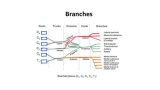
3. Major Motor Function of the Individual Nerves
Nerve Major Motor Function
Axillary(C5,6) Abduction of the shoulder
Musculocutaneous(C5,6,7) Flexion of the elbow
Radial(C5,6,7,8,T1) Extension of the elbow, wrist and finger
Median Flexion of the wrist and finger
Ulnar(C8,T1) Flexion of the wrist and finger
5. Interscalene brachial plexus block
Blockade distribution
Shoulder
and upper
arm
Supraclavicular
branches of
cervical plexus
Sparing of
inferior trunk
(C8-T1)
7. INTERSCALENE BRACHIAL PLEXUS
BLOCK
Indications
• Shoulder surgery
• Rotator cuff repair
• Upper arm surgery
• ORIF for fractures
• Surgery of the clavicle
Contraindications
• Patient’s refusal
• Local infection
• Active bleeding in an anticoagulated patient,
• Proven allergy to local anesthetic
• COPD/ Contralateral paresis of the phrenic nerve
• Previous neurologic deficit of the involved arm.
8. Interscalene brachial plexus block
• Advantages: Additional blockade of supraclavicular nerves/Superficial/Easy to perform
• Disadvantages: Hemidiaphragmatic paralysis/Sparing of inferior trunk/Not suitable for hand sx
Complications:
• Intravascular injection
• Intramuscular injection
• Intrafascicular injection
• LAST
• Total spinal anesthesia
• Horner syndrome
10. Landmark-Guided technique
POSITION:
Semisitting or supine/
head turned away
elevated
NEEDLE INSERTION:
Both fingers in interscalene groove/
needle inserted b/w fingers at C6
DIRECTION:
Perpendicular to skin
LA: 15–20 mL
12. PNS guided interscalene block
• Similar landmarks for guidance/ PNS Nerve stimulation 2.0 mA ; pulse width 100 μs / Response at 0.5mA
13. •Position: similar
• The prevertebral fascia, superficial cervical
plexus, and SCM - superficial to plexus. .
Usg guided interscalene block
1-3 cm deep
Linear probe
kept
transverse
3–4 c m supr
to clavicle
14. Usg guided ISB
Identify: anterior and middle scalene
muscles and brachial plexus
Needle insertion:
In plane towards the brachial plexus
Needle direction:
Lateral to medial direction
Pop is appreciated when the
prevertebral fascia is crossed
Careful aspiration
10-20ml of LA
16. oi
Infection • Astrict aseptic technique is used
Hematoma • Avoid multiple needle insertions, particularly in anticoagulated patients
• Apply steady pressure for 5 min when carotid artery is inadvertently punctured
• Use a smaller gauge needle to localize the brachial plexus in patients with difficult anatomy
Vascular
puncture
• Vascular puncture is not common with this technique
• Apply steady pressure for 5 min when the carotid artery is punctured
Local
anesthetic
toxicity
•Systemic toxicity most commonly occurs during or shortly after injection of local anesthetic; this is most
commonly caused by an inadvertent intravascular injection or channeling of forcefully injected local
anesthetic into small veins or lymphatic channels cut during needle manipulation
• Large volumes of long-acting anesthetic should be reconsidered in older and frail patients.
• Careful and frequent aspiration should be performed during the injection
• Avoid forceful, fast injection of local anesthetic
Nerve injury •Never inject local anesthetic when abnormal pressure on injection is encountered (opening pressure >15
psi)
•Local anesthetic should never be injected when patient complains of severe pain or exhibits a withdrawal
reaction on injection
T
otal spinal
anesthesia
•When stimulation is obtained with current intensity of <0.2 mA, the needle should be pulled back to obtain
the same response with current >0.2 mA before injecting local anesthetic to avoid injection into the dural
sleeves and the consequent epidural or spinal spread
• Never inject local anesthetic when abnormal pressure on injection is encountered
Horner
syndrome
•Occurrence of ipsilateral ptosis, hyperemia of the conjunctiva, and nasal congestion is common, and it is
dependent on the site of injection (less common with the low interscalene approach) and total volume of
local anesthetic injected; patients should be instructed on the occurrence of this syndrome and reassured
about its benign nature
Diaphragmatic • Commonly present; avoid interscalene blockade or a large volume of local anesthetic in patients
20. Supraclavicular brachial plexus block
• Indications
• Elbow and hand surgery.
• Contraindications
• Local infection/Significant coagulation abnormalities
• Inability to cooperate during block placement or surgery.
• Supraclavicular block is not used bilaterally
• Patients with respiratory compromise
• Pneumothorax / phrenic nerve block.
22. TECHNIQUE – LANDMARK GUIDED
Position: Semi sitting position with
head rotated to the opp side
Lateral border of SCM is identified
& followed distally to clavicle
A parasagittal line is drawn at this level - Area
medial to it that is at risk for pneumothorax
Needle insertion - Lateral to this parasagittal plane
SCM 2
Heads
Needle
Insertion
point
Clavicle
23. Technique – landmark guided
Needle direction:
Advanced perpendicular to skin
for 5 m m then turned caudally
parallel to midline
Local anesthetic:
15-25ml
25. Position: similar
Ultrasound guided supraclavicular block
Subclavian artery
Crosses first rib b/w ant & middle scalene,
Post to midpoint of clavicle
Parietal pleura & first rib
Linear hyperechoic structure lat & deep to SA
Brachial plexus
Bundle of hypoechoic round nodules post &
superficial to artery
Transverse on
neck, superior to
clavicle
at midpoint,
caudal tilt
26. Transducer :
Transverse plane proximal to
clavicle,tilted caudally
Needle insertion:
In plane toward the brachial
plexus, lateral-to-medial direction
Local anesthetic: 20-25ml
Ultrasound guided supraclavicular
block
27. TECHNIQUE
• Aspirate to check for blood
• Injection displaces the brachial plexus away
from the needle
• Additional advancement of the needle 1–2
mm closer to the plexus may be required
28. ADVANTAGES
• Anesthesia of arm distal to the shoulder
• Superficial/Easy to perform
• Fast onset
DISADVANTAGES
• Potential for pneumothorax
(comparatively less with USG guidance)
COMPLICATIONS
• Phrenic nerve block with diaphragmatic paralysis
• Sympathetic nerve block with development of horner syndrome
• Intravascular/Interfascicular injection
• Pneumothorax
31. Indications:
• Arm, elbow, forearm, and hand surgery
Advantages
• Reduced possibility of pneumothorax
• Avoidance of cervical vascular structures
• Does not produce a reduction in respiratory function.
• Superior to the axillary nerve block for axillary and musculocutaneous nerve block
Disadvantages
• The axillary vessel puncture
• Vessel compression in this area is difficult.
• Medial sparing of arm
Infraclavicular Brachial Plexus Block
32. Landmark guided technique
Direction:
Palpate AA as proximal in the axilla.
Insert needle at 60° angle from horizontal
Position:
Externally rotate and abduct arm.
Palpate coracoid process
Needle insertion:
2 cm medial & 2 cm caudad from
coracoid process
33. Vertical approach/PNS guided infraclavicular block
The nerve stimulator:
1-1.2 mA. Finger and/or
thumb flexion at 0.5 mA
Needle insertion: Midpoint of
the line from the jugular fossa to
the acromioclavicular joint.
Needle direction:
90-degree needle angle.
34. U L T R A S O U N D G U I D E D
INFRACLAVICULAR B L O C K
Axillary artery : Deep to the pectoralis major and minor
Cords surround AA
Place the probe lateral to the pleura
in order to minimize the risk of
3–5 cm
deep
Parasagittal,
Medial to coracoid,
Inferior to clavicle
35. USG guided infraclavicular block
Identification
Hyperechoic cords of around AA
Needle insertion: In-plane from the
cephalad end of the probe, inferior to
the clavicle
Direction: Toward the posterior
aspect of the axillary artery and
passes through the pectoralis major
and minor.
Local anesthetic : 10-20
m L
Inject 2-3 smaller aliquots at different
locations
36.
37.
38. COSTOCLAVICULAR BLOCK
• Brachial plexus block in the costoclavicular space
• B/w the posterior surface of the clavicle and the second rib
• Probe placed transversely in the infraclavicular fossa and directed cephalad
• All three cords seen lateral to the axillary artery
41. AXILLARY BRACHIAL PLEXUS BLOCK
INDICATIONS:
• Elbow, forearm, and hand surgery
• Chronic pain treatment.
• Cutaneous anesthesia for superficial procedures.
ADVANTAGES
• Safest of the four
• Reduced risk of pneumothorax
• Reduced risk of phrenic nerve blockade
DISADVANTAGES
• Musculocutaneous nerve lies outside the sheath and must be blocked separately
42. Landmark guided technique- Axillary nerve block
• Single-Injection (Stimulation) Technique
• Depending on the surgical site, needle is inserted above the arterial pulse (median nerve) or
below (radial nerve)
• Double-Injection Technique
• Needle is first inserted above the artery, below the coracobrachialis & L A is injected after
medial N stimulation
• Needle is then withdrawn and inserted below the artery and above the triceps. Radial nerve is
located and L A is injected.
• Multiple-Injection Technique
• Median nerve, radial nerve and musculocutaneous nerves are blocked
• Transarterial Technique
• Needle is directed towards the AA, advanced deeper until blood cannot be aspirated, half of
the volume of the L A is injected
43. LANDMARK AND PNS GUIDED AXILLARY NERVE BLOCK
Needle insertion: Above the
artery, below the coracobrachialis
Nerve stimulation: Start at 1-1.5
mA, reduce to 0.3-0.5mA and elicit
motor response
Direction: 30* for ulnar , radial and
median nerves. Inserted further
deep for musculocutaneous
44. LANDMARKS AND PATIENT POSITIONING
Position: Abduction of arm to 90*
Transducer position: short axis to arm, just
distal to pectoralis major insertion
Needle insertion: In-plane from the
anterior aspect and directed toward the
posterior aspect of the AA
45. ULTRASOUND ANATOMY
AA: Superficial in the medial proximal arm
Muscles: biceps, coracobrachialis , conjoined tendon.
Nerves: Median, ulnar, & radial nerves.
Musculocutaneous N.: b/w the biceps and
coracobrachialis
46. Needle insertion:
In-plane from antr aspect & directed
toward postr aspect of AA
Needle direction:
Identify AA 1–3 cm from skin
5–7 ml administered posteriorly, the
needle withdrawn to skin, redirected
toward the median and ulnar nerves,
7–10 ml injected.
Local anesthetic: 15-20 ml
USG guided Axillary Brachial Plexus Block
47. ULTRASOUN D -G U IDED B L O C K S AT T HE
E L B O W
• Indications: Han d and wrist surgery
• Transducer position: Transverse on
the elbow
• Goal: Injection of LA within the
vicinity of individual nerves (radial,
median, and ulnar)
• LA: 4–5 m L per nerve
48. THE RADIAL NERVE
• Lateral aspect of elbow, B/W brachioradialis & brachialis
• Transducer placed transversely on the anterolateral aspect
of the distal arm
• 3–4 cm above elbow crease
• Hyperechoic, triangular or oval structure with honeycomb
appearance
• Nerve divides just distal to the elbow crease into the
superficial (sensory) and deep (motor) branches
49. • Needle insertion: in plane, through the brachioradialis next to radial N.
• After negative aspiration, 4–5 ml of LA is injected
• If the spread is inadequate, slight adjustments can be made and inject 2–3 ml more
50. THE
MEDIAN
N E R V E
At the level of the elbow crease
Transducer is placed just above the crease to visualize brachial artery
Median nerve lies on the medial to the artery as a hyperechoic structure
51. • Arm abducted and the palm facing up.
• Transducer positioned transversely on the antecubital fossa.
• Needle is inserted in-plane from either side of the transducer.
• After negative aspiration, 4–5 ml of local anesthetic is injected.
53. THE ULNAR NERVE
• Posteromedial aspect of the elbow, few centimeters proximal to the crease
• Transducer is positioned more medially until the ulnar nerve is identified.
• The needle is inserted in-plane from either side of the transducer
• After negative aspiration, 4–5 ml of local anesthetic is injected
54. ULTRASOUN D -G U ID ED WRIST B L O C K
• Indications: Han d and finger surgery
• Transducer position: Transverse at wrist crease or distal third of the
forearm
• Goal: L ocal anesthetic injection next to the median, ulnar and radial
nerve
• Local anesthetic: 10–15 m L (total volume)
55. MEDIAN
N E R V E
• MN crosses elbow medial to BA
• Courses toward wrist deep to flexor
digitorum superficialis in the center of
forearm
• Transducer placed transversely at wrist
crease will reveal a cluster of oval
hyperechoic structures
56. U L N A R N E R V E
• Medial to UA from mid-forearm to
wrist
• Transducer at wrist crease
• hyperechoic anterior surface of
ulna
• U N lateral to bone and superficial
• Tendon of F C U superficial to U N
57. RADIAL N E R V E
• A subcutaneous field block
around styloid process of
radius
• USG can be used at elbow or
mid-forearm.
• Nerve is a thin hyperechoic
structure lateral to the radial
artery and superficial to
the radius