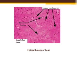
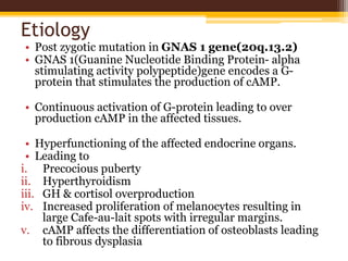
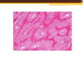
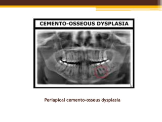
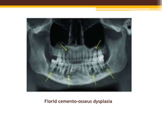
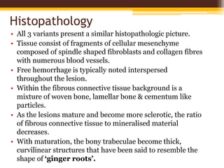
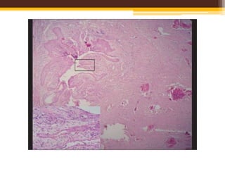
This document discusses fibrous dysplasia and cemento-osseous dysplasia, two fibro-osseous lesions of bone. It covers the classification, etiology, clinical features, radiographic appearance, histopathology, and treatment of these conditions. Specifically, it describes how fibrous dysplasia is caused by a mutation leading to overproduction of cAMP, and can present as monostotic or polyostotic lesions. It also outlines the three types of cemento-osseous dysplasia - focal, periapical, and florid - based on clinical and radiographic characteristics.
Invasive Squamous Cell Carcinoma (SCC)
SCC of the skin is a malignant tumor of keratinocytes, arising in the epidermis.
SCC usually arises in epidermal precancerous lesions and, depending on etiology and level of differentiation, varies in its aggressiveness.
The lesion is a plaque or a nodule with varying degrees of keratinization in the nodule and/or on the surface.
Thumb rule:
Undifferentiated SCC: is soft and has no hyperkeratosis;
Differentiated SCC: is hard on palpation and has hyperkeratosis.
Exposure:
Sunlight. Phototherapy, PUVA (oral psoralen + UVA). Excessive photochemotherapy can lead to promotion of SCC, particularly in patients with skin phototypes I and II or in patients with history of previous exposure to ionizing radiation or methotrexate treatment for psoriasis.
Lesions :
Indurated papule, plaque, or nodule ; adherent thick keratotic scale or hyperkeratosis ; when eroded or ulcerated, the lesion may have a crust in the center and a firm, hyperkeratotic, elevated margin
Clark levels
level I, intra-epidermal;
level II, invades papillary dermis;
level III fills papillary dermis;
level IV, invades reticular dermis;
level V, invades subcutaneous fat.
non-skeletal mesodermal tissues: adipose tissue, fibrous tissue, muscle, blood vessels and peripheral nerves (despite neuroectodermal origin)
benign, malignant and intermediate (low-grade malignant – locally aggressive, can recur, no metastatic potential)
originate from primitive mesenchymal stem cells
classification according to differentiation lines (e.g. liposarcoma is not a tumor arising from adipose tissue but exhibiting lipoblastic differentiation)
This presentation mainly deals with granuloma formation and various factors involved in it. It describes the examples of granulomatous disorders and gives a details on how to seperate them on histopathology.It also describes type 4 hypersensitivty reaction concisely
Definition
Morphology and ultra structure
Types of giant cell
Formation of giant cell
Inclusion bodies of giant cell
Giant cell in detail
It’s a mass formed by the union of several distinct cells (usually macrophage).
And usually arise in response to an infection.
Merriam – Webster - Dictionary
Giant cell as an unusually large cell, especially a large multinucleated often phagocytic cell.
A) Cell wall :
Mature giant cell wall is from five to ten times thicker than the cell wall of the surrounding cells
Cell wall has irregular surface with numerous projections jutting into the cytoplasm.
B) Cytoplasm :
Its dense and granular and contain protein
Contain RNA
Traces of carbohydrate and fat.
Fibrosseous lesions of the jaw
INTRODUCTION
Charles Waldron Classification Of The Fibro-Osseous Lesions Of The Jaws (1985)
1. Fibrous Dysplasia
a. Monostotic
b. Polyostotic
2. Fibro-Osseous (Cemental) Lesions Presumably Arising In The Periodontal Ligament
a. Periapical Cemental Dysplasia
b. Localized Fibro-Osseous-Cemental Lesions (Probably Reactive In Nature)
c. Florid Cement-Osseous Dysplasia (Gigantiform Cementoma)
d. Ossifying & Cemenifying Fibroma
3. Fibro-Osseous Neoplasms Of Uncertain Or Detectable Relationship To Those Arising In The Periodontal Ligament
a. Cemetoblastoma, Osteoblastoma & Osteoid Osteoma
b. Juvenile Active Ossifying Fibroma & Other So Called Aggressive, Active Ossifying /Cementifying Fibromas.
Classification Schemes of Fibro-OsseousLesions
1. Charles Waldron Classification Of The Fibro-Osseous Lesions Of The Jaws
(1985)
2. Working Classification Of Fibro-Osseous Lesions By Mico M. Malek (1987)
3. Peiter J. Slootweg & Hellmuth Muller (1990)
4. WHO Classification (1992)
5. Waldron Modified Classification Of Fibro-Osseous Lesions Of Jaws (1993)
6. Brannon & Fowler Classification (2001)
7. WHO Classification Of Fibro-Osseous Lesions Of Jaws (2005)
8. Paul M. Speight & Roman Carlos Classification (2006)
9. Eversole Classification (2008)
Working Classification Of Fibro-Osseous Lesions By Mico M. Malek (1987)
In 1987 from the viewpoint of
diagnostic pathologist, a
working classification of fibroosseous
lesions was given by
Mico M. Malek which is as
follows
Peiter J. Slootweg & Hellmuth Muller (1990)
In 1990 Peiter. J. Slootweg & Hellmuth Muller gave a classification that laid emphasis primarily on the histopathological features, and they underscore that this classification requires inclusion of adjacent normal bone to make diagnosis. However in the absence of this, the clinical & radiological features have to be taken in to consideration.
Group I: Fibrous Dysplasia
Group II: Juvenile Ossifying Fibroma
Group III: Ossifying Fibroma
Group IV: Periapical Cemental Dysplasia & Florid Osseous Dysplasia
WHO Classification (1992)
Waldron Modified Classification Of Fibro-Osseous Lesions Of Jaws (1993)
Later on, to overcome the demerits of his own classification, Waldron
reviewed the subject of benign fibro-osseous lesions of jaws (BFOL) in
1993 and suggested a modification of his earlier classification.
Brannon & Fowler Classification (2001)
In 2001, Brannon & Fowler gave another classification which was quite different from that of Waldron & WHO classification. This was done to include more number of lesions which were also showing features like FOL.
WHO Classification Of Fibro-Osseous LesionsOf Jaws (2005)
1) Ossifying Fibroma (OF)
2) Fiberous Dysplasia
3) Osseous Dysplasia
a. Periapical Osseous Dysplasia
b. Focal Osseous Dysplasia
c. Florid Osseous Dysplasia
d. Familial Gigantiform Cementoma
4) Central Giant Cell Granuloma
5) Cherubism
6) Aneurismal Bone Cyst
7) Solitary Bone Cyst
Paul M. Speight & Roman Carlos Classification(2006)
The Indian Dental Academy is the Leader in continuing dental education , training dentists in all aspects of dentistry and
offering a wide range of dental certified courses in different formats.
micro teaching on communication m.sc nursing.pdfAnurag Sharma
Microteaching is a unique model of practice teaching. It is a viable instrument for the. desired change in the teaching behavior or the behavior potential which, in specified types of real. classroom situations, tends to facilitate the achievement of specified types of objectives.
Title: Sense of Taste
Presenter: Dr. Faiza, Assistant Professor of Physiology
Qualifications:
MBBS (Best Graduate, AIMC Lahore)
FCPS Physiology
ICMT, CHPE, DHPE (STMU)
MPH (GC University, Faisalabad)
MBA (Virtual University of Pakistan)
Learning Objectives:
Describe the structure and function of taste buds.
Describe the relationship between the taste threshold and taste index of common substances.
Explain the chemical basis and signal transduction of taste perception for each type of primary taste sensation.
Recognize different abnormalities of taste perception and their causes.
Key Topics:
Significance of Taste Sensation:
Differentiation between pleasant and harmful food
Influence on behavior
Selection of food based on metabolic needs
Receptors of Taste:
Taste buds on the tongue
Influence of sense of smell, texture of food, and pain stimulation (e.g., by pepper)
Primary and Secondary Taste Sensations:
Primary taste sensations: Sweet, Sour, Salty, Bitter, Umami
Chemical basis and signal transduction mechanisms for each taste
Taste Threshold and Index:
Taste threshold values for Sweet (sucrose), Salty (NaCl), Sour (HCl), and Bitter (Quinine)
Taste index relationship: Inversely proportional to taste threshold
Taste Blindness:
Inability to taste certain substances, particularly thiourea compounds
Example: Phenylthiocarbamide
Structure and Function of Taste Buds:
Composition: Epithelial cells, Sustentacular/Supporting cells, Taste cells, Basal cells
Features: Taste pores, Taste hairs/microvilli, and Taste nerve fibers
Location of Taste Buds:
Found in papillae of the tongue (Fungiform, Circumvallate, Foliate)
Also present on the palate, tonsillar pillars, epiglottis, and proximal esophagus
Mechanism of Taste Stimulation:
Interaction of taste substances with receptors on microvilli
Signal transduction pathways for Umami, Sweet, Bitter, Sour, and Salty tastes
Taste Sensitivity and Adaptation:
Decrease in sensitivity with age
Rapid adaptation of taste sensation
Role of Saliva in Taste:
Dissolution of tastants to reach receptors
Washing away the stimulus
Taste Preferences and Aversions:
Mechanisms behind taste preference and aversion
Influence of receptors and neural pathways
Impact of Sensory Nerve Damage:
Degeneration of taste buds if the sensory nerve fiber is cut
Abnormalities of Taste Detection:
Conditions: Ageusia, Hypogeusia, Dysgeusia (parageusia)
Causes: Nerve damage, neurological disorders, infections, poor oral hygiene, adverse drug effects, deficiencies, aging, tobacco use, altered neurotransmitter levels
Neurotransmitters and Taste Threshold:
Effects of serotonin (5-HT) and norepinephrine (NE) on taste sensitivity
Supertasters:
25% of the population with heightened sensitivity to taste, especially bitterness
Increased number of fungiform papillae
TEST BANK for Operations Management, 14th Edition by William J. Stevenson, Ve...kevinkariuki227
TEST BANK for Operations Management, 14th Edition by William J. Stevenson, Verified Chapters 1 - 19, Complete Newest Version.pdf
Tom Selleck Health: A Comprehensive Look at the Iconic Actor’s Wellness Journeygreendigital
Tom Selleck, an enduring figure in Hollywood. has captivated audiences for decades with his rugged charm, iconic moustache. and memorable roles in television and film. From his breakout role as Thomas Magnum in Magnum P.I. to his current portrayal of Frank Reagan in Blue Bloods. Selleck's career has spanned over 50 years. But beyond his professional achievements. fans have often been curious about Tom Selleck Health. especially as he has aged in the public eye.
Follow us on: Pinterest
Introduction
Many have been interested in Tom Selleck health. not only because of his enduring presence on screen but also because of the challenges. and lifestyle choices he has faced and made over the years. This article delves into the various aspects of Tom Selleck health. exploring his fitness regimen, diet, mental health. and the challenges he has encountered as he ages. We'll look at how he maintains his well-being. the health issues he has faced, and his approach to ageing .
Early Life and Career
Childhood and Athletic Beginnings
Tom Selleck was born on January 29, 1945, in Detroit, Michigan, and grew up in Sherman Oaks, California. From an early age, he was involved in sports, particularly basketball. which played a significant role in his physical development. His athletic pursuits continued into college. where he attended the University of Southern California (USC) on a basketball scholarship. This early involvement in sports laid a strong foundation for his physical health and disciplined lifestyle.
Transition to Acting
Selleck's transition from an athlete to an actor came with its physical demands. His first significant role in "Magnum P.I." required him to perform various stunts and maintain a fit appearance. This role, which he played from 1980 to 1988. necessitated a rigorous fitness routine to meet the show's demands. setting the stage for his long-term commitment to health and wellness.
Fitness Regimen
Workout Routine
Tom Selleck health and fitness regimen has evolved. adapting to his changing roles and age. During his "Magnum, P.I." days. Selleck's workouts were intense and focused on building and maintaining muscle mass. His routine included weightlifting, cardiovascular exercises. and specific training for the stunts he performed on the show.
Selleck adjusted his fitness routine as he aged to suit his body's needs. Today, his workouts focus on maintaining flexibility, strength, and cardiovascular health. He incorporates low-impact exercises such as swimming, walking, and light weightlifting. This balanced approach helps him stay fit without putting undue strain on his joints and muscles.
Importance of Flexibility and Mobility
In recent years, Selleck has emphasized the importance of flexibility and mobility in his fitness regimen. Understanding the natural decline in muscle mass and joint flexibility with age. he includes stretching and yoga in his routine. These practices help prevent injuries, improve posture, and maintain mobilit
Pulmonary Thromboembolism - etilogy, types, medical- Surgical and nursing man...VarunMahajani
Disruption of blood supply to lung alveoli due to blockage of one or more pulmonary blood vessels is called as Pulmonary thromboembolism. In this presentation we will discuss its causes, types and its management in depth.
HOT NEW PRODUCT! BIG SALES FAST SHIPPING NOW FROM CHINA!! EU KU DB BK substit...GL Anaacs
Contact us if you are interested:
Email / Skype : kefaya1771@gmail.com
Threema: PXHY5PDH
New BATCH Ku !!! MUCH IN DEMAND FAST SALE EVERY BATCH HAPPY GOOD EFFECT BIG BATCH !
Contact me on Threema or skype to start big business!!
Hot-sale products:
NEW HOT EUTYLONE WHITE CRYSTAL!!
5cl-adba precursor (semi finished )
5cl-adba raw materials
ADBB precursor (semi finished )
ADBB raw materials
APVP powder
5fadb/4f-adb
Jwh018 / Jwh210
Eutylone crystal
Protonitazene (hydrochloride) CAS: 119276-01-6
Flubrotizolam CAS: 57801-95-3
Metonitazene CAS: 14680-51-4
Payment terms: Western Union,MoneyGram,Bitcoin or USDT.
Deliver Time: Usually 7-15days
Shipping method: FedEx, TNT, DHL,UPS etc.Our deliveries are 100% safe, fast, reliable and discreet.
Samples will be sent for your evaluation!If you are interested in, please contact me, let's talk details.
We specializes in exporting high quality Research chemical, medical intermediate, Pharmaceutical chemicals and so on. Products are exported to USA, Canada, France, Korea, Japan,Russia, Southeast Asia and other countries.
Explore natural remedies for syphilis treatment in Singapore. Discover alternative therapies, herbal remedies, and lifestyle changes that may complement conventional treatments. Learn about holistic approaches to managing syphilis symptoms and supporting overall health.
These simplified slides by Dr. Sidra Arshad present an overview of the non-respiratory functions of the respiratory tract.
Learning objectives:
1. Enlist the non-respiratory functions of the respiratory tract
2. Briefly explain how these functions are carried out
3. Discuss the significance of dead space
4. Differentiate between minute ventilation and alveolar ventilation
5. Describe the cough and sneeze reflexes
Study Resources:
1. Chapter 39, Guyton and Hall Textbook of Medical Physiology, 14th edition
2. Chapter 34, Ganong’s Review of Medical Physiology, 26th edition
3. Chapter 17, Human Physiology by Lauralee Sherwood, 9th edition
4. Non-respiratory functions of the lungs https://academic.oup.com/bjaed/article/13/3/98/278874
Factory Supply Best Quality Pmk Oil CAS 28578–16–7 PMK Powder in Stockrebeccabio
Factory Supply Best Quality Pmk Oil CAS 28578–16–7 PMK Powder in Stock
Telegram: bmksupplier
signal: +85264872720
threema: TUD4A6YC
You can contact me on Telegram or Threema
Communicate promptly and reply
Free of customs clearance, Double Clearance 100% pass delivery to USA, Canada, Spain, Germany, Netherland, Poland, Italy, Sweden, UK, Czech Republic, Australia, Mexico, Russia, Ukraine, Kazakhstan.Door to door service
Hot Selling Organic intermediates
Lung Cancer: Artificial Intelligence, Synergetics, Complex System Analysis, S...Oleg Kshivets
RESULTS: Overall life span (LS) was 2252.1±1742.5 days and cumulative 5-year survival (5YS) reached 73.2%, 10 years – 64.8%, 20 years – 42.5%. 513 LCP lived more than 5 years (LS=3124.6±1525.6 days), 148 LCP – more than 10 years (LS=5054.4±1504.1 days).199 LCP died because of LC (LS=562.7±374.5 days). 5YS of LCP after bi/lobectomies was significantly superior in comparison with LCP after pneumonectomies (78.1% vs.63.7%, P=0.00001 by log-rank test). AT significantly improved 5YS (66.3% vs. 34.8%) (P=0.00000 by log-rank test) only for LCP with N1-2. Cox modeling displayed that 5YS of LCP significantly depended on: phase transition (PT) early-invasive LC in terms of synergetics, PT N0—N12, cell ratio factors (ratio between cancer cells- CC and blood cells subpopulations), G1-3, histology, glucose, AT, blood cell circuit, prothrombin index, heparin tolerance, recalcification time (P=0.000-0.038). Neural networks, genetic algorithm selection and bootstrap simulation revealed relationships between 5YS and PT early-invasive LC (rank=1), PT N0—N12 (rank=2), thrombocytes/CC (3), erythrocytes/CC (4), eosinophils/CC (5), healthy cells/CC (6), lymphocytes/CC (7), segmented neutrophils/CC (8), stick neutrophils/CC (9), monocytes/CC (10); leucocytes/CC (11). Correct prediction of 5YS was 100% by neural networks computing (area under ROC curve=1.0; error=0.0).
CONCLUSIONS: 5YS of LCP after radical procedures significantly depended on: 1) PT early-invasive cancer; 2) PT N0--N12; 3) cell ratio factors; 4) blood cell circuit; 5) biochemical factors; 6) hemostasis system; 7) AT; 8) LC characteristics; 9) LC cell dynamics; 10) surgery type: lobectomy/pneumonectomy; 11) anthropometric data. Optimal diagnosis and treatment strategies for LC are: 1) screening and early detection of LC; 2) availability of experienced thoracic surgeons because of complexity of radical procedures; 3) aggressive en block surgery and adequate lymph node dissection for completeness; 4) precise prediction; 5) adjuvant chemoimmunoradiotherapy for LCP with unfavorable prognosis.
3. Classification
A). Based on Morphology:
Long
bones
e.g. upper
and lower
limbs;
Have
marrow
cavity filled
with yellow
marrow
Short
bones
e.g. carpels
and tarsals;
Have
marrow but
lack marrow
cavity
Flat
bones
e.g. skull
and facial
bones;
Spongy
bone is
present
between
upper &
lower
layer of
compact
bone
Irregular
bones
e.g. sphenoid
and ethmoid;
Spongy bone
covered with a
thin layer of
compact bone
and has
marrow
without any
marrow cavity
Sesamoid
bones
e.g. patella;
Develop in areas
of pressure or
tension
5. B). Based on Development:
Endochondral
Replacement of hyaline
cartilage with bony
tissue.
e.g. bones of trunk and
extremities
Intramembranous
Replacement of sheet
like connective tissue
with bony tissue
e.g. cranial & facial flat
bones of skull, mandible
and clavicle
6.
7. C). Based on Microscopic Structure:
Mature Bone Immature Bone
-first formed bone
-rarely seen after
birth
-seen in alveolar
bone & during
healing of fractures.
Compact/Lam
ellar/Cortical
Bone
-well formed
concentric
lamellae around
haversian canal
Cancellous/Spongy/Trab
ecular Bone:
-irregular bony spikules
called ‘Trabeculae’ around
marrow spaces.
10. Composition
• Mineral Content
• Hydroxyapatite crystals with
carbonate content & low Ca/P
ratio.
• Bone crystals as ‘leaf like’
structures aligned parallel to
collagen fibres.
• Narrow gaps between crystals
is associated with water and
organic macromolecules.
• Organic Content
• Mainly type I collagen
• Also type V
• Alveolar bone = type I + type
III + type V + type XII
collagen
• Sharpeys fibres contain type
III with type I collagen
• Type III & type XII are
produced by fibroblasts during
production of periodontal
ligament.
• Type I, V and XII are
expressed by osteoclasts.
11. Non Collagenous Proteins
1). Osteocalcin:
-Has amino acid – gamma carboxy glutamic acid
-Demonstrated in alveolar bone
-Carboxy terminal is chemo-attractant to osteoclasts precursors.
2). Osteopontin:
-Present in alveolar bone
-Heavily glycosylated & phosphorylated
-Aspartate is predominant
-Inhibits hydroxyapatite crystal growth
3). Bone Sialoprotein:
-Glutamic acid is predominant
-Initiation of mineral crystal formation
4).Osteonectin:
-Calcium binding glycoprotein
12. Bone Histology
• Periosteum
• Endosteum
• Circumferential lamellae
• Concentric lamellae
• Haversian canal
• Haversian canal + Concentric Lamellae = OSTEON
• Reversal Line: sharply delineated by cement lines which is highly
basophilic(due to rich content of glycoproteins & proteoglycans) and marks
the limit of bone erosion prior to osteon formation.
• Resting Line
• Volksmann canal connects the osteons and the periosteum.
• Lacunae containing osteocytes at the junction of lamellae
• Canaliculi radiating from lacunae; helps in exchange of nutrients and
wastes between lacunae (osteocytes).
• Interstitial lamellae: present between osteons; remnants of osteon, left
behind during remodelling.
Fibrocollagenous layer
(outer)
Inner layer with bone cells with rich
vascular supply
15. • Non heridetary disorder of unknown cause.
• Can occur in any part of the skeleton but the
bones of skull, thigh, ribs, upper arms and pelvis
are most commonly involved.
• It is not a form of cancer.
• Most lesions are monostotic, asymptomatic &
identified incidentally & can be treated with
clinical observation.
16. Etiology
• Post zygotic mutation in GNAS 1 gene(20q.13.2)
• GNAS 1(Guanine Nucleotide Binding Protein- alpha
stimulating activity polypeptide)gene encodes a G-
protein that stimulates the production of cAMP.
• Continuous activation of G-protein leading to over
production cAMP in the affected tissues.
• Hyperfunctioning of the affected endocrine organs.
• Leading to
i. Precocious puberty
ii. Hyperthyroidism
iii. GH & cortisol overproduction
iv. Increased proliferation of melanocytes resulting in
large Cafe-au-lait spots with irregular margins.
v. cAMP affects the differentiation of osteoblasts leading
to fibrous dysplasia
17. Pathophysiology
• Medullary bone is replaced by fibrous tissue which
appears radiolucent on radiographs-”Ground Glass
Appearance ”.
• Trabeculae of woven bone contains fluid filled cysts that
are embedded largely in collagenous fibrous matrix.
• Bony trabeculae are abnormally thin & irregular & often
described as “Chinese Characters”
.
• Fibrous Dyspalsia is characterised by “Shepherd’s
Crook” deformity which refers to coxa varus angulation
of proximal femur.
• Cause of transformation is not completely known,
however levels of transcription factor C-fos are raised
which leads to gene over expression & tumor formation.
18. Types
• Monostotic: Involvement of a single bone.
• Polyostotic: Many bones are involved.
Most severe form is McCune Albright
Syndrome
19. Clinical Features
• Four disease patterns are recognised:
i. Monostotic
ii. Polyostotic
iii. Craniofacial form
iv. Cherubism
Age : 3-15 years
2/3 of patients with polyostotic disease are asymptomatic before
they are aged 10 years.
Monostotic patients as old as 20-30 years are asymptomatic.
Males and females are equally affected.
Clinical findings of increasing pain & an enlarging soft tissue
mass suggest malignant change.
20. Monostotic Fibrous Dysplasia
70-80% of the lesions.
Most frequently occurs in the ribs(28%),
femur(23%) and humerous in decreasing order of
frequency.
The clinical term ‘Leontiasis Ossea’ has often been
applied to cases of fibrous dysplasia which affect the
maxilla or facial bones & give the patient a leonine
appearance.
21. Clinical Features
• Age: children & young adults.
• Sex: equally affects males &
females
• Painless swelling or bulging of
jaws.
(usually involves labial/buccal
plate; when it involves
mandible, sometimes it causes a
protruberant excrescence of
inferior border)
• Malalignment, tipping of teeth
due to progressive expansile
nature of lesion & tenderness
may develop.
• Malocclusion
• Mucosa over the lesion is
invariably intact.
(Fibrous Dysplasia of maxilla is
a serious form of disease since it
has marked predilection for
occurrence in children & is
impossible to eradicate without
radical, mutilating surgery).
• lesions extend locally to involve
maxillary sinus, zygomatic
process & floor of orbit & base of
skull.
• Bulging of canine fossa
• Extreme prominence of
zygomatic process
• Marked facial deformity
22. Radiographic Features
• Has 3 basic patterns:
i. Small, unilocular/large, multilocular with well
circumscribed border containing a network of fine
bony trabeculae.
ii. Increased trabeculations render the lesion more
opaque-Mottled Appearance
iii. Many delicate trabeculations-Ground Glass or peau
d’ orange appearance; it is not well circumscribed
and blends into adjacent normal bone.
• In all types, cortical plate is thinned because of
expansile nature of growth.
24. Histopatholgy
• Lesion is fibrous composed of proliferating
fibroblasts in compact stroma of interlacing
collagen fibres.
• Irregular bony trabeculae are scattered
throughout the lesion without any definite
pattern- ‘Chinese Character’ shaped.
• Trabeculae are of coarse, woven bone.
25.
26. Treatment
• Surgical removal of lesion.
• Lesions with type III radiographic findings
should be block resected.
• Malignant transformation into osteosarcoma.
27. Mc Cune Albright Syndrome
or
Polyostotic Fibrous Dysplasia
Defined as association of polyostotic
fibrous dysplasia, precocious puberty,
café-au-lait spots & other
endocrinopathies due to hyperactivity
various endocrine glands.
28. Etiology
• Post zygotic activation mutation of GS alpha
gene in affected tissues.
• GS alpha subunit is a component of G-protein
complex, which couples hormone receptors to
Adenylyl Cyclase (intracellular second
messenger) in a submembrane site.
29. Clinical Features
• Precocious puberty associated with the condition is gonadotrophin-
independent.
• Acromegaly
• Gonadotrophin-McCune Albright Syndrome
• Hyperprolactinemia
• Some severely affected patients may present with associated
hepatic, cardiac and GI dysfunction.
• Cutaneous pigmentation is the most common extraskeletal
manifestation & is ipsilateral to side of bony lesions.
• Café-au-lait spots are related to amount of melanin in basal cells
of epidermis.
• Pigmentation may occur at birth & may precede the development of
skeletal & endocrine abnormalities.
32. Histopathology
• Areas of fibrous metaplasia within flat and tubular
bones.
• Progressively expanding fibrous lesion of bone
forming mesenchyme.
• Concentric expansion in an outward direction within
the medullary bone.
• Well-defined, non-encapsulated.
• Lesions are rich in spindle shaped fibroblasts with a
swirled appearance within the marrow space.
33. Treatment
• Mild cases: surgical radiation
• Severe cases: X-ray radiation
• Prognosis depends on the degree of skeletal
involvement.
• Malignant transformation into osteosarcoma
can also occur.
35. Pathogenesis
• Periodontal origin; or
• Defect in extraligamentary bone remodelling
that maybe triggered by local factors or possibly
by hormonal imbalance.
36. Types
• Based on clinical and radiographic features, it is
divided into three groups:
i. Focal cemento-osseus dysplasia
ii. Periapical cemento-osseus dysplasia
iii. Florid cemento-osseus dysplasia
37. Focal Cemento-Osseus Dysplasia
• Exhibits a single site of involvement.
• Most commonly in black females with a
predilection for 3-6 decade.
• Posterior mandible is the commonest site.
• Usually asymptomatic with a positive vitality
test of the affected teeth.
• Most of the lesions are smaller than 1.5cm in
diameter.
38. Radiographic Features
• Lesions vary from being completely radiolucent to
densely radiopaque with a thin peripheral
radiolucent rim.
• Most common is a mixed radiolucent and
radiopaque pattern.
• Well defined lesion with a slightly irregular border.
• Occurs in both dentulous and edentulous areas.
40. Periapical Cemento-Osseus Lesions
• Also called as ‘Cementoma’,
‘Fibrocementoma’, ‘Periapical Fibro
Osteoma’.
• Teeth associated with lesion is invariably vital.
• Involves periapical region of anterior mandible.
• Generally occurs between 30-50 yrs of age.
41. Radiographic Features
• Early lesions appear as circumscribed areas of
radiolucency involving periapical areas of tooth.
• Mature lesions create a mixed radiolucent and a
radiopaque appearance.
• Periodontal ligament is intact.
• There is no fusion to the teeth.
43. Florid Cemento-Osseus Lesions
• Most clinically extensive form of cemento-osseus dysplasia,
thus the term – ‘Florid’.
• Common in black females with marked predilection for
middle aged to older adults.
• Multifocal involvement, not limited to anterior areas.
• May involve all the 4 quadrants.
• Asymptomatic.
• Dull pain and an alveolar sinus tract maybe present.
• Some degree of expansion maybe seen.
• Bilateral and symmetric involvement.
44. Radiographic Features
• Initially, lesion is predominantly radiolucent,
with time, becomes mixed and then
predominantly radiopaque with a thin
peripheral rim.
• Maybe totally radiopaque and blend with
adjacent normal appearing bone.
• Both, edentulous and dentulous areas are
affected.
46. Histopathology
• All 3 variants present a similar histopathologic picture.
• Tissue consist of fragments of cellular mesenchyme
composed of spindle shaped fibroblasts and collagen fibres
with numerous blood vessels.
• Free hemorrhage is typically noted interspersed
throughout the lesion.
• Within the fibrous connective tissue background is a
mixture of woven bone, lamellar bone & cementum like
particles.
• As the lesions mature and become more sclerotic, the ratio
of fibrous connective tissue to mineralised material
decreases.
• With maturation, the bony trabeculae become thick,
curvilinear structures that have been said to resemble the
shape of ‘ginger roots’.
47.
48. Treatment
• For asymptomatic patients: regular recall
examinations with prophylaxis and
reinforcement of good home hygiene care.
• For symptomatic patients:
• Antibiotics
• Sequestration of sclerotic cement like masses
and is followed by healing.
• Saucerisation of dead bone.