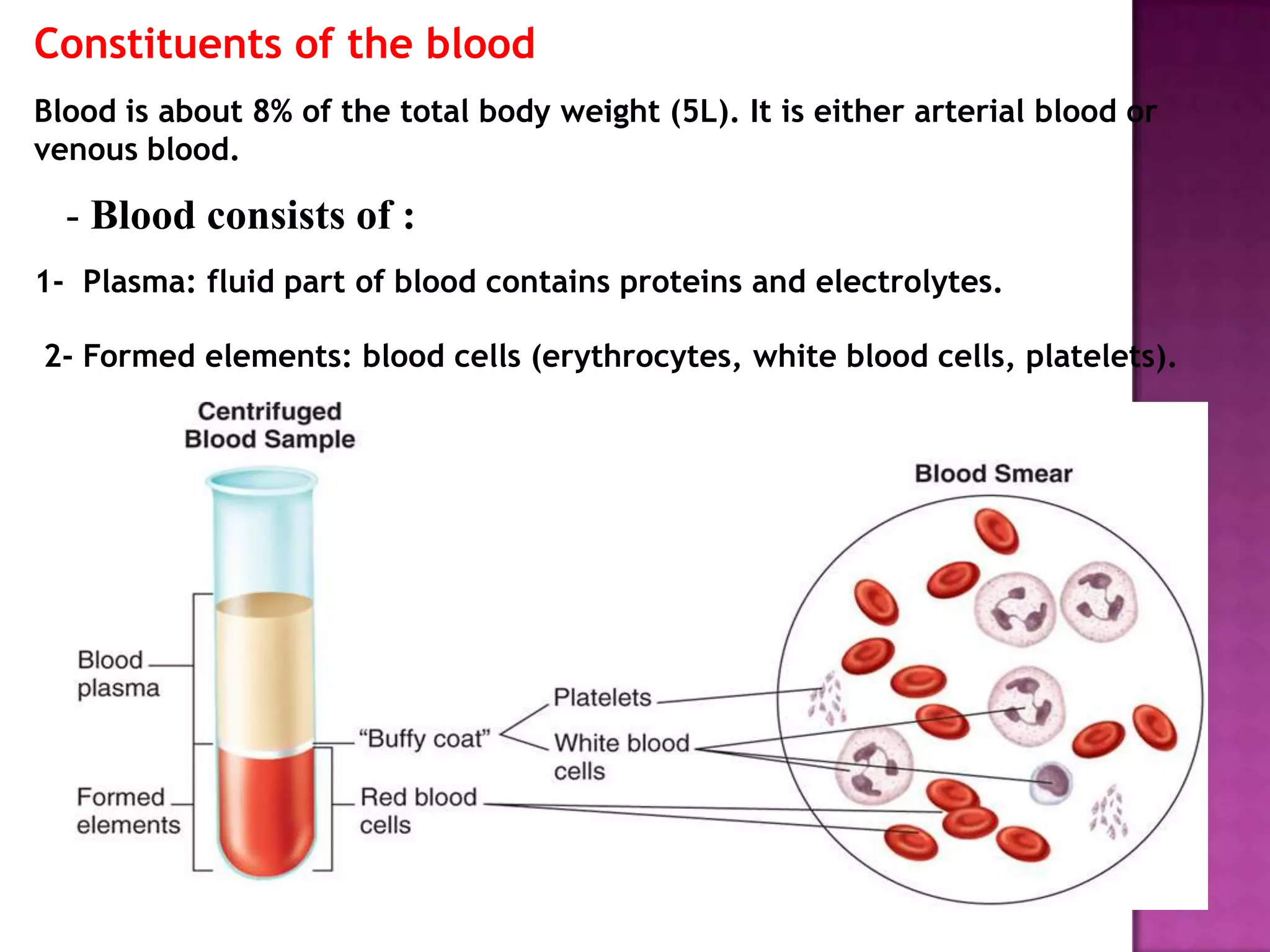
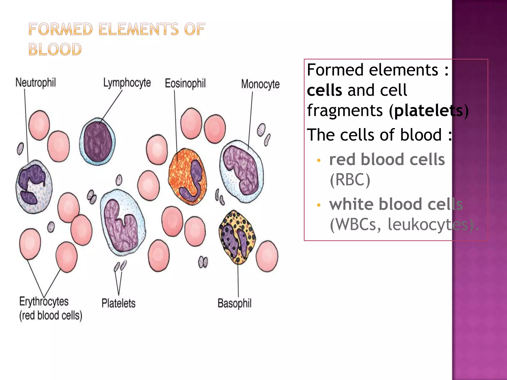
Blood consists of plasma and formed elements. Plasma contains proteins and electrolytes while formed elements are blood cells including erythrocytes, leukocytes, and platelets.
Erythrocytes are biconcave discs that contain hemoglobin and transport oxygen. Leukocytes include granulocytes like neutrophils, eosinophils and basophils and agranulocytes like lymphocytes and monocytes that protect against infection. Platelets are cell fragments involved in blood clotting.
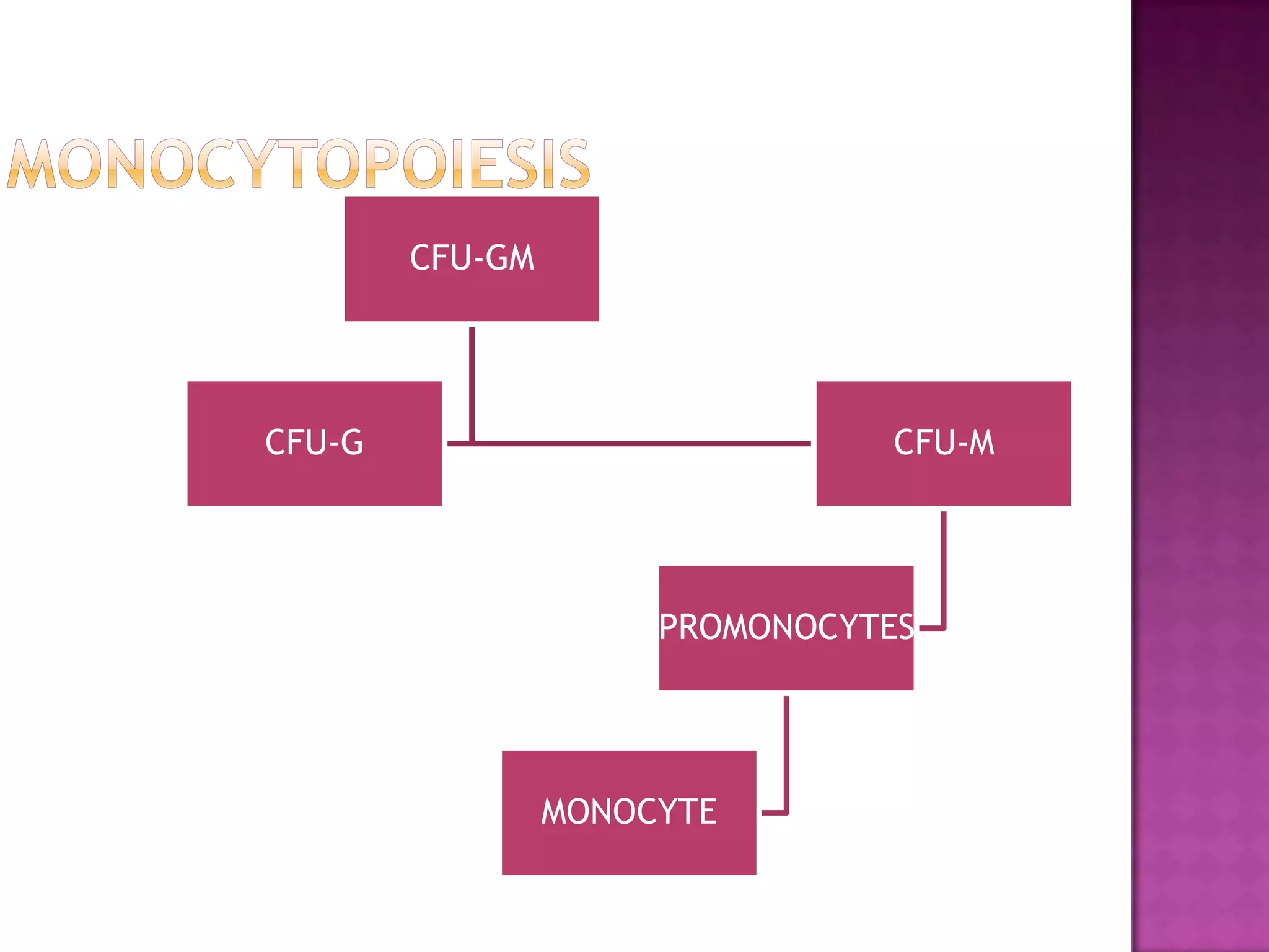
Hematopoiesis occurs in the bone marrow where stem cells differentiate into various blood cell types through the influence of growth factors like erythropoietin and cytokines. Blood groups are determined by