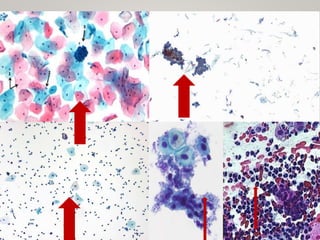
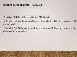
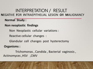
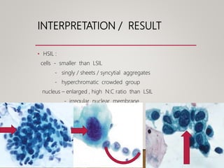
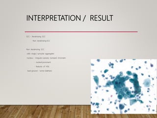
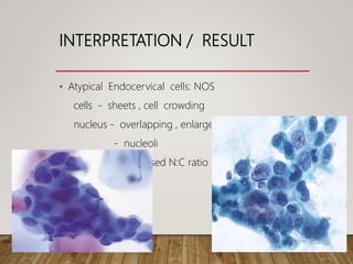
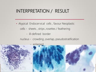
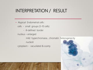
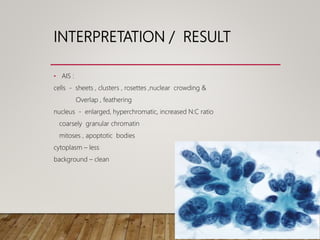
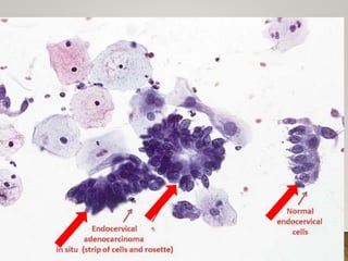
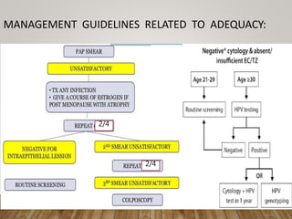
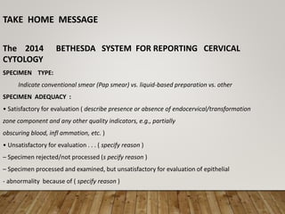
The Bethesda System is a standardized reporting system used for Pap test results. It provides a structured format for reporting cervical and vaginal cytology diagnoses. The system includes sections for specimen type, adequacy, interpretation/result, and optional notes. Interpretation/result categories include negative for intraepithelial lesion or malignancy, epithelial cell abnormalities (such as atypical squamous cells, low-grade squamous intraepithelial lesion, or high-grade squamous intraepithelial lesion), and other malignant neoplasms. Specific cytological features are described for each category. The system aims to improve consistency and communication of Pap test results.
Atlas on bethesda system for reporting cervical cytologyAshish Jawarkar
This is an atlas with more nearly 100 images, authentic taken from NCI web atlas. Useful to understand and report pap smears. The subject has been presented in a way which will help students reproduce in exams.
Atlas on bethesda system for reporting cervical cytologyAshish Jawarkar
This is an atlas with more nearly 100 images, authentic taken from NCI web atlas. Useful to understand and report pap smears. The subject has been presented in a way which will help students reproduce in exams.
This is a presentation on the topic of cytology of the breast, prepared by Dr Ashish Jawarkar, he is MD in pathology and a teacher at Parul institute of Medical sciences and research Vadodara.
Cervical cytology was introduced by George
Papanicolaou into clinical practice in 1940. In 1945,
the Papanicolaou smear received the endorsement of
the American cancer society as an effective method
for the prevention of cervical cancer .
www.drvikramsaraswat.co.in
www.drsaraswatpathlabs.com
Cervical screening is the process of detecting and removing abnormal tissue or cells in the cervix before cervical cancer develops. By aiming to detect and treat cervical neoplasia early on, cervical screening aims at secondary prevention of cervical cancer. Several screening methods for cervical cancer are the Pap test (also known as Pap smear or conventional cytology), liquid-based cytology, the HPV DNA testing and the visual inspection with acetic acid. Pap test and liquid-based cytology have been effective in diminishing incidence and mortality rates of cervical cancer in developed countries but not in developing countries.Prospective screening methods that can be used in low-resource areas in the developing countries are the HPV DNA testing and the visual inspection.
I am working as a pathologist at the department of clinical pathology, Mandalay General Hospital, Mandalay Division, Myanmar.
Dr San Yu Maung (M.B.B.S.) (M.Med.Sc.) (Pathology)
Ozempic: Preoperative Management of Patients on GLP-1 Receptor Agonists Saeid Safari
Preoperative Management of Patients on GLP-1 Receptor Agonists like Ozempic and Semiglutide
ASA GUIDELINE
NYSORA Guideline
2 Case Reports of Gastric Ultrasound
Tom Selleck Health: A Comprehensive Look at the Iconic Actor’s Wellness Journeygreendigital
Tom Selleck, an enduring figure in Hollywood. has captivated audiences for decades with his rugged charm, iconic moustache. and memorable roles in television and film. From his breakout role as Thomas Magnum in Magnum P.I. to his current portrayal of Frank Reagan in Blue Bloods. Selleck's career has spanned over 50 years. But beyond his professional achievements. fans have often been curious about Tom Selleck Health. especially as he has aged in the public eye.
Follow us on: Pinterest
Introduction
Many have been interested in Tom Selleck health. not only because of his enduring presence on screen but also because of the challenges. and lifestyle choices he has faced and made over the years. This article delves into the various aspects of Tom Selleck health. exploring his fitness regimen, diet, mental health. and the challenges he has encountered as he ages. We'll look at how he maintains his well-being. the health issues he has faced, and his approach to ageing .
Early Life and Career
Childhood and Athletic Beginnings
Tom Selleck was born on January 29, 1945, in Detroit, Michigan, and grew up in Sherman Oaks, California. From an early age, he was involved in sports, particularly basketball. which played a significant role in his physical development. His athletic pursuits continued into college. where he attended the University of Southern California (USC) on a basketball scholarship. This early involvement in sports laid a strong foundation for his physical health and disciplined lifestyle.
Transition to Acting
Selleck's transition from an athlete to an actor came with its physical demands. His first significant role in "Magnum P.I." required him to perform various stunts and maintain a fit appearance. This role, which he played from 1980 to 1988. necessitated a rigorous fitness routine to meet the show's demands. setting the stage for his long-term commitment to health and wellness.
Fitness Regimen
Workout Routine
Tom Selleck health and fitness regimen has evolved. adapting to his changing roles and age. During his "Magnum, P.I." days. Selleck's workouts were intense and focused on building and maintaining muscle mass. His routine included weightlifting, cardiovascular exercises. and specific training for the stunts he performed on the show.
Selleck adjusted his fitness routine as he aged to suit his body's needs. Today, his workouts focus on maintaining flexibility, strength, and cardiovascular health. He incorporates low-impact exercises such as swimming, walking, and light weightlifting. This balanced approach helps him stay fit without putting undue strain on his joints and muscles.
Importance of Flexibility and Mobility
In recent years, Selleck has emphasized the importance of flexibility and mobility in his fitness regimen. Understanding the natural decline in muscle mass and joint flexibility with age. he includes stretching and yoga in his routine. These practices help prevent injuries, improve posture, and maintain mobilit
Couples presenting to the infertility clinic- Do they really have infertility...Sujoy Dasgupta
Dr Sujoy Dasgupta presented the study on "Couples presenting to the infertility clinic- Do they really have infertility? – The unexplored stories of non-consummation" in the 13th Congress of the Asia Pacific Initiative on Reproduction (ASPIRE 2024) at Manila on 24 May, 2024.
Factory Supply Best Quality Pmk Oil CAS 28578–16–7 PMK Powder in Stockrebeccabio
Factory Supply Best Quality Pmk Oil CAS 28578–16–7 PMK Powder in Stock
Telegram: bmksupplier
signal: +85264872720
threema: TUD4A6YC
You can contact me on Telegram or Threema
Communicate promptly and reply
Free of customs clearance, Double Clearance 100% pass delivery to USA, Canada, Spain, Germany, Netherland, Poland, Italy, Sweden, UK, Czech Republic, Australia, Mexico, Russia, Ukraine, Kazakhstan.Door to door service
Hot Selling Organic intermediates
These lecture slides, by Dr Sidra Arshad, offer a quick overview of physiological basis of a normal electrocardiogram.
Learning objectives:
1. Define an electrocardiogram (ECG) and electrocardiography
2. Describe how dipoles generated by the heart produce the waveforms of the ECG
3. Describe the components of a normal electrocardiogram of a typical bipolar leads (limb II)
4. Differentiate between intervals and segments
5. Enlist some common indications for obtaining an ECG
Study Resources:
1. Chapter 11, Guyton and Hall Textbook of Medical Physiology, 14th edition
2. Chapter 9, Human Physiology - From Cells to Systems, Lauralee Sherwood, 9th edition
3. Chapter 29, Ganong’s Review of Medical Physiology, 26th edition
4. Electrocardiogram, StatPearls - https://www.ncbi.nlm.nih.gov/books/NBK549803/
5. ECG in Medical Practice by ABM Abdullah, 4th edition
6. ECG Basics, http://www.nataliescasebook.com/tag/e-c-g-basics
Flu Vaccine Alert in Bangalore Karnatakaaddon Scans
As flu season approaches, health officials in Bangalore, Karnataka, are urging residents to get their flu vaccinations. The seasonal flu, while common, can lead to severe health complications, particularly for vulnerable populations such as young children, the elderly, and those with underlying health conditions.
Dr. Vidisha Kumari, a leading epidemiologist in Bangalore, emphasizes the importance of getting vaccinated. "The flu vaccine is our best defense against the influenza virus. It not only protects individuals but also helps prevent the spread of the virus in our communities," he says.
This year, the flu season is expected to coincide with a potential increase in other respiratory illnesses. The Karnataka Health Department has launched an awareness campaign highlighting the significance of flu vaccinations. They have set up multiple vaccination centers across Bangalore, making it convenient for residents to receive their shots.
To encourage widespread vaccination, the government is also collaborating with local schools, workplaces, and community centers to facilitate vaccination drives. Special attention is being given to ensuring that the vaccine is accessible to all, including marginalized communities who may have limited access to healthcare.
Residents are reminded that the flu vaccine is safe and effective. Common side effects are mild and may include soreness at the injection site, mild fever, or muscle aches. These side effects are generally short-lived and far less severe than the flu itself.
Healthcare providers are also stressing the importance of continuing COVID-19 precautions. Wearing masks, practicing good hand hygiene, and maintaining social distancing are still crucial, especially in crowded places.
Protect yourself and your loved ones by getting vaccinated. Together, we can help keep Bangalore healthy and safe this flu season. For more information on vaccination centers and schedules, residents can visit the Karnataka Health Department’s official website or follow their social media pages.
Stay informed, stay safe, and get your flu shot today!
TEST BANK for Operations Management, 14th Edition by William J. Stevenson, Ve...kevinkariuki227
TEST BANK for Operations Management, 14th Edition by William J. Stevenson, Verified Chapters 1 - 19, Complete Newest Version.pdf
Pulmonary Thromboembolism - etilogy, types, medical- Surgical and nursing man...VarunMahajani
Disruption of blood supply to lung alveoli due to blockage of one or more pulmonary blood vessels is called as Pulmonary thromboembolism. In this presentation we will discuss its causes, types and its management in depth.
The prostate is an exocrine gland of the male mammalian reproductive system
It is a walnut-sized gland that forms part of the male reproductive system and is located in front of the rectum and just below the urinary bladder
Function is to store and secrete a clear, slightly alkaline fluid that constitutes 10-30% of the volume of the seminal fluid that along with the spermatozoa, constitutes semen
A healthy human prostate measures (4cm-vertical, by 3cm-horizontal, 2cm ant-post ).
It surrounds the urethra just below the urinary bladder. It has anterior, median, posterior and two lateral lobes
It’s work is regulated by androgens which are responsible for male sex characteristics
Generalised disease of the prostate due to hormonal derangement which leads to non malignant enlargement of the gland (increase in the number of epithelial cells and stromal tissue)to cause compression of the urethra leading to symptoms (LUTS
New Directions in Targeted Therapeutic Approaches for Older Adults With Mantl...i3 Health
i3 Health is pleased to make the speaker slides from this activity available for use as a non-accredited self-study or teaching resource.
This slide deck presented by Dr. Kami Maddocks, Professor-Clinical in the Division of Hematology and
Associate Division Director for Ambulatory Operations
The Ohio State University Comprehensive Cancer Center, will provide insight into new directions in targeted therapeutic approaches for older adults with mantle cell lymphoma.
STATEMENT OF NEED
Mantle cell lymphoma (MCL) is a rare, aggressive B-cell non-Hodgkin lymphoma (NHL) accounting for 5% to 7% of all lymphomas. Its prognosis ranges from indolent disease that does not require treatment for years to very aggressive disease, which is associated with poor survival (Silkenstedt et al, 2021). Typically, MCL is diagnosed at advanced stage and in older patients who cannot tolerate intensive therapy (NCCN, 2022). Although recent advances have slightly increased remission rates, recurrence and relapse remain very common, leading to a median overall survival between 3 and 6 years (LLS, 2021). Though there are several effective options, progress is still needed towards establishing an accepted frontline approach for MCL (Castellino et al, 2022). Treatment selection and management of MCL are complicated by the heterogeneity of prognosis, advanced age and comorbidities of patients, and lack of an established standard approach for treatment, making it vital that clinicians be familiar with the latest research and advances in this area. In this activity chaired by Michael Wang, MD, Professor in the Department of Lymphoma & Myeloma at MD Anderson Cancer Center, expert faculty will discuss prognostic factors informing treatment, the promising results of recent trials in new therapeutic approaches, and the implications of treatment resistance in therapeutic selection for MCL.
Target Audience
Hematology/oncology fellows, attending faculty, and other health care professionals involved in the treatment of patients with mantle cell lymphoma (MCL).
Learning Objectives
1.) Identify clinical and biological prognostic factors that can guide treatment decision making for older adults with MCL
2.) Evaluate emerging data on targeted therapeutic approaches for treatment-naive and relapsed/refractory MCL and their applicability to older adults
3.) Assess mechanisms of resistance to targeted therapies for MCL and their implications for treatment selection
Explore natural remedies for syphilis treatment in Singapore. Discover alternative therapies, herbal remedies, and lifestyle changes that may complement conventional treatments. Learn about holistic approaches to managing syphilis symptoms and supporting overall health.
2. INTRODUCTION
• The Bethesda system (TBS) :
• It is a system of reporting cervical or vaginal cytologic diagnoses
,used for reporting Pap smear results
• Introduced in 1988 , revised in 1991 , 2001 , 2014.
3. THE BETHESDA SYSTEM (TBS)
• SPECIMEN TYPE
• SPECIMEN ADEQUACY
• GENERAL CATEGORIZATION (optional)
• INTERPRETATION / RESULT
• ANCILLARY TESTING
• AUTOMATED REVIEW
• EDUCATIONAL NOTES AND SUGGESTIONS (optional)
4. • The Bethesda system
• SPECIMEN TYPE
Indicate conventional smear (Pap smear)
vs. liquid-based preparation
vs. other.
5. • Conventional Pap
In a conventional Pap smear , samples are smeared directly onto a
microscope slide after collection.
• Liquid based cytology
The sample of (epithelial) cells is taken from the Transitional Zone
Liquid-based cytology uses an arrow-shaped brush
The cells taken are suspended in a bottle of preservative &
transported to the laboratory .
6.
7. SPECIMEN ADEQUACY
• Satisfactory for evaluation
• Unsatisfactory for evaluation …(specify reason)
• Specimen rejected/not processed (specify reason)
• Specimen processed and examined, but unsatisfactory for evaluation of
epithelial abnormality because of (specify reason)
8. • Satisfactory :
Presence / absence of Endocervical / transformation zone
component .
Appropriate labeling & identifying information .
Relevant clinical information .
• Unsatisfactory :
Rejected specimen
Fully evaluated , unsatisfactory
9. • Minimum squamous cellularity criteria :
5000 well visualized / well preserved squamous cells in liquid based
preparation .
8000 – 12000 cells in conventional preparation .
• Endocervical / Transformation Zone component :
10 well preserved endocervical / squamous metaplastic cells singly / in
clusters .
10.
11. GENERAL CATEGORIZATION (optional)
• Negative for Intraepithelial Lesion or Malignancy
• Other: See Interpretation/result (e.g., endometrial cells in a woman >= 40
years of age)
• Epithelial Cell Abnormality: See Interpretation/result (specify ‘squamous’ or
‘glandular’ as appropriate)
12. INTERPRETATION / RESULT
Negative For Intraepithelial Lesion Or Malignancy,
( when there is no cellular evidence of neoplasia , state this in
the General Categorization above and / or in the
Interpretation / Result section of the report , whether or not
there are organisms or other non-neoplastic findings ).
13. INTERPRETATION / RESULT
NEGATIVE FOR INTRAEPITHELIAL LESION OR MALIGNANCY
Normal Study :
Non neoplastic findings
Non Neoplastic cellular variations :
Reactive cellular changes :
Glandular cell changes post hysterectomy
Organisms :
Trichomonas , Candida , Bacterial vaginosis ,
Actinomyces ,HSV ,CMV
19. • Endocervical cells :
Columnar in shape & contain mucin
In cervical smears endocervical cells arranged singly in
layers & in sheets forming a palisade .
Honey comb / picket fence appearance .
nucleus is large ,rounded, placed at basal portion
nucleus is granular evenly distributed chromatin
cytoplasm - eosinophilic / basophilic
20.
21. Endometrial cells :
in smears appear as rounded clusters
central core of connective tissue with elongated cells
(stromal)surrounded by round- oval cells(glandular) - Exodus
cytoplasm - scanty , vacuolated
nucleus - size similar to intermediate cells
- karyorrhexis
cell border - ill defined
histiocytes
22. • In liquid based preparation , exfoliated endometrial
cells may be slightly larger , with more easily
visible nucleoli and enhanced chromatin details
compared to conventional smear preparations .
EXODUS
23. NILM- NON NEOPLASTIC
FINDINGS
• Non neoplastic cellular variations
• Squamous Metaplasia :
nucleus - round to oval
evenly distributed chromatin
in conventional smears – spider cells
48. INTERPRETATION / RESULT
• ASC
• ASC-US:
changes that are suggestive of LSIL but are
insufficient for a definitive interpretation
ASC – 1. Squamous differentiation
2. N:C ratio
3. nucleus – size enlarged hyperchromasia ,
clumped chromatin ,irregular , multinucleated
Seen in atypical parakeratosis , atypical repair , atypia
in postmenopausal woman
66. ANCILLARY TESTING
• Provide a brief description of the test method(s) and report the result so
that it is easily understood by the clinician.
AUTOMATED REVIEW
If case examined by automated device, specify device and result.
EDUCATIONAL NOTES AND SUGGESTIONS
(optional)
Suggestions should be concise and consistent with clinical follow-up guidelines
published by professional organizations (references to relevant publications may
be included).
68. • Colposcopy is recommended (if LSIL)
• Loop electrosurgical excision or colposcopy for HSIL.
• ASCUS….. (HPV testing/ Repeat cytology in 1 year
If HPV negative - cotesting - cotesting at 3 years.
If HPV positive - colposcopy.
If 1 year cytology is negative - routine screening
• NILM – Repeat cotesting in 3 years is preferred.
• NILM – after 65 years – No further screening is required if
prior negative screening.
69. TAKE HOME MESSAGE
The 2014 BETHESDA SYSTEM FOR REPORTING CERVICAL
CYTOLOGY
SPECIMEN TYPE:
Indicate conventional smear (Pap smear) vs. liquid-based preparation vs. other
SPECIMEN ADEQUACY :
• Satisfactory for evaluation ( describe presence or absence of endocervical/transformation
zone component and any other quality indicators, e.g., partially
obscuring blood, infl ammation, etc. )
• Unsatisfactory for evaluation . . . ( specify reason )
– Specimen rejected/not processed (s pecify reason )
– Specimen processed and examined, but unsatisfactory for evaluation of epithelial
- abnormality because of ( specify reason )
70. GENERAL CATEGORIZATION ( optional )
• Negative for Intraepithelial Lesion or Malignancy
• Other: See Interpretation/Result ( e.g., endometrial cells in a woman ≥45 years of
age )
• Epithelial Cell Abnormality: See Interpretation/Result ( specify ‘squamous’ or
‘glandular’ as appropriate )
INTERPRETATION/RESULT
NEGATIVE FOR INTRAEPITHELIAL LESION OR MALIGNANCY
(When there is no cellular evidence of neoplasia, state this in the General
Categorization above and/or in the Interpretation/Result section of the report --
whether or not there are organisms or other non-neoplastic findings )
NON-NEOPLASTIC FINDINGS ( optional to report optional to report; list not
inclusive )
• Non-neoplastic cellular variations
– Squamous metaplasia
– Keratotic changes
– Tubal metaplasia
– Atrophy
– Pregnancy-associated changes
71. Reactive cellular changes associated with:
– Infl ammation (includes typical repair)
• Lymphocytic (follicular) cervicitis
– Radiation
– Intrauterine contraceptive device (IUD)
• Glandular cells status post hysterectomy
ORGANISMS
• Trichomonas vaginalis
• Fungal organisms morphologically consistent with Candida spp.
• Shift in flora suggestive of bacterial vaginosis
• Bacteria morphologically consistent with Actinomyces spp.
• Cellular changes consistent with herpes simplex virus
• Cellular changes consistent with cytomegalovirus
OTHER
• Endometrial cells ( in a woman ≥45 years of age )
( Specify if “negative for squamous intraepithelial lesion” )
72. EPITHELIAL CELL ABNORMALITIES
SQUAMOUS CELL
• Atypical squamous cells
– of undetermined signifi cance (ASC-US)
– cannot exclude HSIL (ASC-H)
• Low-grade squamous intraepithelial lesion (LSIL)
( encompassing: HPV/mild dysplasia/CIN 1 )
• High-grade squamous intraepithelial lesion (HSIL)
( encompassing: moderate and severe dysplasia, CIS; CIN 2 and CIN 3 )
– with features suspicious for invasion (i f invasion is suspected )
• Squamous cell carcinoma
GLANDULAR CELL
• Atypical
– endocervical cells (NOS o r specify in comments )
– endometrial cells (NOS o r specify in comments )
– glandular cells (NOS o r specify in comments )
• Atypical– endocervical cells, favor neoplastic
– glandular cells, favor neoplastic
• Endocervical adenocarcinoma in situ
• Adenocarcinoma– endocervical– endometrial– extrauterine– not otherwise specifi
ed (NOS)
73. OTHER MALIGNANT NEOPLASMS: (specify)
ADJUNCTIVE TESTING
Provide a brief description of the test method(s) and report the result so that it is
easily understood by the clinician.
COMPUTER-ASSISTED INTERPRETATION OF CERVICAL CYTOLOGY
If case examined by an automated device, specify device and result.
EDUCATIONAL NOTES AND COMMENTS APPENDED TO CYTOLOGY
REPORTS ( optional )
Suggestions should be concise and consistent with clinical follow-up guidelines
published by professional organizations (references to relevant publications may be
included).
74. REFERRENCES
• The Bethesda System for Reporting Cervical Cytology
– 3 rd edition – Ritu Nayar , David C.Wilbur
• Gynecological Cytopathology Cervix- 2 nd edition –
Suresh Bhambhani
• Comprehensive Cytopathology – 2 nd edition –
Marluce Bibbo
THANK YOU
Editor's Notes
The Bethesda system (TBS) is a system for reporting cervicalor vaginal cytologic diagnoses,used for reporting Pap smearresults. It was introduced in 1988, and revised in 1991 and 2001. The name comes from the location (Bethesda, Maryland) of the conference that established the system.
Cervical cytology is the most successful cancer prevention programme
Liquid-based cervical cytology was developed to improve the diagnostic reliability of Papanicolaou (Pap) smears. Conventional Pap smears can have false-negative and false-positive results because of inadequate sampling and slide preparation, and errors in laboratory detection and interpretation. However, liquid-based cytology rinses cervical cells in preservatives so that blood and other potentially obscuring material can be separated. It also allows for additional testing of the sample, such as for human papillomavirus (HPV). The comparative accuracy of each technique has been studied extensively and has yielded conflicting results; recent systematic reviews reported that there is no convincing evidence to recommend one technique over the other. Siebers and colleagues designed this prospective study to compare the histologic detection rates and positive predictive values of conventional Pap smears and liquid-based cervical cytology. Routine cytopathologic evaluation
Performed:
Monday - Friday
Reported:
Within 5 days
Use:
A Pap smear examines the cells of the cervix and detects cell abnormalities. Both, cancerous and precancerous cells can be detected. Pap tests are recommended in women ages 21 - 64, every two years with normal Pap results. Vaginal Pap smears can also be performed but must be noted on the requisition.
Result:
Interpretive report
Specimen Requirements:
Collection and Transportation: In general, the ideal time to perform a Pap test is more than 12 days after the last menstrual period and 24 hours or more after douching or sexual intercourse. Perform the test before a bimanual exam. The proper technique for a conventional Pap smear, using the scrape and cytobrush and/or swab, is as follows:
Place the patient in the lithotomy position.
Using an unlubricated vaginal speculum (saline may be used as a lubricant) visualize the cervix as fully aspossible. Be sure the slide being used is already labeled in pencil with the proper patient information and site of sample. Label the slide(s) with the patient’s first and last name, date of birth, and specimen source directly on the frosted end of the glass, in pencil, before beginning the procedure. The laboratory will not accept unlabeled slides. A rare patient may have 2 cervices and then designate as right or left.
If on visual inspection, the cervix is coated with excessive mucus, inflammatory debris, blood or other contaminants, lightly dab the surface with a salinemoistened 4x4 or swab to remove obscuring substances that will make the smear unsatisfactory for interpretation without disturbing the surface epithelium.
After visualization of the cervix is accomplished, insert the cytobrush into the endocervical canal and rotate it half a turn. Withdraw the cytobrush and spread the collected material quickly and evenly onto the half of the slide opposite the frosted end. The endocervical mucus will prevent air-drying during collection of the subsequent cervical component.Using the extended-tip spatula, scrape material with the spatula from the whole circumference of the cervix (in a 360° turning motion). Withdraw the spatula and spread the collected material quickly and evenly onto the half of the slide adjacent to the frosted end. Fix the specimen immediately by dropping the slide into fixative or spraying it with fixative, holding the spray bottle approximately 8 to 12 inches from the slide. Complete the cytology test request form, including relevant clinical information.The importance of immediate and generous fixation with Pap fixative cannot be overstated. The smear should be left on a level surface (not at an angle) and allowed to dry if spray-fixed.If an accurate hormonal assessment is necessary (MI), a lateral vaginal wall scraping should be submitted separately. Do not submit for hormonal assessment if there is clinical evidence of active inflammation.
Avoid using KY Jelly lubricant on the speculum (Use warmed water instead.). Be mindful that powder from gloves doesn’t contaminate the specimen or glass slides. If using powdered gloves rinse them after placing on your hands under running water.Send the smear to the laboratory in a protective slide holder, along with the matched requisition properly inserted in biohazard bag. The laboratory will proceed with the staining, screening and review of the specimen by a cytotechnologist and/or pathologist, according to established guidelines. Do not place thick mucus plugs on the Pap slide, as this interferes with staining and obscures important cellular detail.
Liquid based cytology—
The sample of (epithelial) cells is taken from the Transitional Zone; the squamo-columnar junction of the cervix, between the ecto and endocervix. Liquid-based cytology uses an arrow-shaped brush, rather than the conventional spatula. The cells taken are suspended in a bottle of preservative for transport to the laboratory, where using Pap stains it is analysed.
III.F.1. Collection of cervical/vaginal specimens for conventional smear preparation using the spatula and endocervical brush
III.F.2. Collection of cervical/vaginal specimens for liquid-based preparations using the spatula and endocervical brush
III.F.3. Collection of cervical/vaginal specimens for conventional smear preparation using the “broom-like” device
III.F.4. Collection of cervical/vaginal specimens for liquid-based preparations using the “broom-like” device
II.G. Cell Fixation for Conventional Cervical CytologyTypes of screening[edit]
There are a number of different types of screening method available. In the USA, cervical screening is usually performed using the Pap test (or 'smear test'),[16] though the UK screening programmes changed the screening method to liquid-based cytology in 2008.[17]
Conventional cytology[edit]
Main article: Pap test
In the conventional Pap smear, the physician collecting the cells smears them on a microscope slide and applies a fixative. In general, the slide is sent to a laboratory for evaluation.
Studies of the accuracy of conventional cytology report:[18]
sensitivity 72%
specificity 94%
Liquid-based monolayer cytology[edit]
Since the mid-1990s, techniques based on placing the sample into a vial containing a liquid medium that preserves the cells have been increasingly used. Two of the types are Sure-Path (TriPath Imaging) and Thin-Prep (Cytyc Corp). The media are primarily ethanol-based for Sure-Path and methanol for ThinPrep. Once placed into the vial, the sample is processed at the laboratory into a cell thin-layer, stained, and examined by light microscopy. The liquid sample has the advantage of being suitable for high-risk HPV testing and may reduce unsatisfactory specimens from 4.1% to 2.6%.[19] Proper sample acquisition is crucial to the accuracy of the test, as a cell that is not in the sample cannot be evaluated.
Studies of the accuracy of liquid based monolayer cytology report:
sensitivity 61%[20] to 66%,[18] (although some studies report increased sensitivity from liquid-based smears[19])
specificity 82%[20] to 91%[18]
Human papillomavirus testing[edit]
Human papillomavirus (HPV) infection is a cause of nearly all cases of cervical cancer.[21] Most women will successfully clear HPV infections within 18 months. Those that have a prolonged infection with a high-risk type[22] (e.g. types 16, 18, 31, 45) are more likely to develop Cervical Intraepithelial Neoplasia, due to the effects that HPV has on DNA.
The English National Health Service now includes "HPV triage" in its screening programme. This means that if initial screening test shows borderline results or low-grade abnormal cells, a further test for HPV is made on the sample. If this shows HPV is present, the patient is called for a further examination, but if no HPV is present the patient resumes the usual screening schedule as if no abnormalities had been found.[23]
Studies of the accuracy of HPV testing report:
sensitivity 88% to 91% (for detecting CIN 3 or higher)[20] to 97% (for detecting CIN2+)[24]
specificity 73% to 79% (for detecting CIN 3 or higher)[20] to 93% (for detecting CIN2+)[24]
By adding the more sensitive HPV test, the specificity may decline.[25] If the specificity does decline, the result is increased numbers of false positive tests and, for many women that did not have disease, an increased risk for colposcopy, an invasive procedure[26] and unnecessary treatment. A worthwhile screening test requires a balance between the sensitivity and specificity to ensure that those having a disease are correctly identified as having it and those without the disease are not identified as having it.
Regarding the role of HPV testing, randomized controlled trials have compared HPV to colposcopy. HPV testing appears as sensitive as immediate colposcopy while reducing the number of colposcopies needed.[27] randomized controlled trial have suggested that HPV testing could follow abnormal cytology[20] or could precede cervical cytology examination.[24]
A study published in 2007 suggested that the act of performing a Pap smear produces an inflammatory cytokine response, which may initiate immunologic clearance of HPV, therefore reducing the risk of cervical cancer. Women that had even a single Pap smear in their history had a lower incidence of cancer. "A statistically significant decline in the HPV positivity rate correlated with the lifetime number of Pap smears received."[28]
HPV testing can reduce the incidence of grade 2 or 3 Cervical Intraepithelial Neoplasia or cervical cancer detected by subsequent screening tests among women 32–38 years old according to a randomized controlled trial.[29] The relative risk reduction was 41.3%. For patients at similar risk to those in this study (63.0% had CIN 2-3 or cancer), this leads to an absolute risk reduction of 26%. 3.8 patients must be treated for one to benefit (number needed to treat = 3.8). Click here to adjust these results for patients at higher or lower risk of CIN 2-3.
Satisfactory for evaluation (describe presence or absence of endocervical/transformation zone component and any other quality indicators, e.g., partially obscuring blood, inflammation, etc.)
Unsatisfactory for evaluation …(specify reason)
Specimen rejected/not processed (specify reason)
Specimen processed and examined, but unsatisfactory for evaluation of epithelial abnormality because of (specify reason)
Seen usually in proliferative phase of menstrual cycle & in presence of irritation .nucleus cross sectional area is 10-15 Mm2.abundant cytoplasm,kh granules.superficial or outer most layer of the cervical epithelium
Middle / intermidiate layer of cervical epithelium,in secretory phase of menstrual cycle - both superficial & intermediate.
Intermediate layer is prominent in pregnancy ,with use of progestational agents. Nucleus cross sectional area is 35Mm2.
Nucleus of para basal cell – cross sectional area 50Mm2.cells predominate in post menopausal and post partum status.cytoplasmic area is smaller & the N:C ratio is higher ,cytoplasmic texture is more granular & dense.
Exfoliated endometrial cells are a normal finding in cervical cytology preparations from a woman of reproductive age group and are commonly seen during menses and proliferative phase of menstrual cycle.in post menopausal women presence of endometrial cells are considered abnormal and raise the suspicion of endometrial neoplasia .
In 2014 bethesda system exfoliated endometrial cells should be reported in a women 45 age or older.
Endometrial cells are seen from D1 – D12 of menstrual cycle .
Exodus pattern noted from D6-D10.
In liquid based preparation , exfoliated endometrial cells may be slightly larger , with more easily visible nucleoli and enhanced chromatin details compared to conventional smear preparations .
Histiocytes are often seen in association with exfoliated endometrial cells.their presence has no significance in predicting the presence of endometrial carcinoma .histiocytes have folded,grooved ,kidney shaped nucleus & moderate amount of vacuolated cytoplasm. Some times endometrial cells seen as naked nuclei .
These naked nuclei arranged in clusters must be differentiated from mature small lymphocyte clusters.lymphoid clusters are looser & more irregularly shaped ,& small mature lymphocytes have coarser chromatin than endometrial cells .
Spider cells - cells having spindle shaped cytoplasmic projections ,due to disruption of the cohesion of cellular attachments by the force of smearing procedure ,
The process of metaplasia represents replacement of one type of epithelium with other as a protective response .
N:C ratio 50%, smooth nuclear contours, evenly distributed chromatin .the stimuli for this change - infection , inflammation , trauma
Normally cervix lined by nksse , keratotic changes occur as a protective reactive phenomenon or in association with HPV, HYPERMATURATION of the native squamous epithelium .
TUBAL METAPLASIA is a metaplastic phenomenon in which the normal endocervical epithelium is replaced by an epithelium that recapitulates that of the normal fallopian tube.
This metaplastic epithelium includes several cell types - ciliated cells ,peg cells ,globlet cells.
Tubal metaplasia is a frequent finding in the upper endocervical canal /lower uterine segment, PRESENCE OF CILIA IS CHARACTERISTIC
ATROPHY is a normal aging phenomenon associated with lack of hormonal stimulation leading to thinned epithelium consisting of immature parabasal / basal cells .
Globular collections of basophilic amorphous material – blue blobs- reflect degenerated parabasal cells / inspissated mucus
Altered hormone stimulation – intermediate cell predominates, prominent glycogen with flattened boat like appearance , navicular cells navicular cells have thickened borders & form dense clusters ,
Decidual cells - hormonally stimulated endocervical / endometrial stroma
Criteria cells occur singly & rarely in small clusters
cytoplasm abundant granular / finely vacuolated ,cytoplasmic processes
nuclei - 35 -50 Mm2.,lobulated /multi nucleated
chromatin fine – normo/ hyperchromatic
nuclear membrane - smooth
nucleoli - prominent , basophilic
Cytotrophoblast - placenta derived
criteria - cells occur singly / in clusters ,cytoplasm – scanty with prominent vacuoles ,nucleus - enlarged ,high N:C ratio, hyperchromasia
background – highly inflammed & bloody
SYNCYTIOTROPHOBLAST - late pregnancy & post partum
rare months after delivery
criteria – large multinucleated cells - >=50 nuclei
tapering of granular cytoplasm at one end of the cell
nuclei - normochromatic
ÁRIAS stella reaction
Glandular epithelial cells are involved
Criteria – cells in singles / clusters
cytoplasm vacuolated
N:C ratio- high
nucleus- lare ,hyperchromatic ,irregular,prominent nucleoli,
background - inflammatory -leukophagocytosis
Reparative changes more pronounced in conventional smear as the cells flatten out against the slide,inflammatory background is more pronounced.
It is a form of chronic cervicitis - formation of mature lymphoid follicles in subepithelium
In cytosmear – polymorphous population of lymphocytes
In liquid based preparation – lymphocytes as loosely aggregated clusters / scattered in singles
In conventional preparations - lymphocytes seen in clusters in strands of mucus
radiation associated changes resolve with in 6 months following therapy
signet ring appearance- large vacuoles in cytoplasm displace the nucleus
in liquid based preparation : organisms smaller, nuclear & cytoplasmic eosinophilic granules ,flagella are preserved
in conventional smears : increased neutrophilic infiltrate , flagella are less often identifiable
Clue cells - individual squamous cells covered by a layer of coccobacilli,
absence of lactobacilli
Lactobacilli are GP ,rod shaped bacteria
BV associated with PID , PRETERM BIRTH , post op gynecologic infections
Tangled filamentous organisms with acute angle branching , background – PML’s
Liquid based preparation : strands of actinomycotic organisms tend to be finer & more delicate –as proteineceous material is washed away, neutrophils in the back ground decreased
In conventional preparations : aggregation of proteineceous material tend to form a coating / club at the periphery of actinomyces filaments
HERPES cytopathic effect - multinucleation , molding , margination of chromatin
nucleus – ground glass appearance- due to intra nuclear viral particles & enhancement of nuclear envelope caused by peripheral margination of chromatin
Feathering – cell clusters have a palisading nuclear enlargement with nuclei & cytoplasmic tags protruding from the periphery
ANCILLARY TESTING
Provide a brief description of the test method(s) and report the result so that it is easily understood by the clinician. AUTOMATED REVIEWIf case examined by automated device, specify device and result. EDUCATIONAL NOTES AND SUGGESTIONS (optional)Suggestions should be concise and consistent with clinical follow-up guidelines published by professional organizations (references to relevant publications may be included).
Management guidelines related to adequacy:
women with unsatisfactory – repeat 2-4 months ,
colposcopy if 2 unsatisfactory smears
women as negative without EC/TZ :
>/= 30 - hrHPV
21 – 29 - routine screening