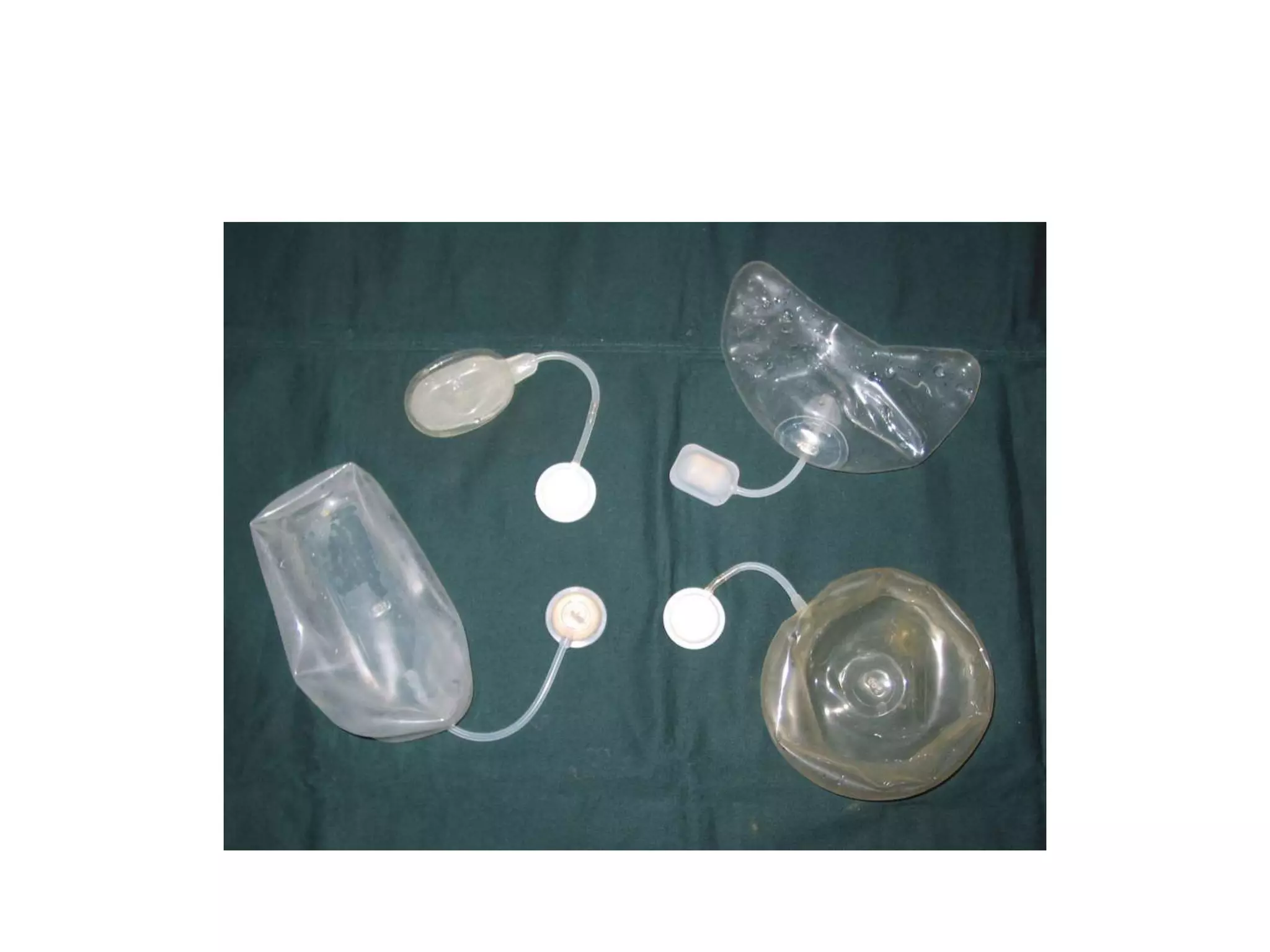
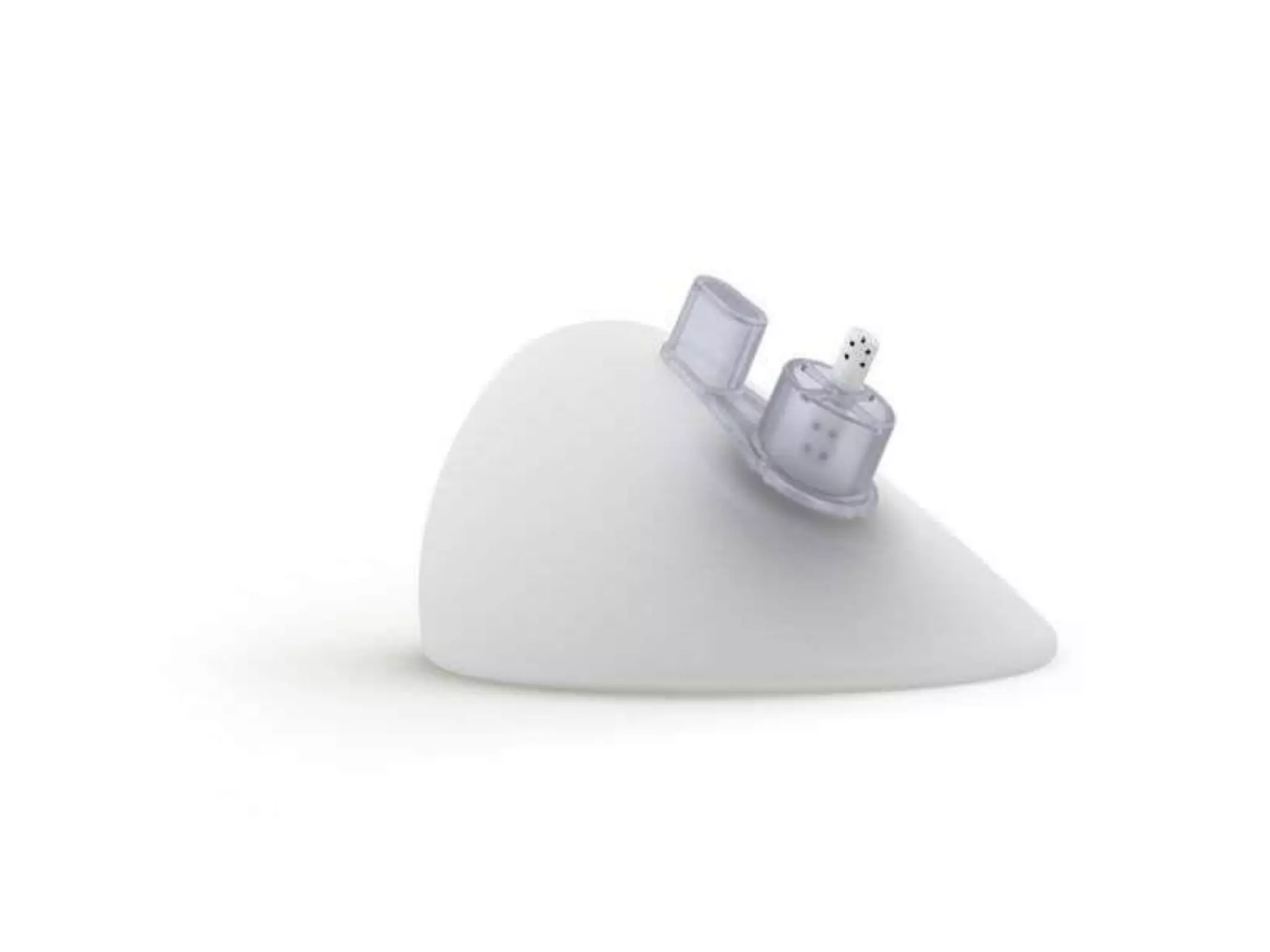
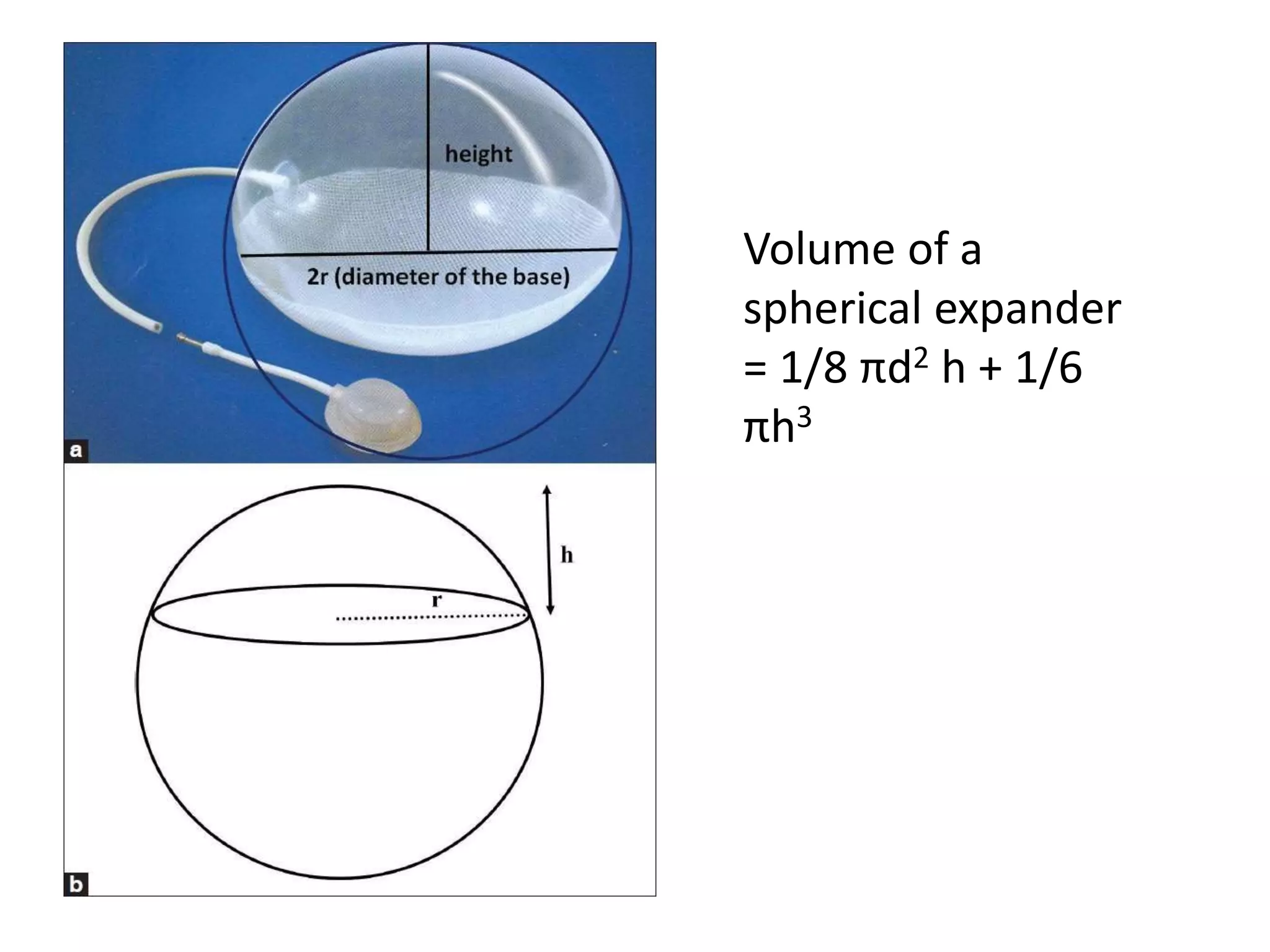
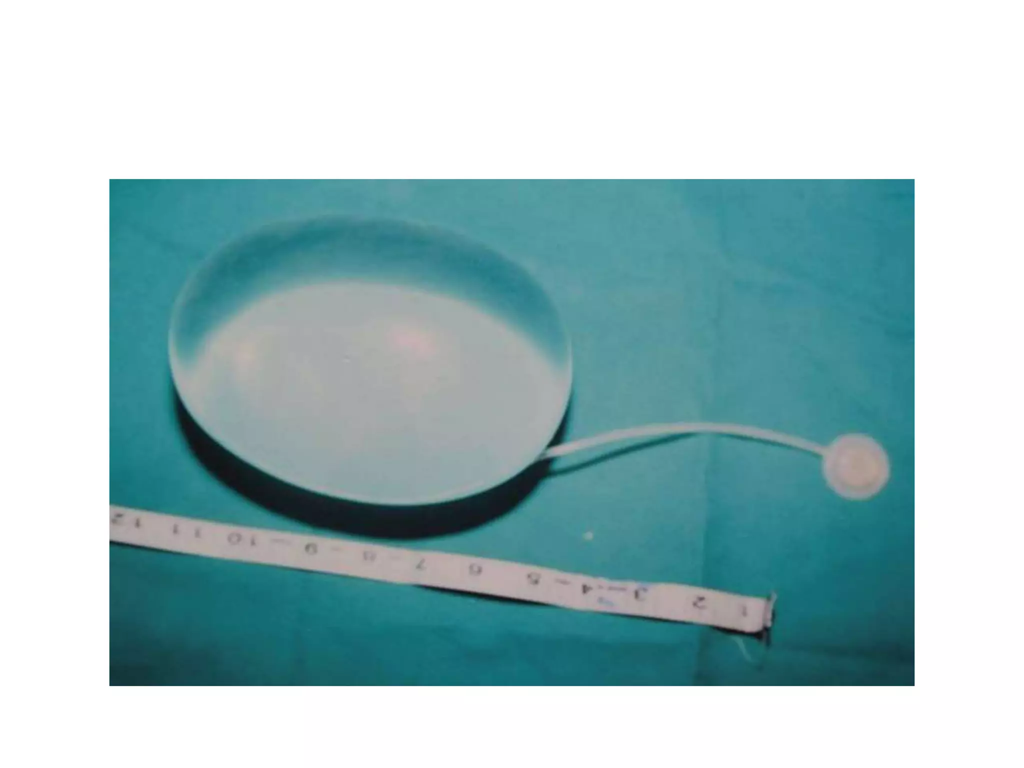
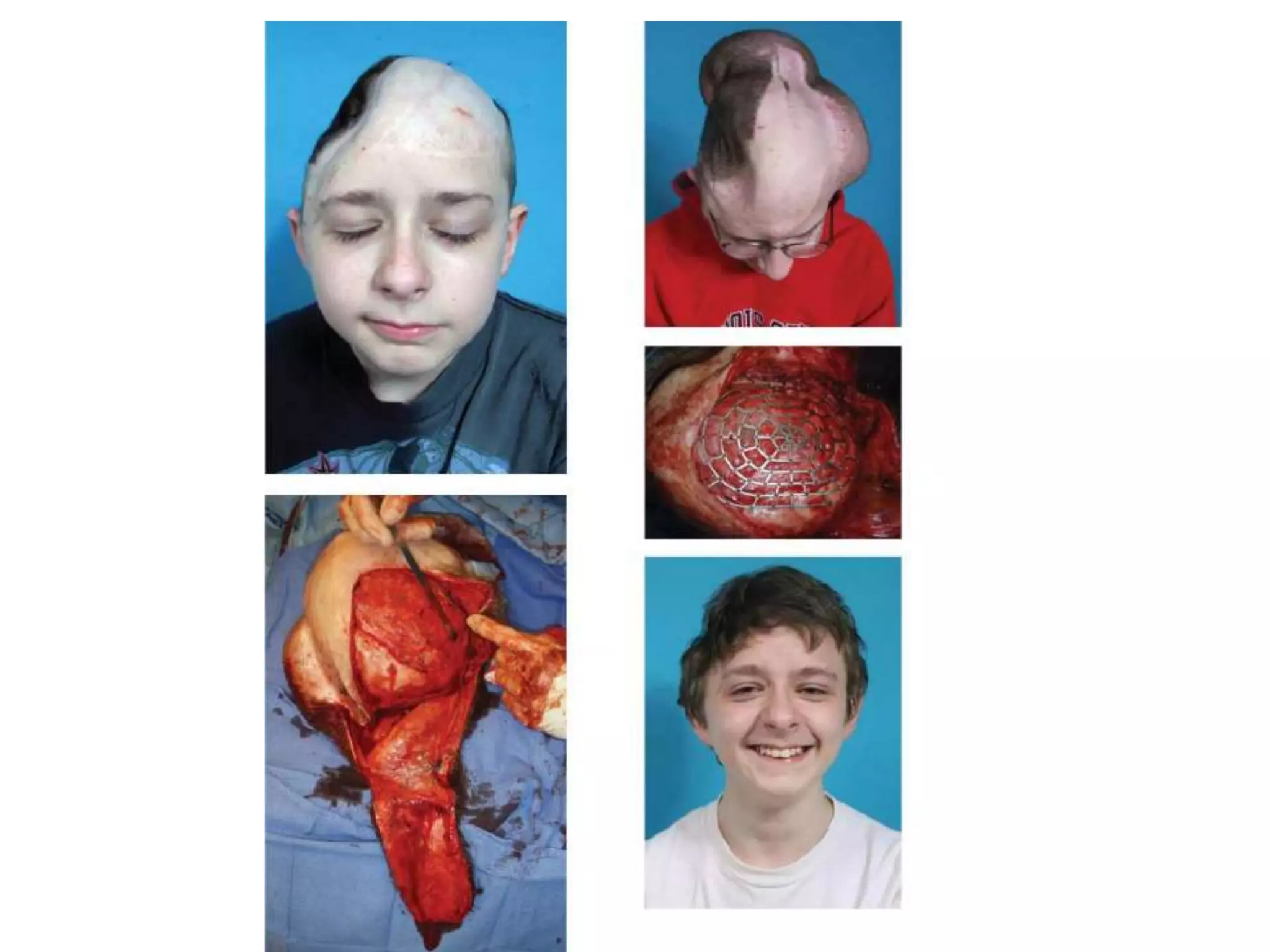
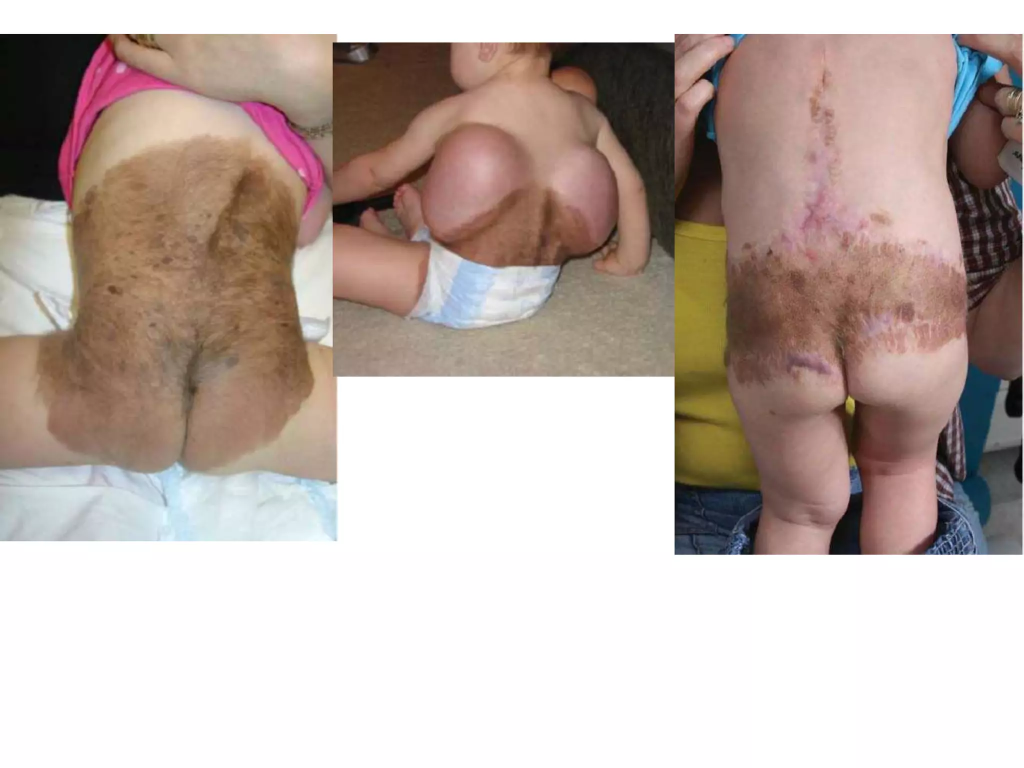
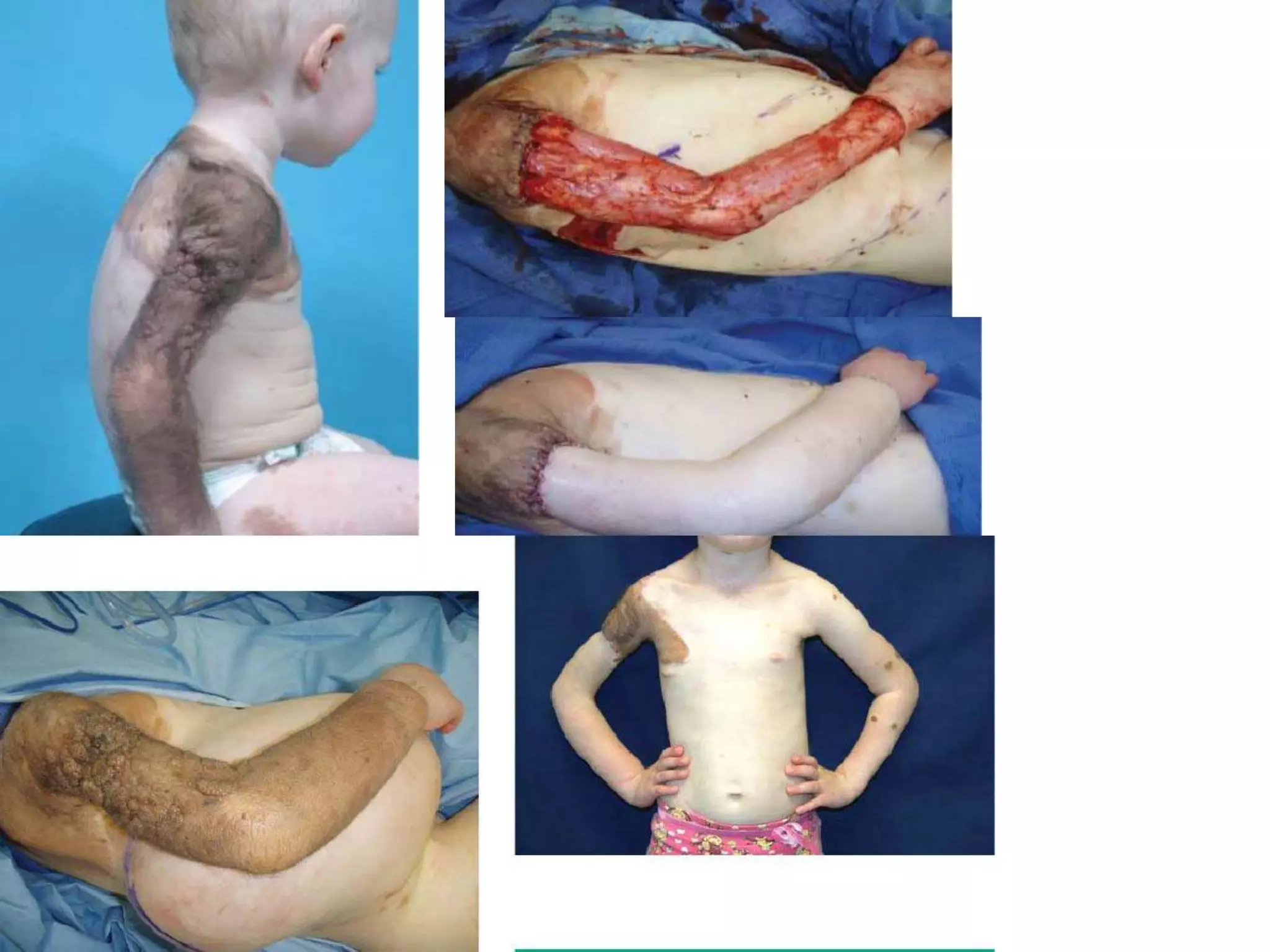
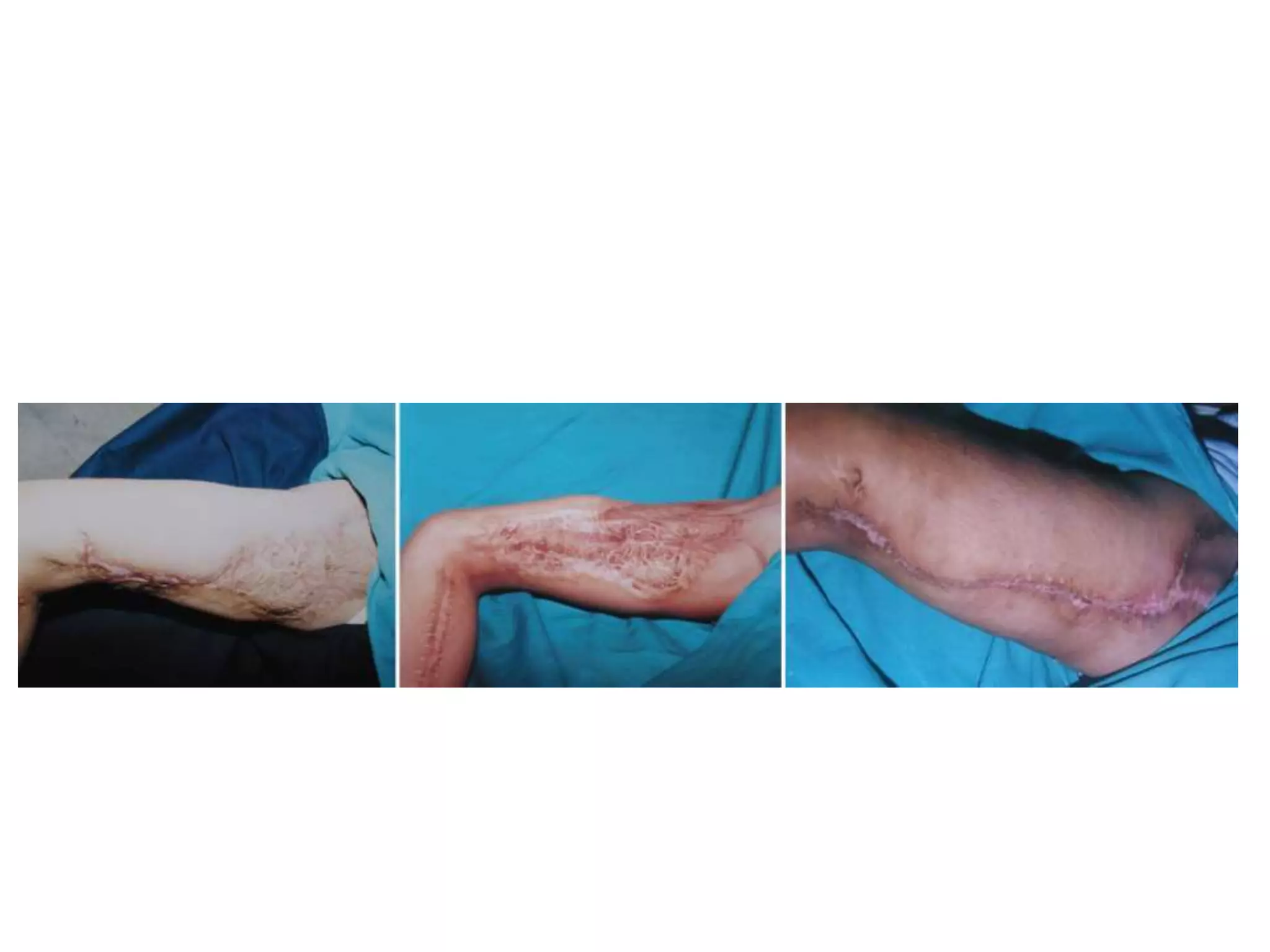
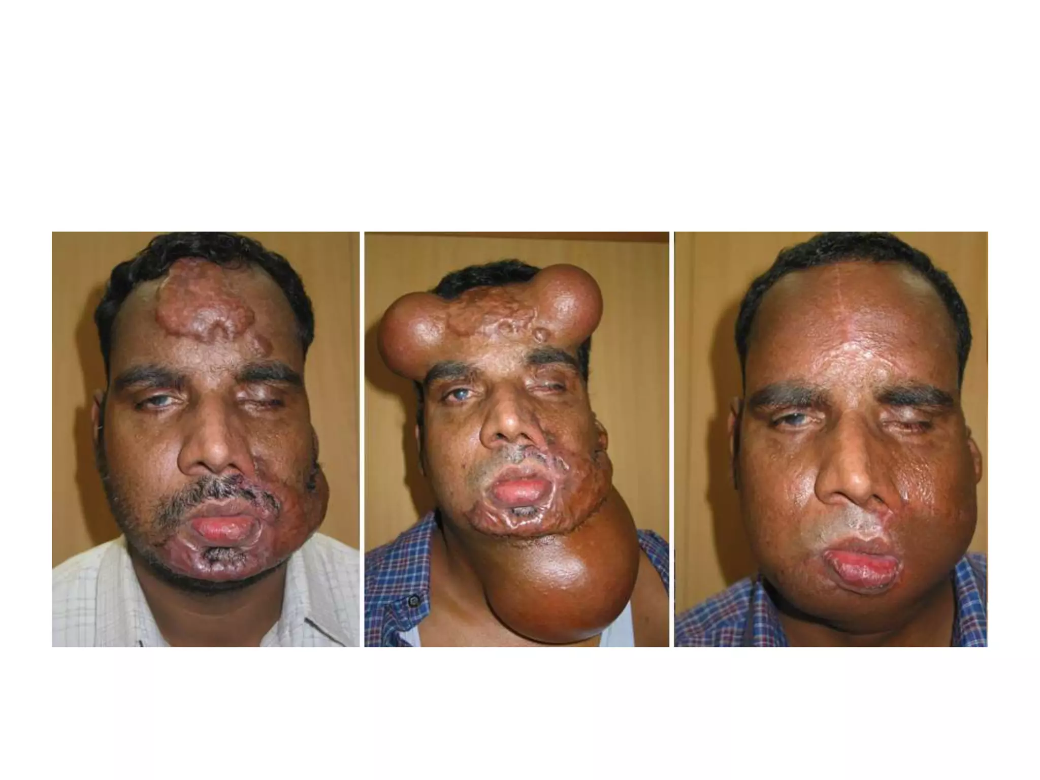
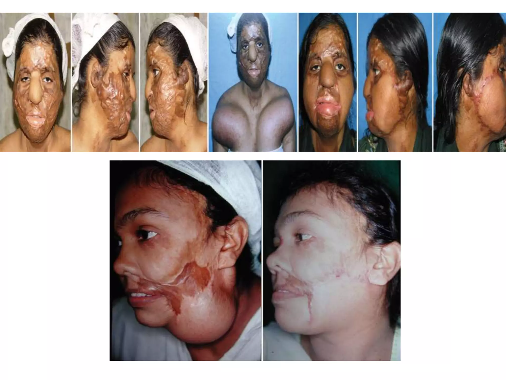
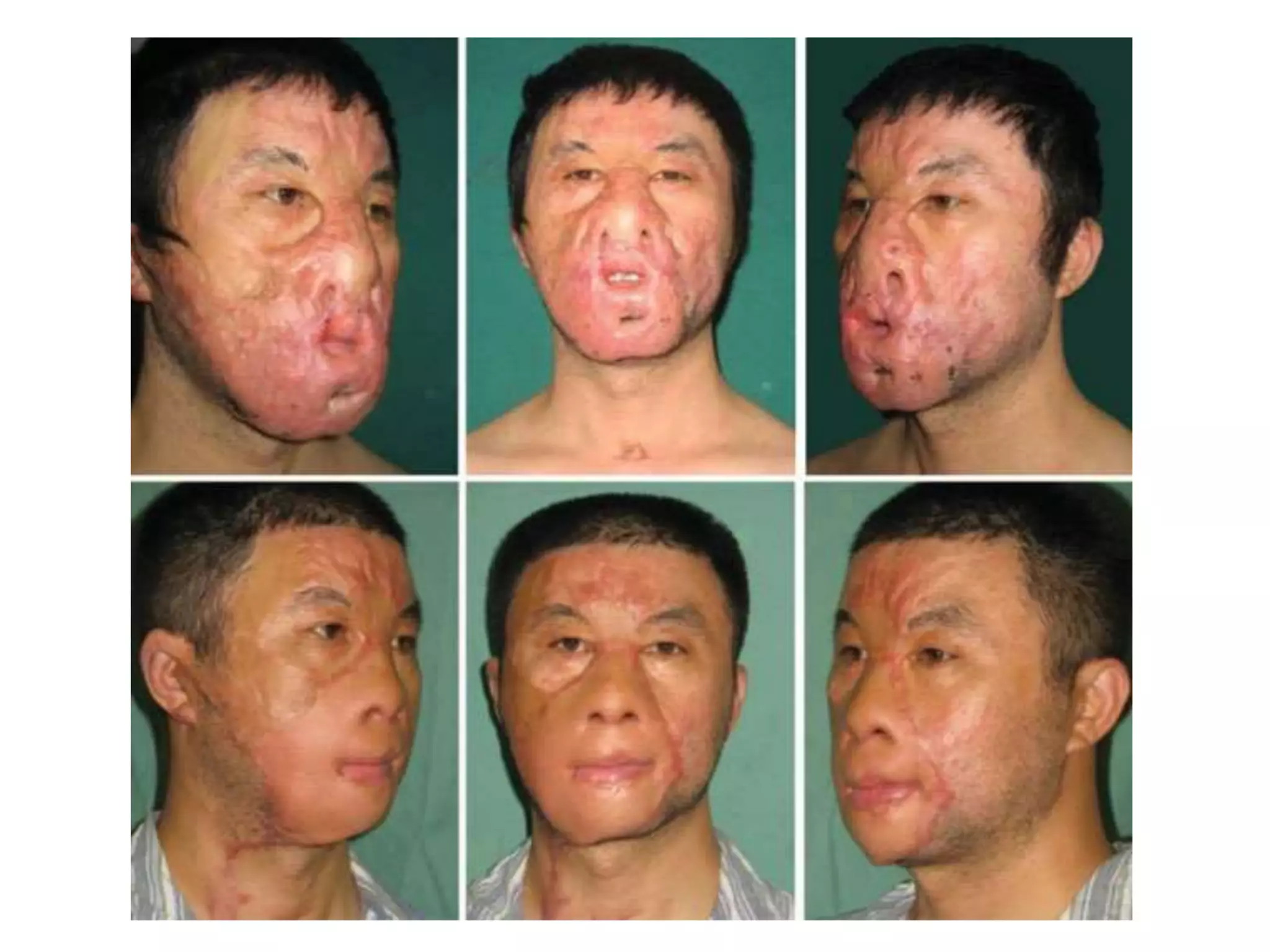
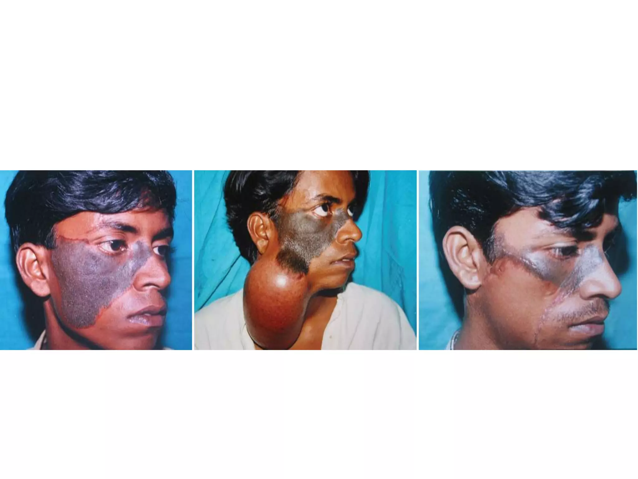
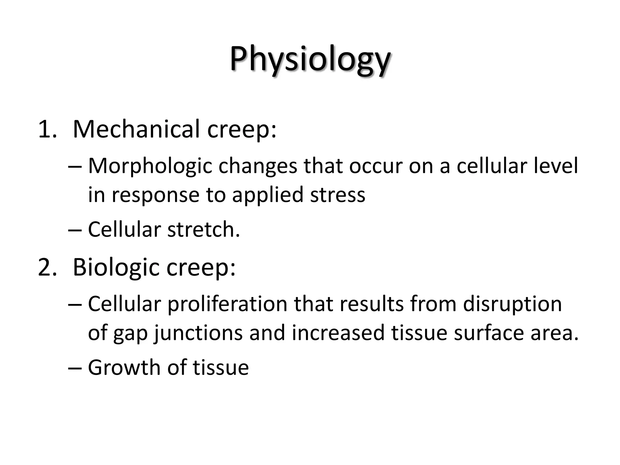
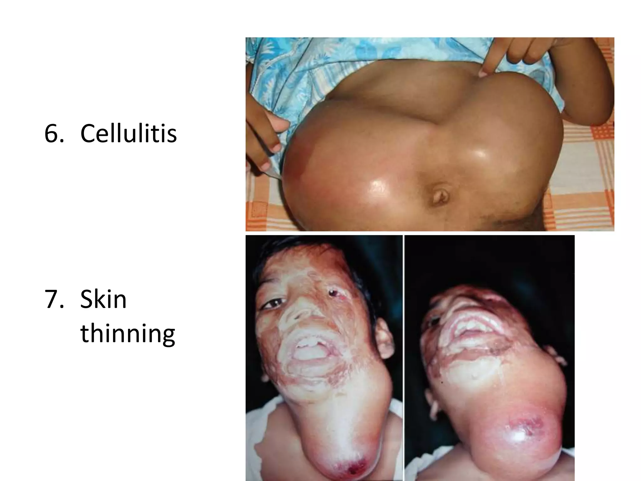
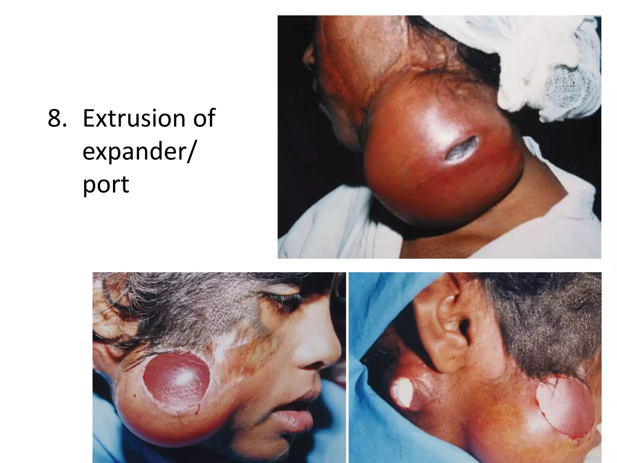
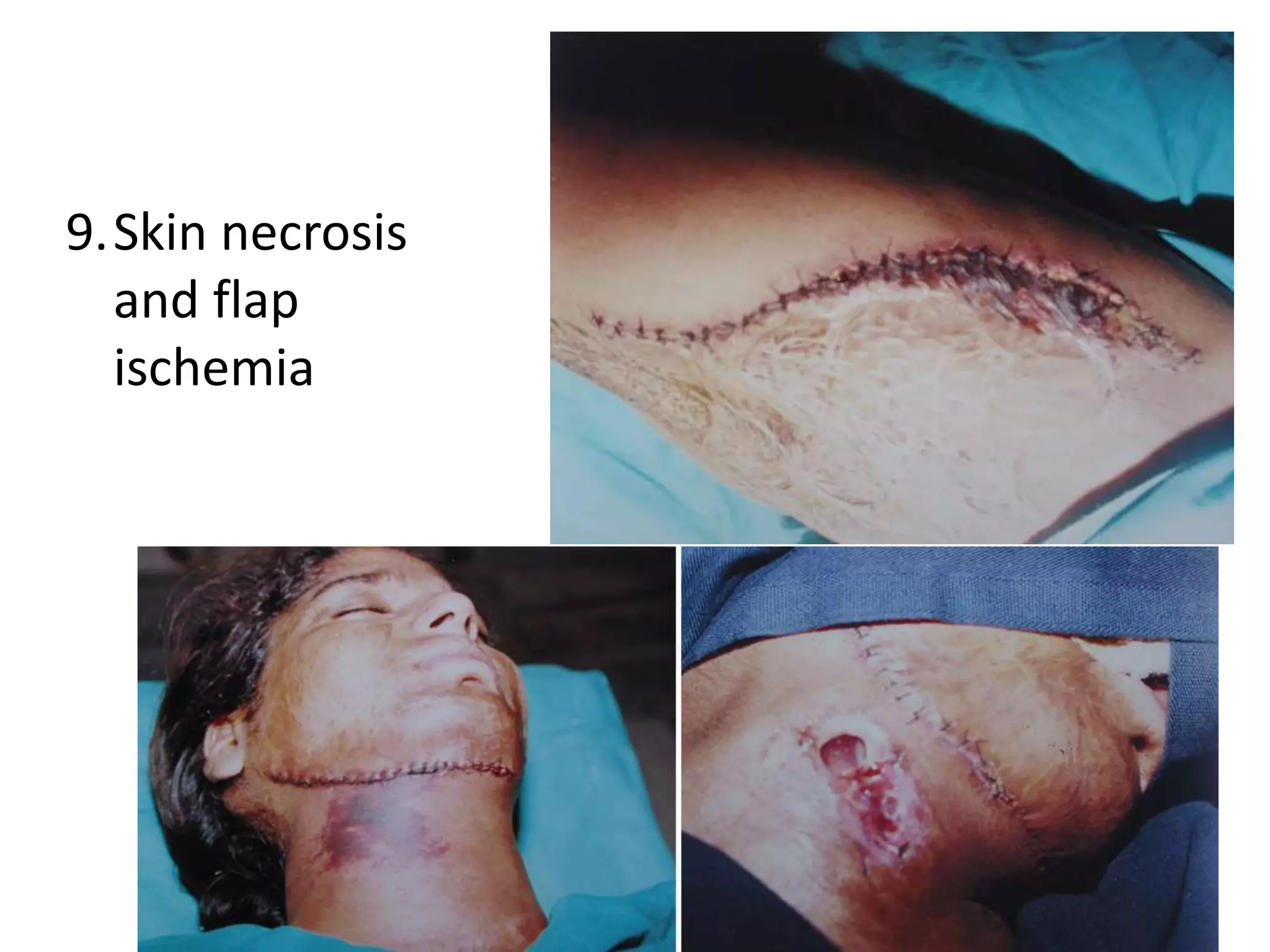
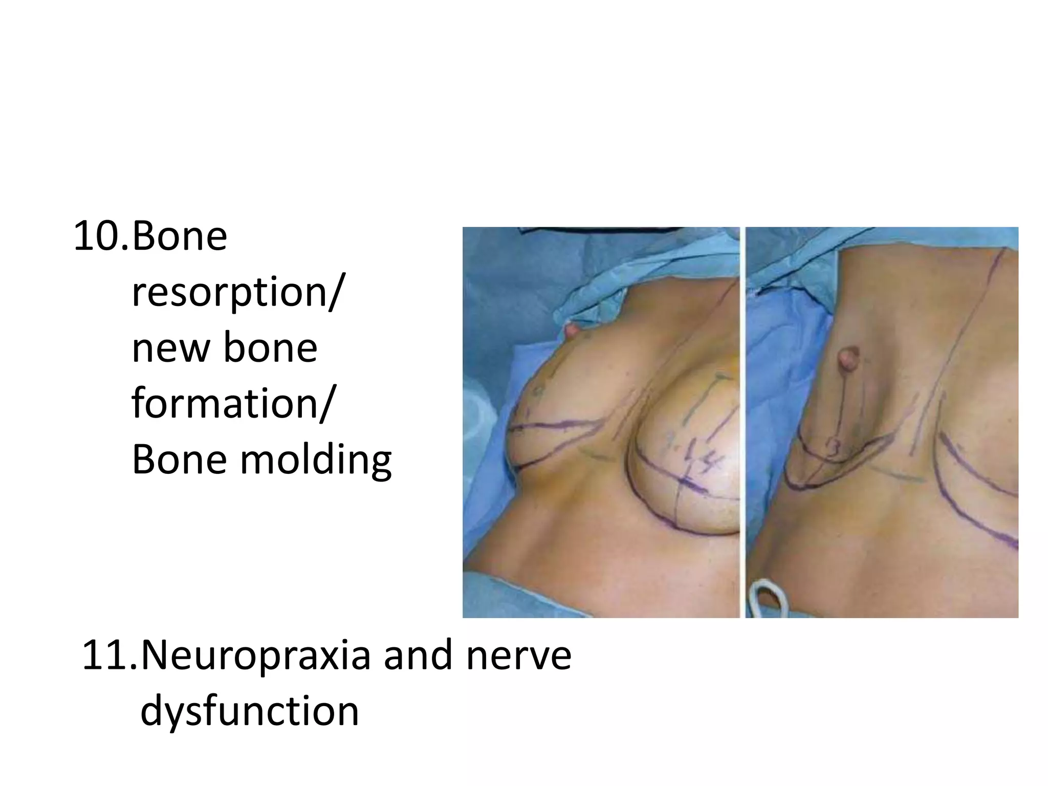
TISSUE EXPANSION
Headings
1. Definition
2. History
3. Types
4. Indications
5. Contraindications
6. Surgical planning
a) Insertion
b) Expansion
c) Reconstruction
7. Physiology
8. Complication
What?
• To promote the growth of healthy
supplementary skin used for the replacement
of damaged skin
History
• In mid-1950s, Neumann‘ ->expansile implant
(latex balloon) to enlarge periauricular skin for
a traumatic ear deformity.
• Dr. Chedomir Radovan at Georgetown
University, in Jan 1976 to resurface an arm
defect
• Dr. Eric Austad -> osmotically driven
self-inflating expander
Types
Shape:
1. Round
2. Rectangular (allow for additional expanded
tissue)
3. Crescent (to minimize dog-ears at the donor
site)
• Most common round and rectangular types.
Valve port:
1. Integrated
a) If only one single pocket is undermined
b) Implant prone to rupture during expansion.
2. Remote ports
a) No inadvertent prosthesis rupture
b) Flipping or migration of the device in vivo
c) Tube obstruction
Volumes/capacities :
• from 50 cc to 1000 cc in increments of 50
cc-100 cc
Indications
• Posttraumatic or postoperative alopecia
• Male pattern baldness
• Forehead skin prior to forehead flap nasal
reconstruction
• Postauricular skin
• Expansion of cheek or neck skin to allow scar
revision, burn excision
Contraindications
• Unwillingness or medical inability to undergo
2 or more operations
• Lack of concern regarding the appearance of a
skin graft or other alternative procedure
• Noncompliance
• Mental disability
• Inability to tolerate the cosmetic deformity
during the expansion process
• Previous removal of a malignancy with a
significant risk of recurrence (covering the site
with an expanded flap makes detection of
recurrence more difficult than in the presence
of a skin graft) or recurrence
• Acute injury
• Poorly vascularized tissues from radiation
therapy (approach with caution because of
increased risk of complications)
• Active infection or open wounds
• Ongoing chemotherapy (expand at a more
gradual rate)
• Expansion in infants and children
(controversial)
Surgical planning
3 stages:
1. Insertion
2. Expansion
3. Reconstruction
• The design for flap expansion should be
planned prior to surgery
• Sequential expansion:
defect is large
partially excised, and expander deflated
expansion initiated again.
third operation to remove expander &
reconstruction.
• For neoplastic lesion:
excise the lesion and apply a temporary skin
graft.
insertion of expanders, expansion, and
reconstruction
• neoplastic cells spread throughout pocket
created to accommodate the expander
Prerequisites:
1. Patient be psychologically stable & accept
temporary aesthetic disfigurement due to
expansion
2. Good quality well-vascularised donor tissue
3. Free of bacterial infection or contamination
Factors:
1. Size of the defect
2. Size and location of the available donor site
3. Expected advancement of a hemispherical
domed flap
• Gibney
Expander base must be at least 2.5-3 times the
defect’s width
• Radovan; Morgan and Edgerton
Expander base be same size as defect to be
closed. So a doubling of dome surface would
cover both defect and donor site
• van Rappard
1. For rectangular or crescentic expander:
surface area of expander base is 2.5 times of
defect
2. For round expanders:
• Diameter of the expander base be 2.5 times
of defect.
• Apical circumference of dome of skin
overlying fully inflated expander is two to 3
times width of the defect (Area gained in a
spherical expander = πh2 )
Volume of a
spherical expander
= 1/8 πd2 h + 1/6
πh3
A c = A d +πr2
V = 1/6 √A d / π (A d + 3πr2)
h = √A d / π ≈ 0.6√A d
Where "A c" is the surface area of a convex
surface of a sphere, A d is the surface area of
the recipient defect, "V" is the volume, "r" is
the radius of the base and "h" is the height of
the spherical tissue expander
• Manders et al:
largest possible expander that will fit at the
donor site be used
tissue stretch-back which is ability of expanded
tissue/tissue stretched over a long period to
contract back immediately after tension is
relieved
• Bhandari, 2009
Total surface area required for resurfacing the
defect = surface area of the defect + surface
area of the donor site + 20% of the defect and
donor site surface areas
20-30% extra tissue for rotation, dog ear and
mechanical creep resulting into "stretch back"
Rectangular expanders 38%
Round expanders 25%
Crescentic expanders 32%
Gain in surface area:
3-D photogrammetry:
• To calculate requirement of tissue expansion
• 3-D photogrammetry has been used for
presurgical estimation of volume deficiency in
a series of craniofacial microsomia patients.
The volume deficiency was calculated by
superimposing the 3-D mirror image of the
normal side in these patients
Guidelines For Insertion Of Expander
Incision:
1. Adjacent to the lesion where it would be the
leading edge of the advancement flap
2. Within the lesion
1. Radial or perpendicular to the expander
– Lessen risk of dehiscence during expansion
– Create additional scars and disturbs relaxed skin
tension lines
2. Tangential incision
– No expansion should be attempted initially
• Incision of 3-5 cm is adequate
• Powder-free gloves
• Avoid use of gauze pieces
• Edge of the expander not within 2 cm of the
suture line
Plane of dissection/insertion:
1. Beneath the subcutaneous tissue but above
the underlying muscle
2. In the scalp and forehead, subgaleally
beneath the frontalis or occipitalis muscles.
3. In the neck, beneath the platysma
4. If overlying skin is scarred and unstable, a
deeper plane (to ensure sufficient skin
vascularity and integrity)
5. If skin is thick and relatively more resistant to
soft-tissue stretch, plane is superficial
• Tight tunnel and space for remote port
• Over a bony prominence, mastoid or over the
ribcage/iliac crest
• Absorbable suture of 4-0 Vicryl to retain the
tubing
• Valve and connector tubing may be kept
externally outside skin for ease of injection, in
the paediatric age-group
After skin closure balloon is inflated (10-20% of
listed vol) to
1. Obliterate any dead space in the pocket
2. Maintain the pocket size
3. Smoothen out any wrinkles and folds in the
expander envelope.
Expansion Process
• 2-3 weeks for uneventful good primary
healing of incision suture line. No expansion
• Broad spectrum antibiotic 5-7 days
• Suture removal is at 10-14 days.
• Weekly expansion
• Prilox an hour prior in paediatric group
• No. 24 scalp vein
• 10 cc/20 cc luer-lock syringe
End of expansion in a session
1. Palpating the expanded dome: If tight and
tense
2. Assessing skin for signs of continued
blanching on pressure at multiple points and
good capillary return on release of pressure
(tissue tolerance)
3. Patient tolerance (pain and discomfort)
• Expansion process lasts for 6-12 weeks
• Good quality expander canbe safely
over-expanded to double the capacity
Removal Of Expander And Flap
Advancement
• 2 weeks allowed for expanded skin to stay
stretched to maximum level
• Incision for advancement flap is usually at
border between expander and lesion/defect
• Saline is withdrawn from port
• Expander removed with port
• Advancement of expanded flap across
lesion/defect
• Absorbable tacking/quilting sutures between
the capsule (proximal to advancing edge) and
base/floor of defect to anchor flap
• Suction drain for 24-48
• Closure done in 2-3 layers
Physiology
1. Mechanical creep:
– Morphologic changes that occur on a cellular level
in response to applied stress
– Cellular stretch.
2. Biologic creep:
– Cellular proliferation that results from disruption
of gap junctions and increased tissue surface area.
– Growth of tissue
Histology
• Epidermis thickens initially, probably because
of postoperative edema, but returns to
baseline within several weeks
• Dermis undergoes rapid decrease in thickness
• Activated fibroblasts
• Thick and compact bundles of collagen
formed, parallel to expander surface
• Increase in vascularity of the dermis
• Hair follicles demonstrate some compression
but no degeneration. New follicles are not
created
• Melanocyte activity increases, returns to
baseline following reconstruction, and the
hyperpigmentation disappears
• Loss of fat (permanent)
• Muscle tissue above or below the expander
demonstrates atrophy but no alteration of
function.
• It resolves following expander removal
• Temporary cranial molding occurs, but
corrects within 3 to 4 months
• Expander causes minimal inflammatory
reaction
• Fibrous capsule forms around the expander,
with myofibroblasts
• Capsule becomes less cellular and more
organized, with bundles of collagen
• New blood vessels
Three layer composition:
• Internal layer: macrophages and fibroblasts. In
some cases, a pseudoepithelial cellular layer
at the implant/capsule interface (synovia-like
metaplasia) is found.
• Middle layer: loosely arranged connective
tissue.
• Outer layer: dense connective tissue with the
external vascular supply
Complications
• range from 5-60%
1. Hematoma
2. Seroma
3. Deflation of the expander, either
spontaneously or iatrogenically during
inflation
4. Migration
5. Scar widening
6. Cellulitis
7. Skin
thinning
8. Extrusion of
expander/
port
9.Skin necrosis
and flap
ischemia
10.Bone
resorption/
new bone
formation/
Bone molding
11.Neuropraxia and nerve
dysfunction
References
• Grabb and Smith's Plastic Surgery, 7th Edition
• Tissue regeneration during tissue expansion and choosing an
expander. Agrawal K, Agrawal S. IJPS. 2012; Vol 45 (1): 7-15
• Tissue expansion: Concepts, techniques and unfavourable
results. Milind S. Wagh, Varun Dixit. Indian Journal of Plastic
Surgery. May-Aug 2013; Vol 46 (2): 333-48.
• Internet sources (Medscape etc)

Tissue expansion- principles and techniques

  • 1.
  • 2.
    Headings 1. Definition 2. History 3.Types 4. Indications 5. Contraindications 6. Surgical planning a) Insertion b) Expansion c) Reconstruction 7. Physiology 8. Complication
  • 3.
    What? • To promotethe growth of healthy supplementary skin used for the replacement of damaged skin
  • 4.
    History • In mid-1950s,Neumann‘ ->expansile implant (latex balloon) to enlarge periauricular skin for a traumatic ear deformity. • Dr. Chedomir Radovan at Georgetown University, in Jan 1976 to resurface an arm defect • Dr. Eric Austad -> osmotically driven self-inflating expander
  • 5.
    Types Shape: 1. Round 2. Rectangular(allow for additional expanded tissue) 3. Crescent (to minimize dog-ears at the donor site) • Most common round and rectangular types.
  • 7.
    Valve port: 1. Integrated a)If only one single pocket is undermined b) Implant prone to rupture during expansion. 2. Remote ports a) No inadvertent prosthesis rupture b) Flipping or migration of the device in vivo c) Tube obstruction
  • 10.
    Volumes/capacities : • from50 cc to 1000 cc in increments of 50 cc-100 cc
  • 11.
    Indications • Posttraumatic orpostoperative alopecia • Male pattern baldness • Forehead skin prior to forehead flap nasal reconstruction • Postauricular skin • Expansion of cheek or neck skin to allow scar revision, burn excision
  • 12.
    Contraindications • Unwillingness ormedical inability to undergo 2 or more operations • Lack of concern regarding the appearance of a skin graft or other alternative procedure • Noncompliance • Mental disability
  • 13.
    • Inability totolerate the cosmetic deformity during the expansion process • Previous removal of a malignancy with a significant risk of recurrence (covering the site with an expanded flap makes detection of recurrence more difficult than in the presence of a skin graft) or recurrence
  • 14.
    • Acute injury •Poorly vascularized tissues from radiation therapy (approach with caution because of increased risk of complications) • Active infection or open wounds • Ongoing chemotherapy (expand at a more gradual rate) • Expansion in infants and children (controversial)
  • 15.
    Surgical planning 3 stages: 1.Insertion 2. Expansion 3. Reconstruction • The design for flap expansion should be planned prior to surgery
  • 16.
    • Sequential expansion: defectis large partially excised, and expander deflated expansion initiated again. third operation to remove expander & reconstruction.
  • 17.
    • For neoplasticlesion: excise the lesion and apply a temporary skin graft. insertion of expanders, expansion, and reconstruction • neoplastic cells spread throughout pocket created to accommodate the expander
  • 18.
    Prerequisites: 1. Patient bepsychologically stable & accept temporary aesthetic disfigurement due to expansion 2. Good quality well-vascularised donor tissue 3. Free of bacterial infection or contamination
  • 19.
    Factors: 1. Size ofthe defect 2. Size and location of the available donor site 3. Expected advancement of a hemispherical domed flap
  • 20.
    • Gibney Expander basemust be at least 2.5-3 times the defect’s width • Radovan; Morgan and Edgerton Expander base be same size as defect to be closed. So a doubling of dome surface would cover both defect and donor site
  • 21.
    • van Rappard 1.For rectangular or crescentic expander: surface area of expander base is 2.5 times of defect
  • 23.
    2. For roundexpanders: • Diameter of the expander base be 2.5 times of defect. • Apical circumference of dome of skin overlying fully inflated expander is two to 3 times width of the defect (Area gained in a spherical expander = πh2 )
  • 24.
    Volume of a sphericalexpander = 1/8 πd2 h + 1/6 πh3
  • 25.
    A c =A d +πr2 V = 1/6 √A d / π (A d + 3πr2) h = √A d / π ≈ 0.6√A d Where "A c" is the surface area of a convex surface of a sphere, A d is the surface area of the recipient defect, "V" is the volume, "r" is the radius of the base and "h" is the height of the spherical tissue expander
  • 28.
    • Manders etal: largest possible expander that will fit at the donor site be used tissue stretch-back which is ability of expanded tissue/tissue stretched over a long period to contract back immediately after tension is relieved
  • 29.
    • Bhandari, 2009 Totalsurface area required for resurfacing the defect = surface area of the defect + surface area of the donor site + 20% of the defect and donor site surface areas 20-30% extra tissue for rotation, dog ear and mechanical creep resulting into "stretch back"
  • 31.
    Rectangular expanders 38% Roundexpanders 25% Crescentic expanders 32% Gain in surface area:
  • 32.
    3-D photogrammetry: • Tocalculate requirement of tissue expansion • 3-D photogrammetry has been used for presurgical estimation of volume deficiency in a series of craniofacial microsomia patients. The volume deficiency was calculated by superimposing the 3-D mirror image of the normal side in these patients
  • 33.
    Guidelines For InsertionOf Expander Incision: 1. Adjacent to the lesion where it would be the leading edge of the advancement flap 2. Within the lesion
  • 34.
    1. Radial orperpendicular to the expander – Lessen risk of dehiscence during expansion – Create additional scars and disturbs relaxed skin tension lines 2. Tangential incision – No expansion should be attempted initially
  • 35.
    • Incision of3-5 cm is adequate • Powder-free gloves • Avoid use of gauze pieces • Edge of the expander not within 2 cm of the suture line
  • 36.
    Plane of dissection/insertion: 1.Beneath the subcutaneous tissue but above the underlying muscle 2. In the scalp and forehead, subgaleally beneath the frontalis or occipitalis muscles. 3. In the neck, beneath the platysma
  • 37.
    4. If overlyingskin is scarred and unstable, a deeper plane (to ensure sufficient skin vascularity and integrity) 5. If skin is thick and relatively more resistant to soft-tissue stretch, plane is superficial
  • 38.
    • Tight tunneland space for remote port • Over a bony prominence, mastoid or over the ribcage/iliac crest • Absorbable suture of 4-0 Vicryl to retain the tubing • Valve and connector tubing may be kept externally outside skin for ease of injection, in the paediatric age-group
  • 39.
    After skin closureballoon is inflated (10-20% of listed vol) to 1. Obliterate any dead space in the pocket 2. Maintain the pocket size 3. Smoothen out any wrinkles and folds in the expander envelope.
  • 40.
    Expansion Process • 2-3weeks for uneventful good primary healing of incision suture line. No expansion • Broad spectrum antibiotic 5-7 days • Suture removal is at 10-14 days. • Weekly expansion • Prilox an hour prior in paediatric group • No. 24 scalp vein • 10 cc/20 cc luer-lock syringe
  • 42.
    End of expansionin a session 1. Palpating the expanded dome: If tight and tense 2. Assessing skin for signs of continued blanching on pressure at multiple points and good capillary return on release of pressure (tissue tolerance) 3. Patient tolerance (pain and discomfort)
  • 43.
    • Expansion processlasts for 6-12 weeks • Good quality expander canbe safely over-expanded to double the capacity
  • 45.
    Removal Of ExpanderAnd Flap Advancement • 2 weeks allowed for expanded skin to stay stretched to maximum level • Incision for advancement flap is usually at border between expander and lesion/defect • Saline is withdrawn from port • Expander removed with port • Advancement of expanded flap across lesion/defect
  • 46.
    • Absorbable tacking/quiltingsutures between the capsule (proximal to advancing edge) and base/floor of defect to anchor flap • Suction drain for 24-48 • Closure done in 2-3 layers
  • 56.
    Physiology 1. Mechanical creep: –Morphologic changes that occur on a cellular level in response to applied stress – Cellular stretch. 2. Biologic creep: – Cellular proliferation that results from disruption of gap junctions and increased tissue surface area. – Growth of tissue
  • 57.
    Histology • Epidermis thickensinitially, probably because of postoperative edema, but returns to baseline within several weeks • Dermis undergoes rapid decrease in thickness • Activated fibroblasts • Thick and compact bundles of collagen formed, parallel to expander surface • Increase in vascularity of the dermis
  • 58.
    • Hair folliclesdemonstrate some compression but no degeneration. New follicles are not created • Melanocyte activity increases, returns to baseline following reconstruction, and the hyperpigmentation disappears • Loss of fat (permanent)
  • 59.
    • Muscle tissueabove or below the expander demonstrates atrophy but no alteration of function. • It resolves following expander removal • Temporary cranial molding occurs, but corrects within 3 to 4 months
  • 60.
    • Expander causesminimal inflammatory reaction • Fibrous capsule forms around the expander, with myofibroblasts • Capsule becomes less cellular and more organized, with bundles of collagen • New blood vessels
  • 61.
    Three layer composition: •Internal layer: macrophages and fibroblasts. In some cases, a pseudoepithelial cellular layer at the implant/capsule interface (synovia-like metaplasia) is found. • Middle layer: loosely arranged connective tissue. • Outer layer: dense connective tissue with the external vascular supply
  • 62.
    Complications • range from5-60% 1. Hematoma 2. Seroma 3. Deflation of the expander, either spontaneously or iatrogenically during inflation 4. Migration 5. Scar widening
  • 63.
  • 64.
  • 65.
  • 66.
  • 67.
    References • Grabb andSmith's Plastic Surgery, 7th Edition • Tissue regeneration during tissue expansion and choosing an expander. Agrawal K, Agrawal S. IJPS. 2012; Vol 45 (1): 7-15 • Tissue expansion: Concepts, techniques and unfavourable results. Milind S. Wagh, Varun Dixit. Indian Journal of Plastic Surgery. May-Aug 2013; Vol 46 (2): 333-48. • Internet sources (Medscape etc)