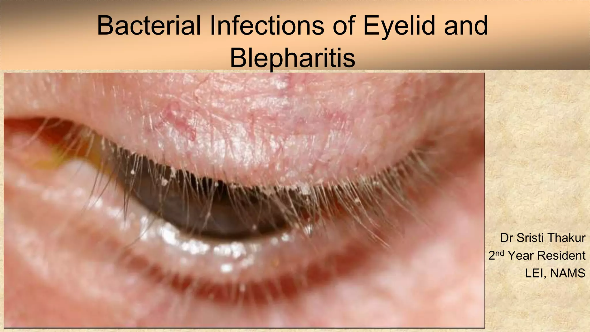
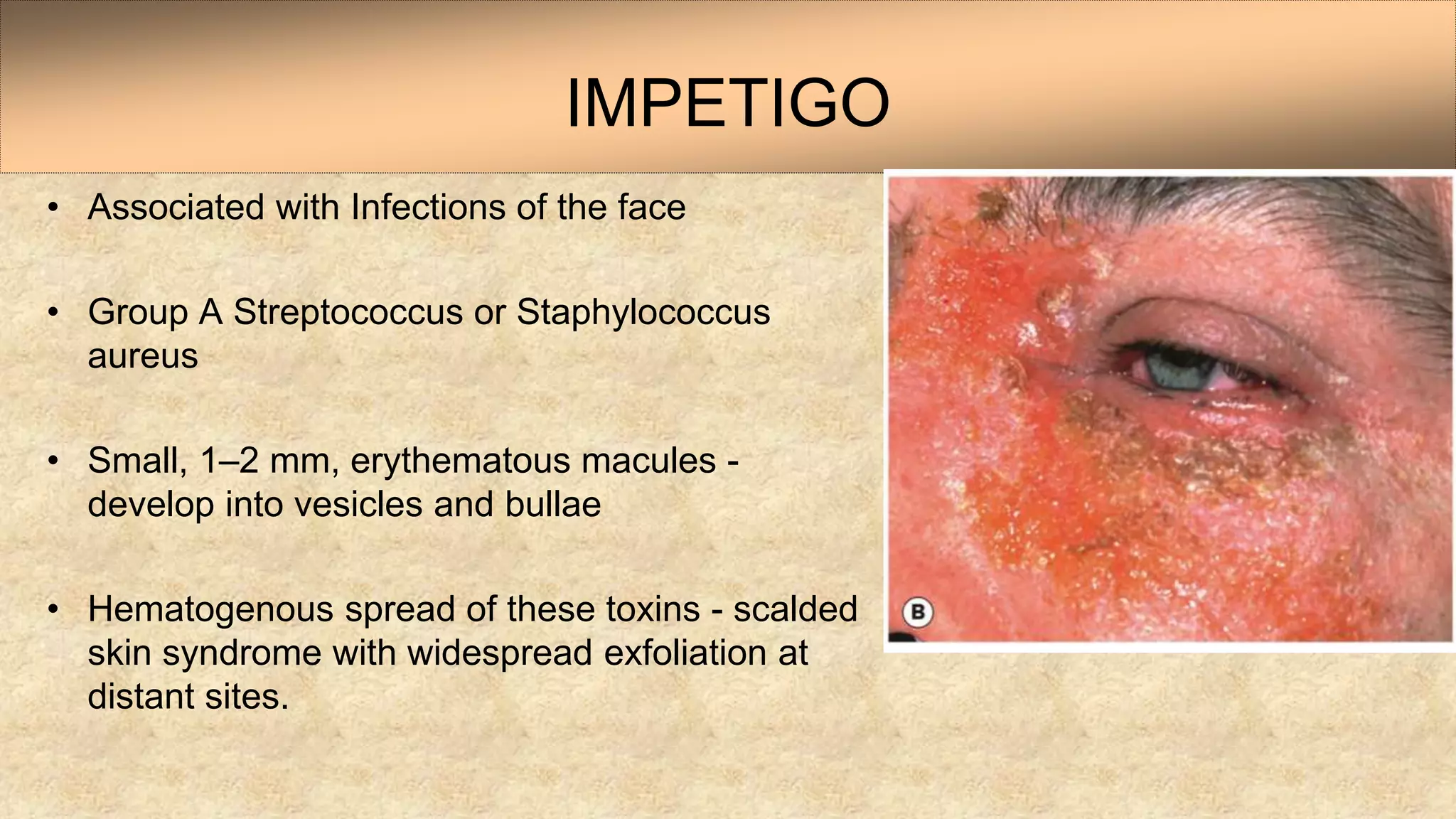
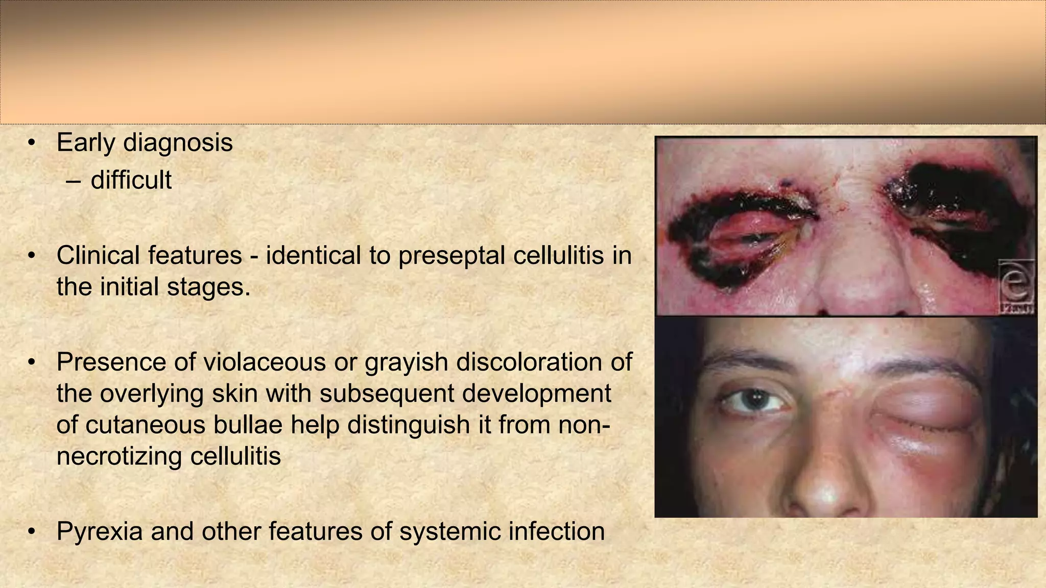
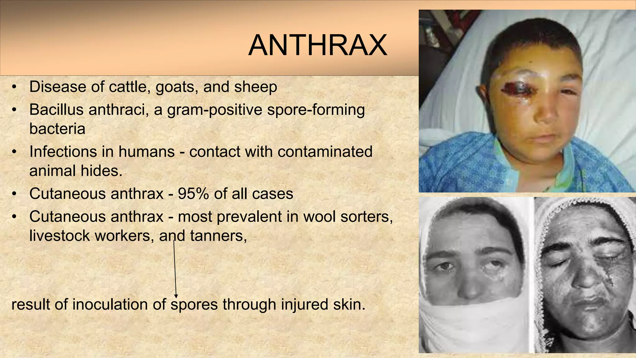
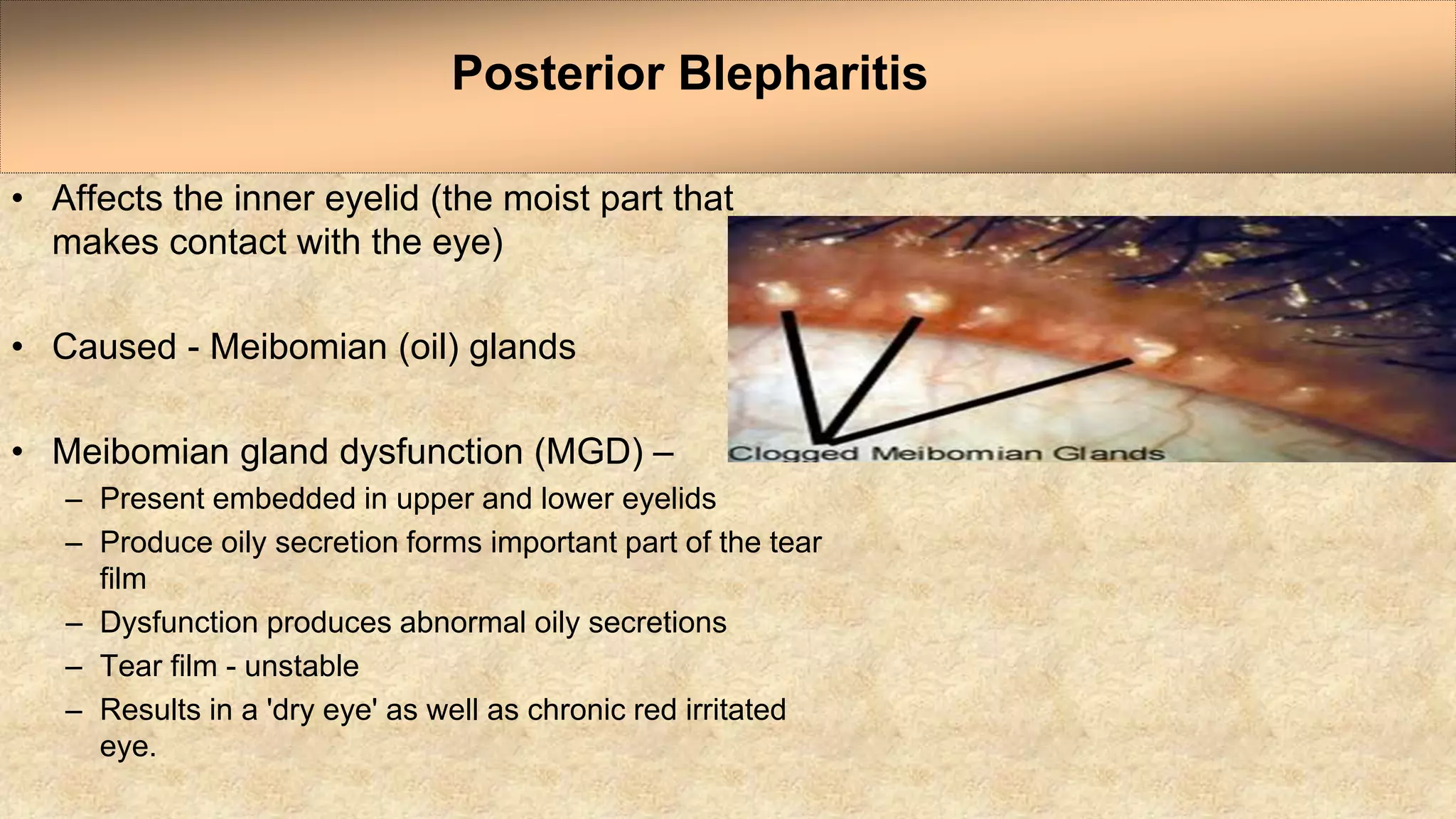
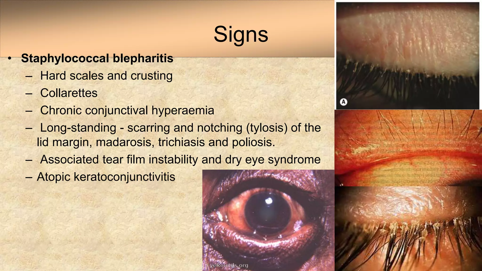
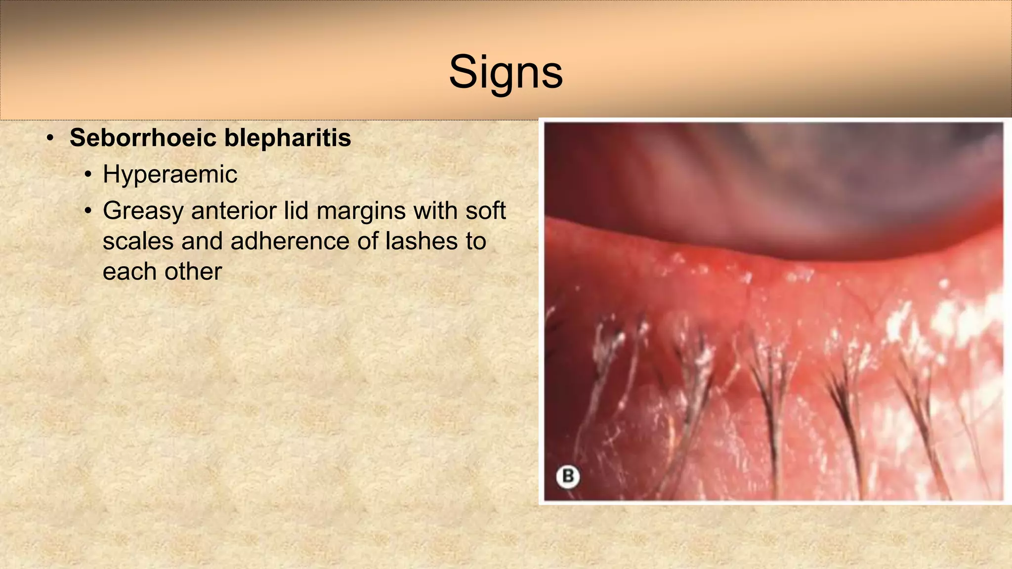
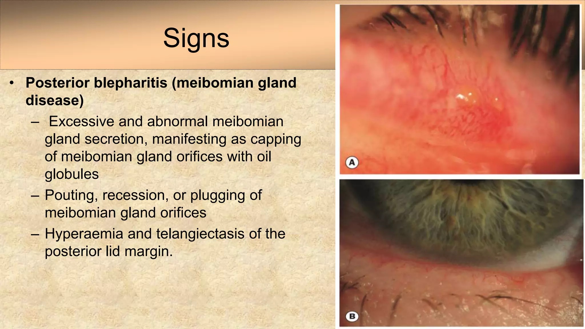
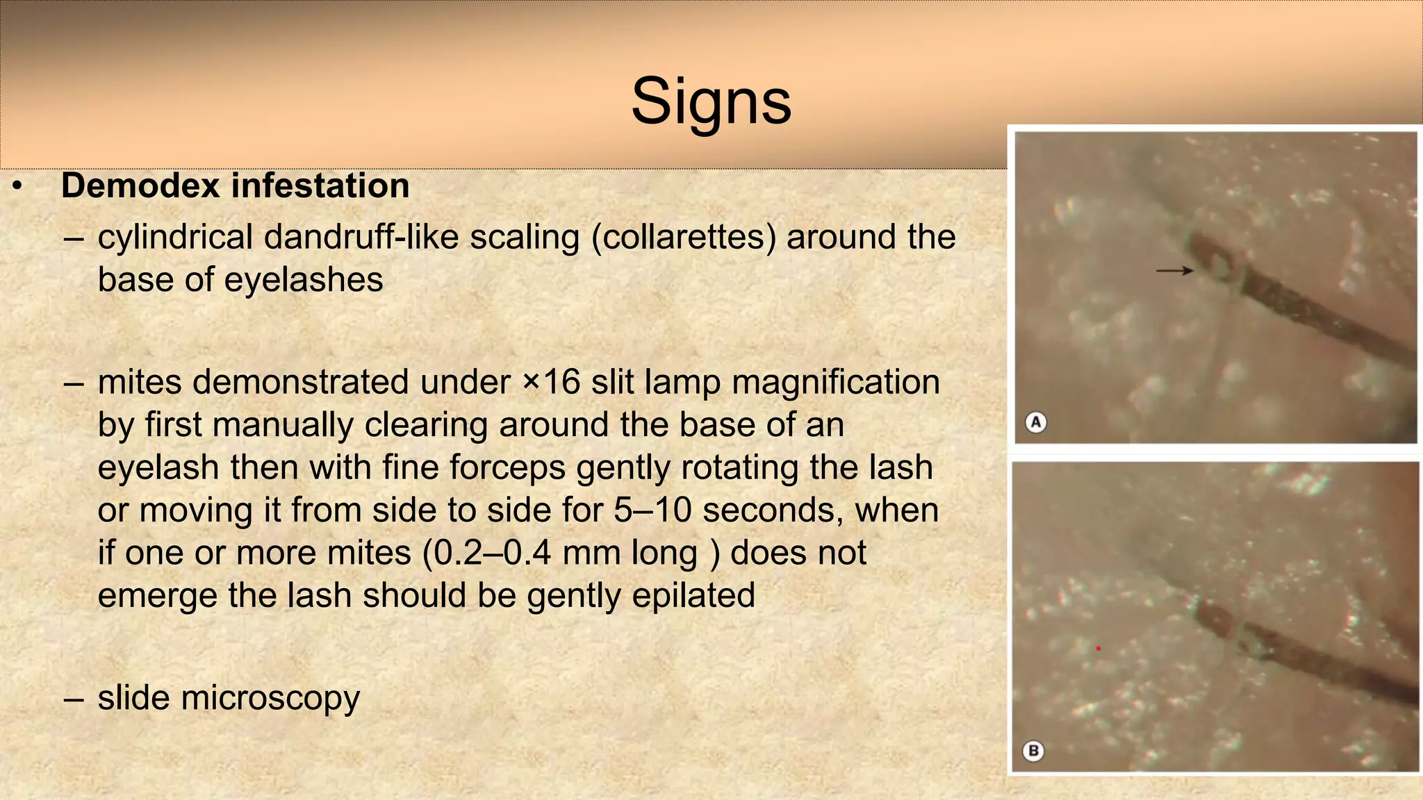
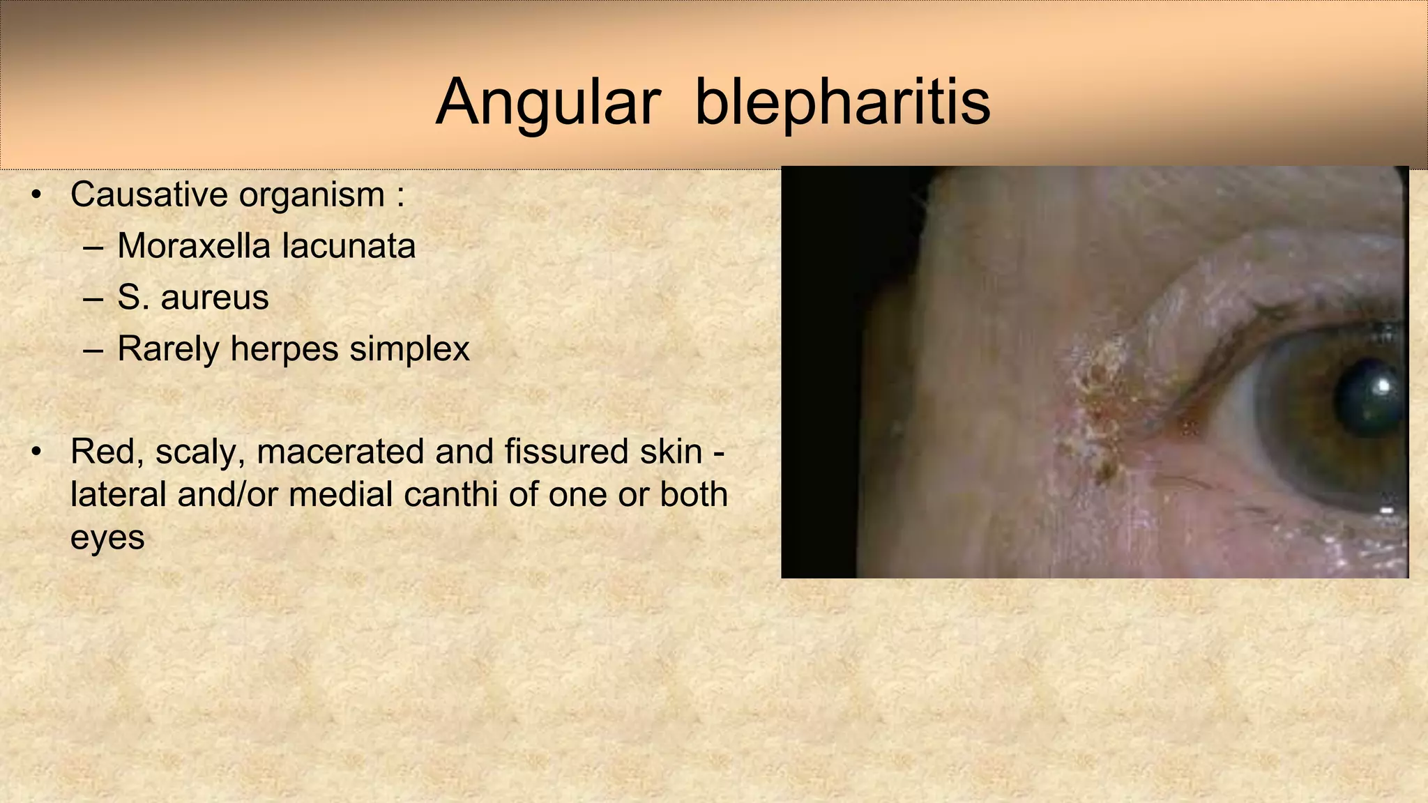
Bacterial Infections of Eyelid and
Blepharitis
Dr Sristi Thakur
2nd Year Resident
LEI, NAMS
Contents
• Anatomy
• External Hordeolum
• Impetigo
• Erysipelas
• Necrotizing fasciitis
• Anthrax
• Syphilis
• Blepharitis
Eyelid Anatomy
External Hordeolum
• Acute staphylococcal abscess of lash
follicle and its associated gland of Zeis
• Common in children and young adults
• Tender swelling in lid margin pointing
anteriorly through the skin, usually with
a lash at its apex
• Multiple lesions
• Occasionally abscesses may involve the
entire lid margin.
Histologically - polymorphonuclear leukocytes,
necrotic cellular debris, edema, and vascular
congestion
Treatment :
- Topical (occasionally oral) antibiotics
- Hot compresses
- Epilation of the associated lash.
IMPETIGO
• Associated with Infections of the face
• Group A Streptococcus or Staphylococcus
aureus
• Small, 1–2 mm, erythematous macules -
develop into vesicles and bullae
• Hematogenous spread of these toxins - scalded
skin syndrome with widespread exfoliation at
distant sites.
• Bullae rapidly progress and
rupture
• form a thin, varnish-like crust in
cases of staphylococcal (bullous)
impetigo
• thick honey-colored crust in cases
of Streptococcus or mixed
infections of streptococci and
staphylococci.
• Infection spread by fingers, towels, or
household utensils, forming satellite lesions.
• Children
• Acute glomerulonephritis - 2–5% of group A
hemolytic streptococcal skin infections
• Highly contagious
• Diagnosis :
- Characteristic clinical appearance
- Specimens - obtained for culture and
sensitivity
Treatment
• Gentle washing of the affected area
• Topical Mupirocin 3 times - 7 days
• Topical Bacitracin or Erythromycin - 7–10 days
• Alternative oral antibiotics - b-lactamase-resistant antibiotics such as Cloxacillin,
Amoxicillin plus Clavulanic acid, Clindamycin, or a Cephalosporin such as
Cephalexin, Cefaclor, or Cefprozil
• Healthy children with community-acquired MRSA infections - Clindamycin or
Trimethoprimsulfamethoxazole.
Erysipelas
• Uncommon acute, potentially severe, dermal and superficial
lymphatic infection
• Streptococcus pyogenes
• Predisposing factors :
- Diabetes
- Obesity
- Alcohol abuse
• Inflamed erythematous plaque develops
• Well-defined raised border distinguishes
erysipelas from other forms of cellulitis
Complications - metastatic infection (rare)
Treatment : oral antibiotics
Recurrence common.
NECROTIZING FASCIITIS
• Rare, fatal soft tissue infection - affects the trunk and
extremities.
• Involvement of the facial and periocular areas rare
• Two microbiologic subtypes :
- Type I - Anaerobes and facultative anaerobes
- Type II - Group A b-hemolytic Streptococcus with or
without S. aureus coinfection
• Periocular necrotizing fasciitis –
– diabetes mellitus
– alchoholism
– immunosuppression
• Associated with :
– Acute Dacryocystitis
– following endoscopic sinus surgery
– Blepharoplasty
– minor trauma
• Early diagnosis
– difficult
• Clinical features - identical to preseptal cellulitis in
the initial stages.
• Presence of violaceous or grayish discoloration of
the overlying skin with subsequent development
of cutaneous bullae help distinguish it from non-
necrotizing cellulitis
• Pyrexia and other features of systemic infection
• Infection spreads along fascial planes
• Subcutaneous involvement - more extensive than
apparent by the cutaneous margins of the infection.
• Preseptal necrotizing fasciitis - spread into the
posterior orbit via the fascial envelopes of the rectus
muscles
• Clinically
• Blood cultures
• Tissue cultures
• Tenderness
- beyond the margins of the clinically apparent infection
• CT scan with contrast
- demonstrate enhancement and thickening of the adjacent fascial planes.
Diagnosis
• Treatment
– Intravenous Penicillin G with appropriate anaerobic coverage.
• Patients
- cardiac monitor and often require intensive care.
• Care of these patients
– collaboration of an intensivist, infectious disease specialist, and a surgeon.
• Surgical debridement
ANTHRAX
• Disease of cattle, goats, and sheep
• Bacillus anthraci, a gram-positive spore-forming
bacteria
• Infections in humans - contact with contaminated
animal hides.
• Cutaneous anthrax - 95% of all cases
• Cutaneous anthrax - most prevalent in wool sorters,
livestock workers, and tanners,
result of inoculation of spores through injured skin.
• Inflammatory pruritic papule after a 1–10-day incubation
period.
• Papule vesicle pustule necrotic ulcer black
eschar
• Progressive lid edema –
– sloughing of the skin of the eyelid
– cicatricial ectropion with corneal exposure
– necessitating secondary reconstruction
• Drug of choice for cutaneous anthrax
– oral potassium Penicillin V
SYPHILIS
• Contagious venereal disease
• Treponema pallidum - bacterial spirochete
• After an average incubation period of 21 days,
chancre begins as a single, painless, small,
firm, red papule or a crusted superficial
erosion
• Regional lymphadenopathy
• Secondary syphilis - maculopapular,
papulosquamous, pustular, follicular,
or nodular lesion following an average
8-week incubation period.
• Eyelid manifestations of tertiary
syphilis
- Typical granulomatous lesion
(gumma)
- Diffuse gummatous ulcerations -
untreated late congenital syphilis
- Tarsitis or lid abscess - Tertiary
syphilis.
• Definitive diagnosis
- Darkfield examinations
- Direct fluorescent antibody tests of exudates and
tissue
Treatment
• Parenteral Penicillin
- Treatment of choice for all stages of acquired syphilis
• Primary, secondary, and early latent syphilis
- Benzathine Penicillin G: 2.4 million units intramuscularly in a single dose.
• Late latent and benign tertiary syphilis
- Benzathine Penicillin G: 7.2 million units in three divided doses of 2.4 million
units intramuscularly administered weekly for three successive weeks
• Neurosyphilis
- 3–4 millions units of aqueous Penicillin administered intravenously every 4
h over 10–14 days.
• Allergic to penicillin
- Tetracycline or Doxycycline.
BLEPHARITIS
• Types
• Associated conditions
• Symptoms
• Signs
• Treatment
• Complications
• Inflammation of the eyelash follicles, along the edge of the eyelid
• Word "blepharitis" derived - Greek word blepharos, which means "eyelid," and
the Greek suffix itis, which is typically used to denote an inflammation
BLEPHARITIS
CLASSIFICATION
37
Staphylococcal Blepharitis
• More common young patients
• Chronic infection of base of lashes
• Staphylococcus Aureus
• Associated with styes
• Secondary
– Papillary conjunctivitis
– Punctate corneal erosions
– Marginal keratitis
Seborrhoeic Blepharitis
• More common older patients
• Excessive lipid secretion meibomian
glands
– Meibomitis/MGD
• Lid commensals break down to free
fatty acids
• Shiny waxy lids with greasy lashes
• Secondary
– Papillary conjunctivitis
– Punctate corneal erosions
Posterior blepharitis
• Meibomian gland dysfunction and alterations in meibomian gland secretions
• Loss of the tear film phospholipids that act as surfactants results in increased
tear evaporation and osmolarity, and an unstable tear film
• More persistent and chronic inflammatory condition than anterior blepharitis
• Association with acne rosacea
• Reaction to the extremely common hair follicle and sebaceous gland -dwelling
mite Demodex and other microorganisms
• Demodex folliculorum longus in anterior blepharitis and Demodex folliculorum
brevis in posterior blepharitis –
Acne Rosacea
• Strongly associated with
seborrhoeic blepharitis
• Mild forms not diagnosed
• Telangiectasis, papules,
pustules, and sebaceous gland
hypertrophy
• Affects the inner eyelid (the moist part that
makes contact with the eye)
• Caused - Meibomian (oil) glands
• Meibomian gland dysfunction (MGD) –
– Present embedded in upper and lower eyelids
– Produce oily secretion forms important part of the tear
film
– Dysfunction produces abnormal oily secretions
– Tear film - unstable
– Results in a 'dry eye' as well as chronic red irritated
eye.
Posterior Blepharitis
Mechanism of Action
• Alterations in the composition of the meibomian secretions occur in patients with
chronic blepharitis.
• Alteration in nonpolar lipids raises the melting point of the meibum, leading to
thickening of the meibum and stagnation
• Decreased amounts of polar lipids result in uneven spreading of tears
• Staphylococcus epidermidis, Propionibacterium acnes, corynebacteria, and
Staphylococcus aureus
• S. epidermidis and S. aureus produce lipolytic enzymes
• disruption of the tear film integrity
• In seborrheic blepharitis, the increased amount of low viscosity meibum favors the
growth of bacteria and leads to inflammation of the lids.
Diagnosis
• Bilateral and symmetrical
• Burning
• Grittiness
• Mild photophobia
• Crusting
• Redness of the lid margins with remissions and
exacerbations are characteristic.
• Symptoms are usually worse in the mornings although
in patients with associated dry eye they may increase
during the day.
• Contact lens wear may be poorly tolerated
SYMPTOMS
Signs
• Staphylococcal blepharitis
– Hard scales and crusting
– Collarettes
– Chronic conjunctival hyperaemia
– Long-standing - scarring and notching (tylosis) of the
lid margin, madarosis, trichiasis and poliosis.
– Associated tear film instability and dry eye syndrome
– Atopic keratoconjunctivitis
Signs
• Seborrhoeic blepharitis
• Hyperaemic
• Greasy anterior lid margins with soft
scales and adherence of lashes to
each other
Signs
• Posterior blepharitis (meibomian gland
disease)
– Excessive and abnormal meibomian
gland secretion, manifesting as capping
of meibomian gland orifices with oil
globules
– Pouting, recession, or plugging of
meibomian gland orifices
– Hyperaemia and telangiectasis of the
posterior lid margin.
– Turbid or toothpaste-like
– Severe cases - secretions become so inspissated
that expression is impossible.
– Lid transillumination - gland loss and cystic
dilatation of meibomian ducts.
– Tear film oily and foamy and often unstable, and
froth may accumulate on the lid margins or inner
canthi.
Signs
• Demodex infestation
– cylindrical dandruff-like scaling (collarettes) around the
base of eyelashes
– mites demonstrated under ×16 slit lamp magnification
by first manually clearing around the base of an
eyelash then with fine forceps gently rotating the lash
or moving it from side to side for 5–10 seconds, when
if one or more mites (0.2–0.4 mm long ) does not
emerge the lash should be gently epilated
– slide microscopy
Secondary changes
– papillary conjunctivitis
– inferior corneal punctate epithelial
erosions
– corneal scarring and vascularization
including Salzmann nodular degeneration
and advancing wave-like epitheliopathy-
type changes
– stye formation
– marginal keratitis and occasionally
bacterial keratitis (especially in contact
lens wearers)
– phlyctenulosis
Treatment
• Lid hygiene - once or twice daily initially; compliance and technique is highly
variable.
• warm compress should first be applied for several minutes to soften crusts at the
bases of the lashes.
• Lid cleaning - performed to mechanically remove crusts and other debris,
scrubbing the lid margins with a cotton bud or clean facecloth dipped in a warm
dilute solution of baby shampoo or sodium bicarbonate.
• Expression of accumulated meibum by rolling the finger anteriorly over the
margin.
• Putative action of lid hygiene against Demodex is via prevention of
reproduction.
• Antibiotics
– Topical sodium fusidic acid, erythromycin, bacitracin, azithromycin or
chloramphenicol - active folliculitis in anterior disease
– Following lid hygiene the ointment should be rubbed onto the anterior lid
margin with a cotton bud or clean finger
• Oral antibiotic regimens
- Doxycycline (50–100 mg twice daily for 1 week and then daily for 6–24
weeks)
- Other tetracyclines, or azithromycin (500 mg daily for 3 days for three cycles
at 1-week intervals)
– Tetracyclines - treatment of posterior disease
– Azithromycin - anterior
– Erythromycin 250 mg once or twice daily is an alternative.
• Plant and fish oil supplements
• Topical steroid - fluorometholone 0.1% or loteprednol four times daily for 1
week
• Tear substitutes
• Tea tree oil
• Topical permethrin and topical (1% cream) or oral (two doses of 200 µg/kg 1 week
apart) ivermectin
• High temperature cleaning of bedding, the use of tea tree shampoo and facial soap,
and treating the patient’s partner may all help to reduce recurrences.
• Topical Ciclosporin
• Pulsed light application
• Purpose-designed devices to probe, heat and/or express the meibomian glands (e.g.
Lipiflow ™) in posterior disease.
• Complications are treated specifically
Complications
• Stye
• Chalazion
• Chronic conjuctivitis
• Corneal ulceration
56
Angular blepharitis
• Causative organism :
– Moraxella lacunata
– S. aureus
– Rarely herpes simplex
• Red, scaly, macerated and fissured skin -
lateral and/or medial canthi of one or both
eyes
• Skin chafing secondary to
tear overflow
• Associated papillary and
follicular conjunctivitis
• Treatment
– topical chloramphenicol,
– bacitracin
– erythromycin.
Phthiriasis palpebrarum
• Crab louse Phthirus pubis - adapted to living
in pubic hair, but is also commonly found in
other hair-covered body areas such as the
chest, axillae and eyelids (phthiriasis
palpebrarum).
• Symptoms
– chronic irritation
– itching of the lids, but the lice are often an
incidental discovery.
– Conjunctivitis - uncommon..
• Lice are readily visible anchored to lashes - lice have six
legs rather than the eight possessed by ticks
• Ova and their empty shells appear as oval, brownish,
opalescent pearls adherent to the base of the cilia
• Treatment
– mechanical removal of the lice and their attached lashes
with fine forceps.
– If necessary, topical yellow mercuric oxide 1% or
petroleum jelly can be applied to the lashes and lids
twice a day for 10 days
– Delousing of the patient, family members, clothing and
bedding is important to prevent recurrence
Tick infestation of the eyelid
• Ticks - attach themselves to the eyelid
• should be removed at the earliest opportunity in
order to minimize the risk of contracting a tick-
borne zoonosis such as Lyme disease, Rocky
Mountain fever or tularaemia.
• Insect repellent - pyrethrin or a pyrethroid
should be sprayed on the tick twice at intervals
of a minute
• alternatively a scabies cream containing
permethrin
• Tick should be detached as close to its skin
attachment as possible in order to remove its head
and mouthparts
• In areas endemic for Lyme disease - routine
antibiotic prophylaxis with doxycycline (in the
absence of contraindications) following a confirmed
deer tick bite.
• Lyme disease transmission - require attachment of
the tick for at least 36 hrs
Bibliography
• Jack j kanski, brad bowling, Clinical ophthalmology
• Yanoff and Duker ophthalmology
• Albert and Jakobiee Volume 3
• AAO SERIES: Orbit, Eyelids and Lacrimal system
• Parsons’ Diseases of Eye 22nd edition
Bacterial eyelid infections and blepharitis.

Bacterial eyelid infections and blepharitis.

  • 1.
    Bacterial Infections ofEyelid and Blepharitis Dr Sristi Thakur 2nd Year Resident LEI, NAMS
  • 2.
    Contents • Anatomy • ExternalHordeolum • Impetigo • Erysipelas • Necrotizing fasciitis • Anthrax • Syphilis • Blepharitis
  • 3.
  • 6.
    External Hordeolum • Acutestaphylococcal abscess of lash follicle and its associated gland of Zeis • Common in children and young adults • Tender swelling in lid margin pointing anteriorly through the skin, usually with a lash at its apex • Multiple lesions • Occasionally abscesses may involve the entire lid margin.
  • 7.
    Histologically - polymorphonuclearleukocytes, necrotic cellular debris, edema, and vascular congestion Treatment : - Topical (occasionally oral) antibiotics - Hot compresses - Epilation of the associated lash.
  • 8.
    IMPETIGO • Associated withInfections of the face • Group A Streptococcus or Staphylococcus aureus • Small, 1–2 mm, erythematous macules - develop into vesicles and bullae • Hematogenous spread of these toxins - scalded skin syndrome with widespread exfoliation at distant sites.
  • 9.
    • Bullae rapidlyprogress and rupture • form a thin, varnish-like crust in cases of staphylococcal (bullous) impetigo • thick honey-colored crust in cases of Streptococcus or mixed infections of streptococci and staphylococci.
  • 10.
    • Infection spreadby fingers, towels, or household utensils, forming satellite lesions. • Children • Acute glomerulonephritis - 2–5% of group A hemolytic streptococcal skin infections • Highly contagious • Diagnosis : - Characteristic clinical appearance - Specimens - obtained for culture and sensitivity
  • 11.
    Treatment • Gentle washingof the affected area • Topical Mupirocin 3 times - 7 days • Topical Bacitracin or Erythromycin - 7–10 days • Alternative oral antibiotics - b-lactamase-resistant antibiotics such as Cloxacillin, Amoxicillin plus Clavulanic acid, Clindamycin, or a Cephalosporin such as Cephalexin, Cefaclor, or Cefprozil • Healthy children with community-acquired MRSA infections - Clindamycin or Trimethoprimsulfamethoxazole.
  • 12.
    Erysipelas • Uncommon acute,potentially severe, dermal and superficial lymphatic infection • Streptococcus pyogenes • Predisposing factors : - Diabetes - Obesity - Alcohol abuse
  • 13.
    • Inflamed erythematousplaque develops • Well-defined raised border distinguishes erysipelas from other forms of cellulitis Complications - metastatic infection (rare) Treatment : oral antibiotics Recurrence common.
  • 14.
    NECROTIZING FASCIITIS • Rare,fatal soft tissue infection - affects the trunk and extremities. • Involvement of the facial and periocular areas rare • Two microbiologic subtypes : - Type I - Anaerobes and facultative anaerobes - Type II - Group A b-hemolytic Streptococcus with or without S. aureus coinfection
  • 15.
    • Periocular necrotizingfasciitis – – diabetes mellitus – alchoholism – immunosuppression • Associated with : – Acute Dacryocystitis – following endoscopic sinus surgery – Blepharoplasty – minor trauma
  • 16.
    • Early diagnosis –difficult • Clinical features - identical to preseptal cellulitis in the initial stages. • Presence of violaceous or grayish discoloration of the overlying skin with subsequent development of cutaneous bullae help distinguish it from non- necrotizing cellulitis • Pyrexia and other features of systemic infection
  • 17.
    • Infection spreadsalong fascial planes • Subcutaneous involvement - more extensive than apparent by the cutaneous margins of the infection. • Preseptal necrotizing fasciitis - spread into the posterior orbit via the fascial envelopes of the rectus muscles
  • 18.
    • Clinically • Bloodcultures • Tissue cultures • Tenderness - beyond the margins of the clinically apparent infection • CT scan with contrast - demonstrate enhancement and thickening of the adjacent fascial planes. Diagnosis
  • 19.
    • Treatment – IntravenousPenicillin G with appropriate anaerobic coverage. • Patients - cardiac monitor and often require intensive care. • Care of these patients – collaboration of an intensivist, infectious disease specialist, and a surgeon. • Surgical debridement
  • 20.
    ANTHRAX • Disease ofcattle, goats, and sheep • Bacillus anthraci, a gram-positive spore-forming bacteria • Infections in humans - contact with contaminated animal hides. • Cutaneous anthrax - 95% of all cases • Cutaneous anthrax - most prevalent in wool sorters, livestock workers, and tanners, result of inoculation of spores through injured skin.
  • 21.
    • Inflammatory pruriticpapule after a 1–10-day incubation period. • Papule vesicle pustule necrotic ulcer black eschar • Progressive lid edema – – sloughing of the skin of the eyelid – cicatricial ectropion with corneal exposure – necessitating secondary reconstruction • Drug of choice for cutaneous anthrax – oral potassium Penicillin V
  • 22.
    SYPHILIS • Contagious venerealdisease • Treponema pallidum - bacterial spirochete • After an average incubation period of 21 days, chancre begins as a single, painless, small, firm, red papule or a crusted superficial erosion • Regional lymphadenopathy
  • 23.
    • Secondary syphilis- maculopapular, papulosquamous, pustular, follicular, or nodular lesion following an average 8-week incubation period. • Eyelid manifestations of tertiary syphilis - Typical granulomatous lesion (gumma) - Diffuse gummatous ulcerations - untreated late congenital syphilis - Tarsitis or lid abscess - Tertiary syphilis.
  • 24.
    • Definitive diagnosis -Darkfield examinations - Direct fluorescent antibody tests of exudates and tissue
  • 25.
    Treatment • Parenteral Penicillin -Treatment of choice for all stages of acquired syphilis • Primary, secondary, and early latent syphilis - Benzathine Penicillin G: 2.4 million units intramuscularly in a single dose. • Late latent and benign tertiary syphilis - Benzathine Penicillin G: 7.2 million units in three divided doses of 2.4 million units intramuscularly administered weekly for three successive weeks
  • 26.
    • Neurosyphilis - 3–4millions units of aqueous Penicillin administered intravenously every 4 h over 10–14 days. • Allergic to penicillin - Tetracycline or Doxycycline.
  • 27.
    BLEPHARITIS • Types • Associatedconditions • Symptoms • Signs • Treatment • Complications
  • 28.
    • Inflammation ofthe eyelash follicles, along the edge of the eyelid • Word "blepharitis" derived - Greek word blepharos, which means "eyelid," and the Greek suffix itis, which is typically used to denote an inflammation BLEPHARITIS
  • 29.
  • 30.
    Staphylococcal Blepharitis • Morecommon young patients • Chronic infection of base of lashes • Staphylococcus Aureus • Associated with styes • Secondary – Papillary conjunctivitis – Punctate corneal erosions – Marginal keratitis
  • 31.
    Seborrhoeic Blepharitis • Morecommon older patients • Excessive lipid secretion meibomian glands – Meibomitis/MGD • Lid commensals break down to free fatty acids • Shiny waxy lids with greasy lashes • Secondary – Papillary conjunctivitis – Punctate corneal erosions
  • 32.
    Posterior blepharitis • Meibomiangland dysfunction and alterations in meibomian gland secretions • Loss of the tear film phospholipids that act as surfactants results in increased tear evaporation and osmolarity, and an unstable tear film • More persistent and chronic inflammatory condition than anterior blepharitis • Association with acne rosacea • Reaction to the extremely common hair follicle and sebaceous gland -dwelling mite Demodex and other microorganisms • Demodex folliculorum longus in anterior blepharitis and Demodex folliculorum brevis in posterior blepharitis –
  • 33.
    Acne Rosacea • Stronglyassociated with seborrhoeic blepharitis • Mild forms not diagnosed • Telangiectasis, papules, pustules, and sebaceous gland hypertrophy
  • 34.
    • Affects theinner eyelid (the moist part that makes contact with the eye) • Caused - Meibomian (oil) glands • Meibomian gland dysfunction (MGD) – – Present embedded in upper and lower eyelids – Produce oily secretion forms important part of the tear film – Dysfunction produces abnormal oily secretions – Tear film - unstable – Results in a 'dry eye' as well as chronic red irritated eye. Posterior Blepharitis
  • 35.
    Mechanism of Action •Alterations in the composition of the meibomian secretions occur in patients with chronic blepharitis. • Alteration in nonpolar lipids raises the melting point of the meibum, leading to thickening of the meibum and stagnation • Decreased amounts of polar lipids result in uneven spreading of tears • Staphylococcus epidermidis, Propionibacterium acnes, corynebacteria, and Staphylococcus aureus • S. epidermidis and S. aureus produce lipolytic enzymes • disruption of the tear film integrity • In seborrheic blepharitis, the increased amount of low viscosity meibum favors the growth of bacteria and leads to inflammation of the lids.
  • 36.
    Diagnosis • Bilateral andsymmetrical • Burning • Grittiness • Mild photophobia • Crusting • Redness of the lid margins with remissions and exacerbations are characteristic. • Symptoms are usually worse in the mornings although in patients with associated dry eye they may increase during the day. • Contact lens wear may be poorly tolerated SYMPTOMS
  • 37.
    Signs • Staphylococcal blepharitis –Hard scales and crusting – Collarettes – Chronic conjunctival hyperaemia – Long-standing - scarring and notching (tylosis) of the lid margin, madarosis, trichiasis and poliosis. – Associated tear film instability and dry eye syndrome – Atopic keratoconjunctivitis
  • 38.
    Signs • Seborrhoeic blepharitis •Hyperaemic • Greasy anterior lid margins with soft scales and adherence of lashes to each other
  • 39.
    Signs • Posterior blepharitis(meibomian gland disease) – Excessive and abnormal meibomian gland secretion, manifesting as capping of meibomian gland orifices with oil globules – Pouting, recession, or plugging of meibomian gland orifices – Hyperaemia and telangiectasis of the posterior lid margin.
  • 40.
    – Turbid ortoothpaste-like – Severe cases - secretions become so inspissated that expression is impossible. – Lid transillumination - gland loss and cystic dilatation of meibomian ducts. – Tear film oily and foamy and often unstable, and froth may accumulate on the lid margins or inner canthi.
  • 41.
    Signs • Demodex infestation –cylindrical dandruff-like scaling (collarettes) around the base of eyelashes – mites demonstrated under ×16 slit lamp magnification by first manually clearing around the base of an eyelash then with fine forceps gently rotating the lash or moving it from side to side for 5–10 seconds, when if one or more mites (0.2–0.4 mm long ) does not emerge the lash should be gently epilated – slide microscopy
  • 42.
    Secondary changes – papillaryconjunctivitis – inferior corneal punctate epithelial erosions – corneal scarring and vascularization including Salzmann nodular degeneration and advancing wave-like epitheliopathy- type changes – stye formation – marginal keratitis and occasionally bacterial keratitis (especially in contact lens wearers) – phlyctenulosis
  • 43.
    Treatment • Lid hygiene- once or twice daily initially; compliance and technique is highly variable. • warm compress should first be applied for several minutes to soften crusts at the bases of the lashes. • Lid cleaning - performed to mechanically remove crusts and other debris, scrubbing the lid margins with a cotton bud or clean facecloth dipped in a warm dilute solution of baby shampoo or sodium bicarbonate.
  • 44.
    • Expression ofaccumulated meibum by rolling the finger anteriorly over the margin. • Putative action of lid hygiene against Demodex is via prevention of reproduction. • Antibiotics – Topical sodium fusidic acid, erythromycin, bacitracin, azithromycin or chloramphenicol - active folliculitis in anterior disease – Following lid hygiene the ointment should be rubbed onto the anterior lid margin with a cotton bud or clean finger
  • 45.
    • Oral antibioticregimens - Doxycycline (50–100 mg twice daily for 1 week and then daily for 6–24 weeks) - Other tetracyclines, or azithromycin (500 mg daily for 3 days for three cycles at 1-week intervals) – Tetracyclines - treatment of posterior disease – Azithromycin - anterior – Erythromycin 250 mg once or twice daily is an alternative. • Plant and fish oil supplements • Topical steroid - fluorometholone 0.1% or loteprednol four times daily for 1 week
  • 46.
    • Tear substitutes •Tea tree oil • Topical permethrin and topical (1% cream) or oral (two doses of 200 µg/kg 1 week apart) ivermectin • High temperature cleaning of bedding, the use of tea tree shampoo and facial soap, and treating the patient’s partner may all help to reduce recurrences. • Topical Ciclosporin • Pulsed light application • Purpose-designed devices to probe, heat and/or express the meibomian glands (e.g. Lipiflow ™) in posterior disease. • Complications are treated specifically
  • 47.
    Complications • Stye • Chalazion •Chronic conjuctivitis • Corneal ulceration 56
  • 49.
    Angular blepharitis • Causativeorganism : – Moraxella lacunata – S. aureus – Rarely herpes simplex • Red, scaly, macerated and fissured skin - lateral and/or medial canthi of one or both eyes
  • 50.
    • Skin chafingsecondary to tear overflow • Associated papillary and follicular conjunctivitis • Treatment – topical chloramphenicol, – bacitracin – erythromycin.
  • 51.
    Phthiriasis palpebrarum • Crablouse Phthirus pubis - adapted to living in pubic hair, but is also commonly found in other hair-covered body areas such as the chest, axillae and eyelids (phthiriasis palpebrarum). • Symptoms – chronic irritation – itching of the lids, but the lice are often an incidental discovery. – Conjunctivitis - uncommon..
  • 52.
    • Lice arereadily visible anchored to lashes - lice have six legs rather than the eight possessed by ticks • Ova and their empty shells appear as oval, brownish, opalescent pearls adherent to the base of the cilia • Treatment – mechanical removal of the lice and their attached lashes with fine forceps. – If necessary, topical yellow mercuric oxide 1% or petroleum jelly can be applied to the lashes and lids twice a day for 10 days – Delousing of the patient, family members, clothing and bedding is important to prevent recurrence
  • 53.
    Tick infestation ofthe eyelid • Ticks - attach themselves to the eyelid • should be removed at the earliest opportunity in order to minimize the risk of contracting a tick- borne zoonosis such as Lyme disease, Rocky Mountain fever or tularaemia. • Insect repellent - pyrethrin or a pyrethroid should be sprayed on the tick twice at intervals of a minute • alternatively a scabies cream containing permethrin
  • 54.
    • Tick shouldbe detached as close to its skin attachment as possible in order to remove its head and mouthparts • In areas endemic for Lyme disease - routine antibiotic prophylaxis with doxycycline (in the absence of contraindications) following a confirmed deer tick bite. • Lyme disease transmission - require attachment of the tick for at least 36 hrs
  • 55.
    Bibliography • Jack jkanski, brad bowling, Clinical ophthalmology • Yanoff and Duker ophthalmology • Albert and Jakobiee Volume 3 • AAO SERIES: Orbit, Eyelids and Lacrimal system • Parsons’ Diseases of Eye 22nd edition

Editor's Notes

  • #4 Skin & Subcutaneous tissue 2. Muscles of protraction – orbicularis oculi3. Orbital septum & tarsal plates 4. Orbital fat 5. Muscle of retraction- UL- LPS and muler and LL-capsulopalpebral and inferior tarsus 6. tarsus 7. Conjunctiva
  • #5 Glands of eye lid Meibomian glands: • Present in stroma of tarsal plate arranged vertically. About 30-40 in upper & 20-30 in lower lid. They are modified sebaceous glands & their ducts opens at lid
  • #6 margin Glands of zeis: • Sebaceous glands opens into eyelashe follicles ..Accessory lacrimal glands of Wolfring: • Present near upper border of tarsal plate Glands of Moll: • Modified sweat gland situated near hair follicles or into duct’s of Zeis glands. They do not open directly into skin surface
  • #8 :i) meibomian glands–in tarsal plate.Their secretion forms the oily part of the tearfilm .ii) glands of Zeis–sebaceous glands that open into the follicles of the eyelashes .iii)glands of Moll– modified sweat glands that also open into the eye lash follicles .iv)glands of Wolfring–these are accessory lacrimal or tear glands
  • #9 superficial apocrine or sebaceous glands.. . It usually presents as an elevated, superficial, erythematous, painful, warm papule.
  • #10 Cefazolin, cefuroxin, cephalexin, nafcillin, pxacilli, clindamycin, rifampcin, penicillin, cephalosporin, vanco, macrolides, chloramphenicol
  • #11 , as exfoliative toxins are expressed
  • #13 to guide the antimicrobial therapy. This is especially important with the current emergence of Methicillin-resistant S. aureus (MRSA), even in the community setting. ; thus, precautions should be taken to prevent transmission.
  • #14 Topical Mupirocin tid for 7 days - treatment of choice for localized uncomplicated cases Topical Bacitracin or Erythromycin 7–10 days - effective treatment except in communities where S. aureus resistance b-lactamase-resistant antibiotics such as Cloxacillin, Amoxicillin plus Clavulanic acid, Clindamycin, or a Cephalosporin such as Cephalexin, Cefaclor, or Cefprozil
  • #15 (St Anthony’s fire) ..
  • #16 Penicillin, amoxi, ampi, clarithyo, clinda
  • #17 - rare, due to the excellent blood supply to this region
  • #20 Vascular thrombosis and expression of proteolytic exotoxins contribute to necrosis of the involved tissue.
  • #21 Diagnosis -clinically after frank necrosis has commenced…Blood cultures.. - confirm the infection but is not always positive…Tissue cultures.. - at the time of debridement.
  • #22 Penicillin resistant – amoxy clavulanta, clindamycin, clarithyomycin Due to the compromised vasculature, intravenous antibiotics cannot adequately penetrate the infected area, and surgical debridement is often necessary.
  • #23 The average number of cases of anthrax reported annually in the United States declined from 127 (1916–25) to 0.7 (1977–86), largely as a result of immunization of livestock.20 Anthrax has recently received a great deal of attention as a result of its potential as an agent for bioterrorism. In a World Health Organization (WHO) model of the hypothetical effects of the release of 50 g of aerosolized anthrax spores upwind from a population center of 500 000, it was estimated that the agent would concentrate in excess of 20 km downwind and that nearly 85 000 people would be killed or incapacitated by the event.21
  • #24 Organisms proliferate at the site of inoculation, causing an inflammatory pruritic papule after a 1–10-day incubation period. Papule progresses to a vesicle, then a pustule, and finally, to a necrotic ulcer, forming a black eschar
  • #25 Care should be taken to reduce the risk of transmission to health providers
  • #28 Fig-tarsitis
  • #30 Treponemal serologic tests are positive in ~90% of cases of syphilis and remain positive regardless of disease activity, whereas non-treponemal tests are indicative of disease activity and can become negative with treatment. Furthermore, falsepositive non-treponemal tests may result from a number of medical conditions.
  • #34 clinical appearance of cutaneous malakoplakia showing a hyperkeratotic, dome-shaped, umbilicated mass on the right medial canthal region. .. from Greek Malako "soft" + Plako "plaque")
  • #35 It is difficult to diagnose this lesion clinically because there have been very few case reports. which penetrates well into macrophages, Fig 10. High-power view of cytoplasm of histiocyte de-picting short bacilli, each surrounded by a double membrane. Degenerated bacilli, some of which con-tain curvilinear profiles with flocculent electron-dense deposits are present (asterisks). Large lysosomal bodies appear to have incor-porated degenerated bacte-ria (arrow) (uranyl acetate and lead citrate; original magnfication
  • #37 very common cause of ocular discomfort and irritation…
  • #38 anterior and posterior, both types are often present (mixed blepharitis). .. • Anterior blepharitis affects the area surrounding the bases of the eyelashes and may be staphylococcal or seborrhoeic. more to chronic infective elements and hence more amenable to treatment and remission than the posterior form. Anterior –staphylococcal/dandruff Yellow flakes on lid margin..Posterior seborrhoeic.Inflamed red oily lid edges Anterior blepharitis affects the outside front of the eyelid, where the eyelashes are attached. The two most common causes of anterior blepharitis are bacteria (Staphylococcus) and scalp dandruff
  • #39 Staphylococcus blepharitis is a contagious form, caused by a germ called staphylococci.. may be an abnormal cell-mediated response to components of the cell wall of S. aureus, which may also be responsible for the red eyes and peripheral corneal infiltrates seen in some patients; it is more common and more marked in patients with atopic dermatitis.
  • #40 Seborrhea blepharitis---It is not an infection, but is caused by improper function of the oil glands, causing greasy, waxy scales to accumulate along the eyelid margins. Seborrhea may be a part of an overall skin disorder that affects other areas of the body. Hormones, nutrition, general physical condition and stress are factors in seborrhea. Patients have seborrhea dermatitis as well. .. Seborrhoeic blepharitis is strongly associated with generalized seborrhoeic dermatitis that characteristically involves the scalp, nasolabial folds, skin behind the ears and the sternum. •
  • #41 Bacterial lipases may result in the formation of free fatty acids. This increases the melting point of the meibum, preventing its expression from the glands, contributing to ocular surface irritation and possibly enabling growth of S. aureus. though the mite can be found normally in a majority of older patients, most of whom do not develop symptomatic blepharitis. It has been proposed that circumstances such as overpopulation or hypersensitivity (perhaps to a bacillus carried symbiotically by Demodex) may lead to symptoms. Demodex mites are a major cause of the animal disease mange.
  • #42 Acne rosacea is a relatively common chronic skin disease characterized by persistent erythema, telangiectasis, papules, pustules, and sebaceous gland hypertrophy, predominantly affecting the forehead, cheeks, and nose. Though the pathogenesis is still unclear, recent studies suggest that it is primarily due to an altered innate immune response in those with a genetic predisposition. Certain reactive oxygen species and infectious agents such as Demodex folliculorum and H. pylori have also been implicated.
  • #43 Posterior blepharitis affects the inner eyelid …Posterior blepharitis affects the inner eyelid (the moist part that makes contact with the eye) and is caused by problems with the meibomian (oil) glands in this part of the eyelid..Meibomian gland dysfunction (MGD) Meibomian glands are present embedded in upper and lower eyelids. They produce an oily secretion which forms an important part of the tear film. Dysfunction of the meibomian glands produces abnormal oily secretions. The tear film becomes unstable and results in a 'dry eye' as well as chronic red irritated eye.
  • #44 instability of the tear film and hyperosmolarity, increased bacterial growth, evaporative dry eye, and ocular surface inflammation, including keratinization, scarring, and retraction of the gland orifices, thus further exacerbating MGD. ..Several factors can aggravate MGD such as increasing age, contact lens wear, and hormonal imbalance. S. epidermidis and S. aureus produce lipolytic enzymes such as triglyceride lipase, cholesterol esterase, and wax esterase, which hydrolyze wax and sterol esters with the release of highly irritating free fatty acids, resulting in the disruption of the tear film integrity. In seborrheic blepharitis, the increased amount of low viscosity meibum favors the growth of bacteria and leads to inflammation of the lids.. associated with acne rosacea, seborrheic dermatitis, psoriasis, and atopy.
  • #45 Symptoms are caused by disruption of normal ocular surface function and reduction in tear stability, and are similar in all forms of blepharitis, though stinging may be more common in posterior disease.
  • #46 mainly located around the bases of the lashes; .. Collarettes are cylindrical collections around lash bases Mild papillary conjunctivitis.. Tylosis is a thickening of the tarsal border of the eyelid. In the case of the photo , one might describe the condition as slight ectropion with epidermalization of the tarsal conjunctiva
  • #49 Pressure on the lid margin results in expression of meibomian fluid that may be turbid or toothpaste-like
  • #50 Reaction to the extremely common hair follicle and sebaceous gland -dwelling mite Demodex and other microorganisms Demodex folliculorum longus in anterior blepharitis and Demodex folliculorum brevis in posterior blepharitis can be performed on the mites or lashes if necessary. .
  • #52 There is limited evidence to support any particular treatment protocol for blepharitisPatients should be advised that a permanent cure is unlikely, but control of symptoms is usually possible. . The treatment of anterior and posterior disease is broadly similar for both types, particular given that they commonly co-exist, but some treatments are fairly specific for one or the other…Commercially produced soap/alcohol impregnated pads for lid scrubbing are available and are often highly effective, but care should be taken not to induce mechanical irritation.
  • #53 When substantial meibomian gland disease is present, the regimen may include expression of accumulated meibum by rolling the finger anteriorly over the margin.
  • #54 ; antibiotics are thought to reduce bacterial colonization and may also exert other effects such as a reduction in staphylococcal lipase production with tetracyclinesTetracyclines should not be used in children under the age of 12 years or in pregnant or breast-feeding women because they are deposited in growing bone and teeth; patients should also be aware of the possibility of increased sun sensitivity,,, low potency preparation such as
  • #55 and other dry eye treatments are typically helpful for associated tear insufficiency and instability.. Tea tree oil has been suggested as a treatment, based primarily on its likely activity against Demodex infestation;
  • #57 Corneal ulceration…constant rubbing of corneal by misdirected cilia
  • #60 Skin chafing secondary to tear overflow, especially at the lateral canthus, can cause a similar clinical picture, and may also predispose to infection
  • #63 the tick is attached some distance from the eye such that spray can safely be applied, an insect repellent containing pyrethrin or a pyrethroid should be sprayed on the tick twice at intervals of a minute; alternatively a scabies cream containing permethrin can be applied. These have a toxic effect that prevents the tick from injecting saliva, and after 24 hours it should drop off or can be removed with fine-tipped forceps at the slit lamp (blunt-tipped needle-holders are an alternative in restrained small children).
  • #64 should be detached as close to its skin attachment as possib, but as a minimum patients should be told to seek medical advice urgently at the onset of suspicious symptoms, particularly erythema migrans, over the subsequent few weeksle in order to remove its head and mouthparts, following which it might be retained in sealed packaging to permit identification if necessary.. , but as a minimum patients should be told to seek medical advice urgently at the onset of suspicious symptoms, particularly erythema migrans, over the subsequent few weeks