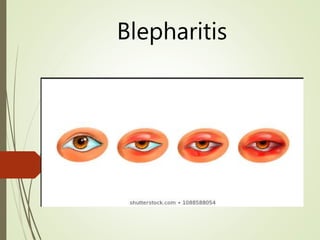
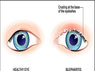
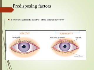
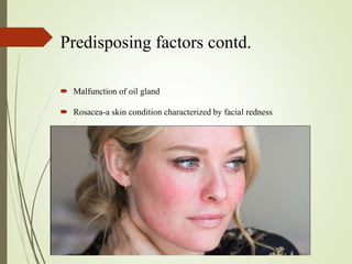
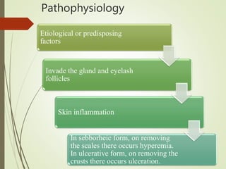
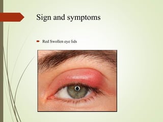
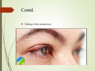
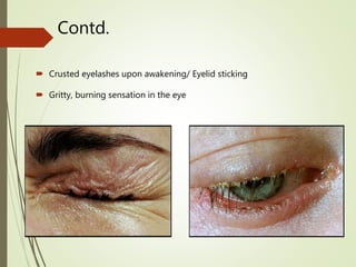
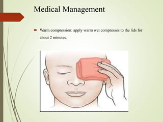
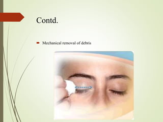
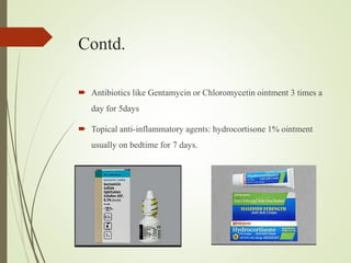
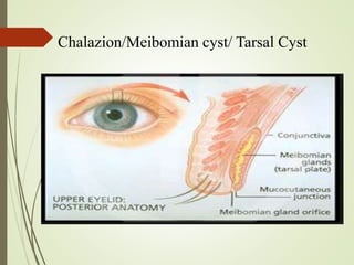
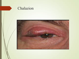
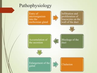
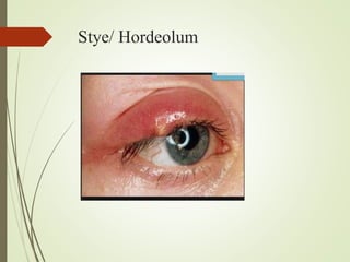
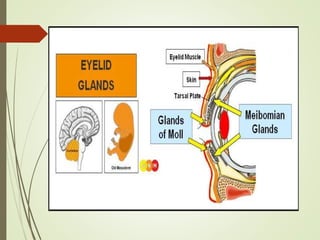
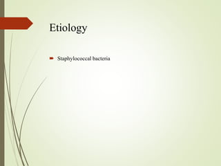
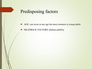
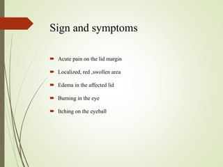
Disorders of Eye
 Review anatomy and physiology
Blepharitis
Definition
 It is a common eye condition characterized by chronic
inflammation of the eyelid generally the part where eyelashes
grow.
 It is an inflammation which occurs when tiny oil glands located
near the base of the eyelashes malfunction.
Etiology
 Commonest causes are dirt, staphylococcal infection and allergy.
 Infection with coagulase positive staphylococcus.
Predisposing factors
 Seborrheic dermatitis-dandruff of the scalp and eyebrow
Predisposing factors contd.
 Malfunction of oil gland
 Rosacea-a skin condition characterized by facial redness
Contd.
 Allergies-allergic reaction to eye medication, contact lens
solution, eye make up
 Eyelash mites or lice
 Certain medications-acne medicine ISOTRETINOIN can lead
to increase in bacteria in eyelids and can affect the tear
production.
Types
Clinically two types of blepharitis
a)Squamous/Sebborheic blepharitis:
 More common than ulcerative form. It is not essentially an infective
condition.
 Metabolic causes, hygienic factors, eye strain and sebborhoea of the scalp
usually leads to the development of this condition.
 In this case, there is accumulation of white scales like dandruff on the lid
margin. On removing this scales, the lid margins appears hyperemic but there
is no ulceration.
Contd.
b)Ulcerative/Infective blepharitis:
 This condition is due to infection of lid margin with coagulase
positive staphylococcus.
 There is suppurative inflammation of ciliary follicles along with
gland of zeis and moll.
 Yellow crusts are deposited at the roots of eyelash.
 On removing the crusts, small ulcers appear around the base of
the eyelashes which bleed freely.
Pathophysiology
Etiological or predisposing
factors
Invade the gland and eyelash
follicles
Skin inflammation
In sebborheic form, on removing
the scales there occurs hyperemia.
In ulcerative form, on removing the
crusts there occurs ulceration.
Sign and symptoms
 Red Swollen eye lids
Contd.
 Flaking of skin around eyes
Contd.
 Crusted eyelashes upon awakening/ Eyelid sticking
 Gritty, burning sensation in the eye
Contd.
 Frequent blinking of eyes
Contd.
 Sensitivity to light (Photophobia)
Sign and symptoms
 Loss of eyelashes
 Misdirected eyelashes inward
Diagnosis
 History and physical examination(eye examination)
 Slit lamp examination
 Culture and sensitivity: Collect the sample of oil or crust on the
eyelid for bacteria, fungi or allergic reaction.
Contd.
 Slit lamp examination
Medical Management
 Warm compression: apply warm wet compresses to the lids for
about 2 minutes.
Contd.
 Maintain hygiene: Hygiene maintenance by cleaning the scalp,
eyebrows and lid margins daily with medicated shampoo (baby
shampoo) to control sebborheic dermatitis.
Contd.
 Mechanical removal of debris
Contd.
 Antibiotics like Gentamycin or Chloromycetin ointment 3 times a
day for 5days
 Topical anti-inflammatory agents: hydrocortisone 1% ointment
usually on bedtime for 7 days.
Preventive measures
 Teach the client to,
 avoid cosmetics.
 separate towel for face and hair and use it individually.
 avoid using the eye frequently with uncleaned hands.
Nursing management
 Emphasize the importance of keeping the scalp, eyebrows and eyelid
margins clean.
 Instruct the patient to remove eyelid crusts with wash cloth or wet
cotton.
 Crusts are easier to remove when warm moist compresses are applied
for10 to 15 minutes.
 Hand washing before and after the care of the eyelids
 Tell the patient to avoid rubbing or touching the eyes
 Tell the patient to trim the nails.
 Avoid cosmetics which may cause irritation.
Chalazion/Meibomian cyst/ Tarsal Cyst
Chalazion
Definition
 It is a cyst in the eyelid that is caused by inflammation of blocked
meibomian gland, usually on the upper eyelid.
 It is a painless inflammation of the meibomian gland
characterized by localized swelling.
Risk Factors
 History of chalazion
 Unclean hands
 Person with blepharitis, stye, rosacea
Pathophysiology
Entry of
microorganism
into the
meibomian gland
Infiltration and
proliferation of
leucocytes on the
wall of the duct
Blockage of the
duct
Accumulation of
the secretion
Enlargement of the
galnd
Chalazion
Sign and Symptoms
 Painless, hard lump on eyelid
 Mild heaviness in the lid
 Mild irritation
 Sensitivity to light
 Eyelid tenderness
Diagnosis
 History and physical examination
 Test for vision
Management
 Warm compression: for 10 to 20 minutes at least four times a day
which
 may soften the hardened oil blocking the duct and promote drainage
and healing.
 Topical antibiotic eye drops or ointment: like Chloramphenicol or
Fusidic acid 3 times a day for 5 days.
 If such therapy fails,
 Incision and curettage under local anesthesia
 Pressure eye patch
 Teaching on proper lid hygiene.
Stye/ Hordeolum
Definition
 It is an infection of the sebaceous glands of zeis or glands of moll
at the base of the eyelashes.
 It is a suppurative inflammation of the follicles of eyelashes along
the edge of the eyelid margin.
Etiology
 Staphylococcal bacteria
Predisposing factors
 AGE: can occur at any age but most common in young adults
 MEATBOLIC FACTORS: diabetes,debility
Sign and symptoms
 Acute pain on the lid margin
 Localized, red ,swollen area
 Edema in the affected lid
 Burning in the eye
 Itching on the eyeball
Contd.
 Irritation on the eye
 Tearing
 Discomfort during blinking
 Foreign body sensation
Diagnosis
 History and physical examination
 Culture and sensitivity: Take sample of the purulent material from
abscess
Medical Management
 Warm compression
 Antibiotics
 If conservative treatment fails, incision and curettage
Nursing management
 Advise the patient to use clean cloth for warm compression and
dispose it separately to prevent from spreading.
 Avoid squeezing the stye which spreads the infection along the
eyelid margin.
 Advise the patient with recurrent or persistent stye to see the
doctor.

Blepharitis, chalazion, stye

  • 1.
    Disorders of Eye Review anatomy and physiology
  • 4.
  • 6.
    Definition  It isa common eye condition characterized by chronic inflammation of the eyelid generally the part where eyelashes grow.  It is an inflammation which occurs when tiny oil glands located near the base of the eyelashes malfunction.
  • 7.
    Etiology  Commonest causesare dirt, staphylococcal infection and allergy.  Infection with coagulase positive staphylococcus.
  • 8.
    Predisposing factors  Seborrheicdermatitis-dandruff of the scalp and eyebrow
  • 9.
    Predisposing factors contd. Malfunction of oil gland  Rosacea-a skin condition characterized by facial redness
  • 10.
    Contd.  Allergies-allergic reactionto eye medication, contact lens solution, eye make up  Eyelash mites or lice  Certain medications-acne medicine ISOTRETINOIN can lead to increase in bacteria in eyelids and can affect the tear production.
  • 11.
    Types Clinically two typesof blepharitis a)Squamous/Sebborheic blepharitis:  More common than ulcerative form. It is not essentially an infective condition.  Metabolic causes, hygienic factors, eye strain and sebborhoea of the scalp usually leads to the development of this condition.  In this case, there is accumulation of white scales like dandruff on the lid margin. On removing this scales, the lid margins appears hyperemic but there is no ulceration.
  • 12.
    Contd. b)Ulcerative/Infective blepharitis:  Thiscondition is due to infection of lid margin with coagulase positive staphylococcus.  There is suppurative inflammation of ciliary follicles along with gland of zeis and moll.  Yellow crusts are deposited at the roots of eyelash.  On removing the crusts, small ulcers appear around the base of the eyelashes which bleed freely.
  • 13.
    Pathophysiology Etiological or predisposing factors Invadethe gland and eyelash follicles Skin inflammation In sebborheic form, on removing the scales there occurs hyperemia. In ulcerative form, on removing the crusts there occurs ulceration.
  • 14.
    Sign and symptoms Red Swollen eye lids
  • 15.
    Contd.  Flaking ofskin around eyes
  • 16.
    Contd.  Crusted eyelashesupon awakening/ Eyelid sticking  Gritty, burning sensation in the eye
  • 17.
  • 18.
    Contd.  Sensitivity tolight (Photophobia)
  • 19.
    Sign and symptoms Loss of eyelashes  Misdirected eyelashes inward
  • 20.
    Diagnosis  History andphysical examination(eye examination)  Slit lamp examination  Culture and sensitivity: Collect the sample of oil or crust on the eyelid for bacteria, fungi or allergic reaction.
  • 21.
  • 22.
    Medical Management  Warmcompression: apply warm wet compresses to the lids for about 2 minutes.
  • 23.
    Contd.  Maintain hygiene:Hygiene maintenance by cleaning the scalp, eyebrows and lid margins daily with medicated shampoo (baby shampoo) to control sebborheic dermatitis.
  • 24.
  • 25.
    Contd.  Antibiotics likeGentamycin or Chloromycetin ointment 3 times a day for 5days  Topical anti-inflammatory agents: hydrocortisone 1% ointment usually on bedtime for 7 days.
  • 26.
    Preventive measures  Teachthe client to,  avoid cosmetics.  separate towel for face and hair and use it individually.  avoid using the eye frequently with uncleaned hands.
  • 27.
    Nursing management  Emphasizethe importance of keeping the scalp, eyebrows and eyelid margins clean.  Instruct the patient to remove eyelid crusts with wash cloth or wet cotton.  Crusts are easier to remove when warm moist compresses are applied for10 to 15 minutes.  Hand washing before and after the care of the eyelids  Tell the patient to avoid rubbing or touching the eyes  Tell the patient to trim the nails.  Avoid cosmetics which may cause irritation.
  • 28.
  • 29.
  • 30.
    Definition  It isa cyst in the eyelid that is caused by inflammation of blocked meibomian gland, usually on the upper eyelid.  It is a painless inflammation of the meibomian gland characterized by localized swelling.
  • 31.
    Risk Factors  Historyof chalazion  Unclean hands  Person with blepharitis, stye, rosacea
  • 32.
    Pathophysiology Entry of microorganism into the meibomiangland Infiltration and proliferation of leucocytes on the wall of the duct Blockage of the duct Accumulation of the secretion Enlargement of the galnd Chalazion
  • 33.
    Sign and Symptoms Painless, hard lump on eyelid  Mild heaviness in the lid  Mild irritation  Sensitivity to light  Eyelid tenderness
  • 34.
    Diagnosis  History andphysical examination  Test for vision
  • 35.
    Management  Warm compression:for 10 to 20 minutes at least four times a day which  may soften the hardened oil blocking the duct and promote drainage and healing.  Topical antibiotic eye drops or ointment: like Chloramphenicol or Fusidic acid 3 times a day for 5 days.  If such therapy fails,  Incision and curettage under local anesthesia  Pressure eye patch  Teaching on proper lid hygiene.
  • 36.
  • 37.
    Definition  It isan infection of the sebaceous glands of zeis or glands of moll at the base of the eyelashes.  It is a suppurative inflammation of the follicles of eyelashes along the edge of the eyelid margin.
  • 39.
  • 40.
    Predisposing factors  AGE:can occur at any age but most common in young adults  MEATBOLIC FACTORS: diabetes,debility
  • 41.
    Sign and symptoms Acute pain on the lid margin  Localized, red ,swollen area  Edema in the affected lid  Burning in the eye  Itching on the eyeball
  • 42.
    Contd.  Irritation onthe eye  Tearing  Discomfort during blinking  Foreign body sensation
  • 43.
    Diagnosis  History andphysical examination  Culture and sensitivity: Take sample of the purulent material from abscess
  • 44.
    Medical Management  Warmcompression  Antibiotics  If conservative treatment fails, incision and curettage
  • 45.
    Nursing management  Advisethe patient to use clean cloth for warm compression and dispose it separately to prevent from spreading.  Avoid squeezing the stye which spreads the infection along the eyelid margin.  Advise the patient with recurrent or persistent stye to see the doctor.