Detailed presentation on Blepharitis, types (bacterial blepharitis / posterior blepharitis or meibomitis / parasitic blepharitis), signs & symptoms, complications, treatment. This presentation is a part of series on Eyelid diseases.
Exposure keratopathy develops when the eyelids do not adequately cover and protect the cornea during blinking. This can be caused by factors like Bell's palsy or exposure proptosis that limit eyelid closure. Without protection, the corneal epithelium dries out and is invaded by infectious organisms, leading to symptoms of irritation, redness, and corneal ulceration or perforation. Treatment focuses on managing the underlying cause through artificial tears, ointments, or contact lenses and may require suturing the eyelids shut to protect the cornea.
This document discusses the management of dry eye. It begins by defining dry eye as a multifactorial disease resulting in ocular discomfort and potential damage due to increased tear film osmolarity and ocular surface inflammation. Dry eye is commonly seen in clinical practice and can range from mild to severe. The document then discusses the anatomy and physiology of tear production, the tear film layers, contributing factors, classifications, signs and symptoms, diagnostic tests, and treatment strategies including artificial tears and anti-inflammatory therapies. Treatment is tailored based on dry eye severity levels with the goal of alleviating symptoms and preventing complications.
This document summarizes different types of allergic conjunctivitis including simple allergic conjunctivitis, vernal keratoconjunctivitis, atopic keratoconjunctivitis, giant papillary conjunctivitis, phlyctenular keratoconjunctivitis, and contact dermoconjunctivitis. It describes the etiology, pathology, symptoms, signs, and management of each type. For simple allergic conjunctivitis, common allergens include pollens and treatments involve reducing allergen exposure, cold compresses, tear substitutes, vasoconstrictors, mast cell stabilizers, and antihistamines. Vernal keratoconjunctivitis causes cob
1. The document discusses various degenerations, cysts, and tumors that can affect the conjunctiva, including pterygium, pinguecula, conjunctival cysts, and both benign and malignant conjunctival tumors.
2. Pterygium is a degenerative condition caused by prolonged UV exposure that involves fibrovascular proliferation invading the cornea. Recurrence after surgery is common.
3. Treatment for pterygium involves surgical excision with conjunctival autograft or amniotic membrane graft to prevent recurrence. Amniotic membrane is harvested from the placenta.
This document discusses dry eye, including its causes, diagnostic tests, and treatment options. It provides an overview of dry eye as one of the most common conditions seen by eye practitioners. Key points include:
- Dry eye is caused by dysfunction of the lacrimal functional unit, which includes the tear glands, tear film, and ocular surface.
- Common diagnostic tests include tear break-up time, Schirmer test, and staining of the ocular surface with dyes. Newer tests measure tear osmolarity.
- Treatment focuses on managing symptoms, treating underlying causes, and stabilizing the tear film through lubricating eye drops, ointments, or gels. Preservative
This document discusses various viral and protozoal causes of corneal ulcers, including herpes simplex virus (HSV), herpes zoster virus, and acanthamoeba. It describes the etiology, clinical features, diagnosis, and treatment of these conditions. Primary and recurrent HSV keratitis present with punctate epithelial lesions and dendritic ulcers. Herpes zoster ophthalmicus causes vesicular skin lesions following reactivation of varicella zoster virus in the trigeminal ganglion. Acanthamoeba keratitis is an opportunistic infection associated with contact lens use that presents with epithelial lesions and stromal infiltrates. Treatment involves antiviral medications for viruses and anti-am
This document discusses trichiasis and entropion. Trichiasis is the misdirection of eyelash growth without inversion of the lid. Entropion is the inversion of the eyelid, usually the lower lid. There are several types of entropion including involutional (due to lid laxity and orbicularis overriding), cicatricial (due to scarring), and congenital. Treatment depends on the underlying cause but may include epilation, electrolysis, laser ablation, cryotherapy, or surgical procedures like lateral tarsal strip, transverse everting sutures, or tarsal fracture. Abnormal lid laxity is diagnosed if the lid can be drawn away from the globe by
Viral keratitis is commonly caused by herpes simplex virus types 1 and 2 and varicella zoster virus. HSV keratitis typically presents as dendritic ulcers or geographic ulcers on the cornea. It can also cause stromal keratitis. Varicella zoster virus causes herpes zoster infection which presents with a painful rash that follows dermatomal patterns, and can also lead to keratitis. Adenovirus is another common cause and presents as punctate epithelial keratitis that may develop into nummular opacities. Treatment involves topical antivirals and steroids depending on the type and severity of infection.
Dr. Ajai Agrawal's presentation defines primary angle closure glaucoma (PACG) as a type of glaucoma characterized by occludable anterior chamber angles that lead to increased intraocular pressure and optic nerve damage. The presentation covers the epidemiology, risk factors, pathogenesis, classification, diagnosis and management of PACG. Treatment may involve medical management, laser peripheral iridotomy to open the angle, or filtering surgeries in advanced cases. Regular follow up is important as PACG can progress to cause vision loss if not properly treated.
The document provides information about the sclera, including its structure, thickness, apertures, nerve supply, inflammation conditions like episcleritis and scleritis, and other related topics. The sclera forms the posterior five-sixths of the outer fibrous tunic of the eyeball. It varies in thickness from 1mm posteriorly to 0.3mm at the muscle insertions. It contains three layers and is pierced by blood vessels and nerves. Inflammation of the sclera can cause episcleritis or scleritis, with the latter being a more serious condition sometimes associated with underlying systemic diseases.
SIANG KLINIK \'DRY EYE\', 27 FEBRUARI 2011Perdami Bekasi
Dry eye is a common condition that causes eye discomfort. It occurs when the tear film is unstable or the eyes do not produce enough tears. Symptoms include dryness, irritation, and blurred vision. Diagnosis involves examining the eyes for signs of damage and testing tear production and stability. Treatment focuses on replacing tears through artificial tears, gels, or ointments. For severe cases, punctal plugs may be placed to block tear drainage, and anti-inflammatory therapies are used to reduce symptoms and prevent further damage to the eyes. The goals of treatment are to relieve discomfort and protect the ocular surface.
Ectropion
It is an outward turning of the eyelid margin . This more frequently affects the lower eyelid.Upper eyelid ectropion is uncommon.Classified in 5 types
1)Congenital 2) Involutional 3) Paralytic 4) Cicatricial 5) Mechanical
Involutional ectropion is more common.Congenital ectropion is very rare.
Symptoms Epiphora :- excessive tearing.Excessive dryness.
Foreign body sensation Irritation.Burning.Redness.Chronic conjunctivitis KeratinizationCorneal exposure
Grading
Lid margin is out rolled and depending on out rolling ectropion can be classified as under:
Grade I –only punctum is everted
Grade II –lid margin is everted and palpebral conjunctiva is visible
Grade III –fornix is also visible
Etiological factors
Horizontal lid laxity:-can be demonstrated by pulling the central part of the lid 8 mm or more from the globe, with a failure to snap back to its normal position on release without the patient first blinking.
Medial canthal tendon laxity
demonstrated by pulling the lower lid laterally and observing the position of the inferior punctum If the lid is normal the punctum should not be displaced more than 1–2 mm
Lateral canthal tendon laxity
characterized by a rounded appearance of the lateral canthus and the ability to pull the lower lid medially more than 2 mm.
>Normally, the displacement should only be 0-2 mm.
Treatment
1 medical therapy
2 surgical therapy
The document discusses corneal hydration and edema. It begins by explaining that the cornea's water content is normally around 78% to maintain transparency. Corneal edema occurs when water content increases above this level. Factors that influence corneal hydration are then described, including stromal swelling pressure, intraocular pressure, and the sodium-potassium pump in the endothelium. Causes of corneal edema are outlined as either primary, such as endothelial dystrophies, or secondary due to trauma, glaucoma, contact lenses or metabolic disorders. Symptoms, evaluation and management of corneal edema are also summarized.
This document provides information on chemical eye burns, including:
- Alkali burns are more dangerous than acid burns as alkalis penetrate rapidly into the eye.
- Immediate irrigation with saline or water for at least 20 minutes is essential to remove the chemical from the eye.
- The Dua classification system is useful for assessing prognosis based on limbal and conjunctival involvement.
- Medical management focuses on removing the chemical, promoting healing, controlling inflammation and infection, and managing intraocular pressure.
Techniques of tear film evaluation by Raju KaitiRaju Kaiti
The document summarizes techniques for evaluating the tear film, which has three layers: an outer lipid layer, intermediate aqueous layer, and inner mucous layer. Non-invasive techniques discussed include tear break-up time tests, lipid layer evaluation using interferometry, and inferior tear meniscus height measurements. Invasive techniques involve Schirmer's tests to evaluate tear secretion, fluorescein and rose bengal staining of the ocular surface, and conjunctival impression cytology to examine goblet cell density. The document provides details on procedures and normal results for each evaluation method.
This document describes different types of ectropion and entropion conditions. It discusses involutional, cicatricial, paralytic, congenital, and mechanical causes. Treatment options are provided depending on the severity and include procedures like conjunctivoplasty, wedge resection, tarsorrhaphy, and skin grafts. Assessment of horizontal lid laxity and canthal tendon positioning helps determine the best surgical approach.
Dr. TP Chhangte discusses anterior uveitis, including its definition, epidemiology, classification, pathology, clinical features, complications, and differential diagnosis. Some key points:
- Anterior uveitis is inflammation of the iris and anterior part of the ciliary body. Its prevalence has been increasing and it commonly affects people aged 20-50.
- Uveitis can be classified anatomically, clinically, etiologically, and pathologically. The anatomical classification divides uveitis into anterior, intermediate, posterior, and panuveitis based on location of inflammation.
- Clinical features of anterior uveitis include eye pain, redness, photophobia, and decreased vision. On examination
This document discusses various causes of corneal edema. It begins by explaining that corneal edema can be caused by endothelial dysfunction or situations exceeding the endothelium's barrier and pump capacity. Specific conditions that can lead to corneal edema are then outlined, including persistent epithelial defects, increased intraocular pressure, primary endothelial diseases, Fuch's dystrophy, posterior polymorphous dystrophy, congenital hereditary endothelial dystrophy, iridocorneal endothelial syndrome, and corneal edema induced by cataract surgery or other ocular procedures and injuries. Treatment options are mentioned for selected conditions.
This document provides an overview of different types of corneal dystrophies, including their classification, clinical features, histopathology, and management. It discusses epithelial and subepithelial dystrophies, corneal dystrophies of Bowman's layer, stromal corneal dystrophies, and Descemet membrane and endothelial dystrophies. The key points are that corneal dystrophies are inherited, bilateral, and slowly progressive disorders that begin early in life and are characterized by corneal opacification without relationship to environmental factors. Diagnosis involves classification based on the anatomical layer affected and treatment typically involves managing symptoms although surgery may be needed if vision is impaired.
Dry eye is a disease characterized by unstable tear film and ocular surface inflammation. It results in eye discomfort, visual disturbance, and potential ocular surface damage. Dry eye is caused by factors that interrupt normal tear production or function like lacrimal gland damage, meibomian gland dysfunction, or neurological issues. Diagnosis involves evaluating symptoms, ocular surface staining, and tear film breakup time. Management includes artificial tears, punctal plugs, cyclosporine drops, and procedures like LipiFlow that target meibomian glands.
This document discusses microbial keratitis, which is a type of corneal inflammation caused by microorganisms. It can be characterized by corneal edema, cellular infiltration, and ciliary congestion. Microbial keratitis is one of the leading causes of preventable blindness. The document describes the different types of microbial keratitis such as ulcerative keratitis and non-ulcerative keratitis. It also discusses the various etiological agents that can cause microbial keratitis including bacteria, fungi, viruses, and parasites. The pathogenesis and clinical features of different microbial keratitis are explained. Predisposing factors, examination findings, and special features of bacterial, fungal, and parasitic keratitis are outlined in the summary.
This document discusses primary angle closure glaucoma (PACG), a type of glaucoma where the iris occludes the drainage angle of the eye, obstructing aqueous outflow. PACG is a leading cause of glaucoma worldwide and is particularly common in East Asian populations. It is classified based on the degree of iris occlusion and presence of optic nerve damage and vision loss. Risk factors include older age, female sex, Asian ethnicity, family history, hypermetropia, and shorter axial length. Symptoms include blurred vision, halos around lights, eye pain, and headache. Signs include elevated eye pressure, shallow anterior chamber, iris changes, and optic nerve damage. The document outlines methods for diagnosing
Pterygium is a wing-shaped growth of conjunctiva that encroaches on the cornea. It is more common in people living in hot, dusty climates who are exposed to sun and ultraviolet rays. Pterygium is a degenerative condition where subconjunctival tissue proliferates as vascularised granulation tissue under the epithelium, destroying the corneal epithelium, Bowman's layer and superficial stroma. It presents as a triangular fold of conjunctiva on the cornea, usually on the nasal side. Surgical excision is required if it encroaches the pupil or causes diplopia. Recurrence after excision is common, ranging from 30-50%, but use of
Lacrimation refers to watering of the eye due to excessive tear production from the lacrimal gland, while epiphora is watering caused by obstruction of the tear drainage system. Differentiating the two is important to provide proper treatment. The document discusses the anatomy and physiology of tear production, drainage, and evaluates causes of watering eye including lacrimation, dry eye, blepharitis, and epiphora due to punctal stenosis or nasolacrimal duct obstruction. Treatment depends on the underlying cause and may include punctal dilation, dacryocystorhinostomy, Jones tube insertion, or endoscopic lacrimal surgery.
The document discusses various diseases and conditions of the eyelids including hordeolum externum (stye), hordeolum internum, chalazion, blepharitis, and entropion. Stye is a localized infection of the eyelash follicle causing pain and swelling at the lid margin. Chalazion is a chronic inflammatory granuloma of the meibomian gland presenting as a painless swelling away from the lid margin. Blepharitis is inflammation of the lid margin that can be squamous, ulcerative, or posterior. Entropion is an in-rolling of the eyelid margin that can be involutional, cicatricial, spastic, or congen
Bullous keratopathy refers to corneal swelling caused by insufficient pumping of fluid out of the cornea. It results in the formation of fluid-filled blisters beneath the surface of the eye. It can occur secondary to endothelial dysfunction from trauma, inflammation, dystrophies or other conditions that damage the corneal endothelial pump. Symptoms include blurred vision, glare, pain from ruptured blisters, and potentially scarring. Management involves reducing fluid buildup through hypertonic drops or lenses, lowering eye pressure if high, and corneal transplantation if vision is significantly decreased or pain becomes intolerable.
Blepharitis- inflammation of lid margins
CLINICAL TYPES:
ANTERIOR BLEPHARITIS
$ Bacterial ulcerative blepharitis :
▪︎ also called as chronic anterior blepharitis or staphylococcal blepharitis
ETIOLOGY
Coagulase positive Staphylococci (most common)
Rarely : Streptococci, Propionibacterium acnes, Moraxella
PATHOGENESIS
Bacteria are carried to lid margins by rubbing with contaminated fingers → inflammation
CLINICAL FEATURES
Symptoms: (worse in the morning) irritation, itching, lacrimation, gluing of cilia, photophobia
Signs :
▪︎ Yellow crusts at the root of cilia
▪︎ Small ulcers on removing the crusts
▪︎ Red, thickened lid margins
▪︎ Mild papillary conjunctivitis and conjunctival hyperemia
TREATMENT
1.Lid hygiene : warm compresses, crust removal and lid margins cleaning (with cotton buds dipped in dilute baby shampoo), avoid rubbing of eyes
2. Antibiotics :
⊙ Eye ointment - applied at lid margin after crust removal
⊙ Eye drops - 3 to 4 times a day
⊙ Oral antibiotics - Erythromycin or Doxycyclin in unresponsive patients
3. Topical Steroids with low potency : Fluorometholone
4. Ocular lubricants ︎
$ Seborrhoeic blepharitis:
▪︎ primarily anterior blepharitis associated with dandruff
ETIOLOGY AND PATHOGENESIS
Glands of Zeis secrete abnormal excessive neutral lipids
Corynebacterium acne splits lipids into irritating fatty acids causing inflammation
SYMPTOMS
Deposition of soft scales at lid margin, discomfort, irritation, watering (occasionally), history of falling of eyelashes
SIGNS
▪︎ Accumulation of white dandruff like scales on lid margin
▪︎ Hyperemic greasy surface found on removal of scales - no ulcer
▪︎ Lashes fall out easily and replace quickly
▪︎ Thickened lid margin
COMPLICATIONS - similar to bacterial blepharitis
TREATMENT
General measures - improvement of health and balanced diet
Treat dandruff adequately
Local measures - removal of scales with lukewarm solution of 3% sodium bicarbonate or baby shampoo
Antibiotics - in mixed blepharitis
POSTERIORBLEPHARITIS (MEIBOMITIS) :
▪︎ Meibomian gland dysfunction
▪︎ Common in middle aged individuals having seborrheic dermatitis
▪︎ Bacterial lipases (producing free fatty acids) play a role in pathogenesis
SYMPTOMS
Irritation, burning, itching, grittiness, watering
SIGNS
▪︎ White frothy secretions on lid margins, oily tear film
▪︎ Secretions with toothpaste appearance over Meibomian gland orifices when lids are pressed
▪︎ Vertical yellow streaks in conjunctiva
▪︎ Secondary changes in conjunctiva and cornea
TREATMENT
Lid hygiene : warm compresses, milking of secretions by vertical massage of lids
Topical antibiotics : Eye ointment after massage, eye drops (3 to 4 times a day)
Systemic antibiotics : Doxycycline 100 mg b.d. for 1 week followed by 100 mg o.d for 6 to 12 weeks
Ocular lubricants
Topical Steroids : Fluorometholone
This document discusses dry eye, including its definition, causes, symptoms, and treatment approaches. It begins by defining dry eye as a multifactorial disease that results in discomfort, visual disturbance, and tear film instability. Various etiological factors that can contribute to dry eye are then outlined, including age, gender, medications, environmental conditions, and ocular surface diseases. Treatment is tailored based on the severity of dry eye and may include artificial tears, anti-inflammatory therapies, punctal plugs, and procedures to improve meibomian gland function. Modern technologies like LipiFlow are also mentioned for managing meibomian gland dysfunction.
The eyelids are mobile tissue curtains placed in front of the eyeballs. These act as shutters protecting the eyes from injuries and excessive light. These also perform an important function of spreading the tear film over the cornea and conjunctiva and also help in drainage of tears by lacrimal pump system.
This document provides an overview of blepharitis, including:
- Blepharitis is inflammation of the eyelid margins and is one of the most common and difficult to treat eye conditions. It can be caused by bacterial infection, dry eye, or skin conditions like rosacea.
- There are two main types - anterior blepharitis affecting the base of the eyelashes, and posterior blepharitis involving the meibomian glands. Anterior blepharitis has staphylococcal and seborrheic variants.
- Symptoms include burning, grittiness, dryness, and worsening in the morning. Signs include scales, debris, hyp
Blepharitis is a common eyelid inflammation that can be caused by bacterial infections or dry eye conditions. It is characterized by scaling or crusting of the eyelashes and eyelid margins. There are two main types - anterior blepharitis, which involves inflammation at the base of the eyelashes, and posterior blepharitis, which affects the glands of the eyelid. Symptoms include burning, irritation, tearing and worsening in the morning. Treatment focuses on eyelid hygiene through warm compresses and lid scrubs to remove scales and debris. Blepharitis can exacerbate dry eye and may cause long-term eyelid changes if left untreated. It may also increase risks of certain inflammatory
Soft contact lens complications can include ocular discomfort, inflammation, infection, and other issues. Ocular discomfort is the most common complication and has many potential causes, both physical and physiological. Inflammation like bulbar redness, corneal infiltrates, and contact lens peripheral ulcers can occur due to factors like tight lenses, deposits, hypoxia, or infection. Proper lens care and frequent replacement are important to prevent complications, along with addressing any underlying causes like dryness or infection. Complications generally require discontinuing lens wear until signs and symptoms resolve.
blepharitis is an eye disorder , this is divided into two types. one anterior blepharitis another is posterior blepharitis .this caused by other organisms like staphylococcus and dryness of the eyes, avoid touching of the eye .advice to take the medication as prescribed by the doctor.
This document provides information on managing acute painless red eye. It discusses taking a thorough history and performing an examination to determine the underlying cause, which can include blepharitis, trichiasis, subtarsal foreign bodies, pinguecula, pterygium, episcleritis, viral conjunctivitis, allergic conjunctivitis, and bacterial conjunctivitis. For each condition, it describes symptoms, causes, and treatment approaches, which may involve lid hygiene, lubricants, antibiotics, anti-inflammatories, or surgery. Proper diagnosis and management are important to resolve the condition and relieve symptoms.
Dry eye management involves evaluating a patient's history, symptoms, and signs of dry eye through examination and tests. Treatment aims to relieve discomfort, maintain a smooth optical surface, and prevent damage. It includes lifestyle modifications, medical treatments like tear substitutes, anti-inflammatory agents, and procedures like punctal occlusion. Regular follow up and counseling are important for monitoring response and providing support.
The document provides an overview of common eye presentations to the emergency department, outlining 10 case studies that demonstrate a range of conditions including infected corneal ulcers, corneal abrasions, uveitis, glaucoma, hyphema, retinal detachments, and orbital cellulitis, and provides guidance on examination findings, differential diagnoses, and management for each case.
The document discusses several common eye conditions - blepharitis, hordeolum, chalazion, ectropion, and entropion. It provides details on the causes, clinical features, diagnosis, and management of each condition. Blepharitis is a chronic inflammatory reaction of the eyelid margin that can be staphylococcal or seborrheic in nature. Hordeolum is an acute suppurative infection of the eyelid glands caused by Staphylococcus aureus. Chalazion is a non-infective inflammation of the meibomian gland, presenting as a painless eyelid lump. Ectropion involves inward rolling of the eyelid
Congenital ptosis, coloboma, epicanthus, and distichiasis are common congenital eyelid anomalies. 
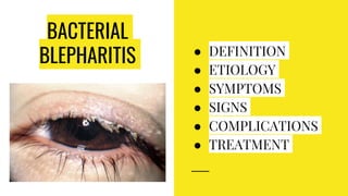
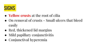
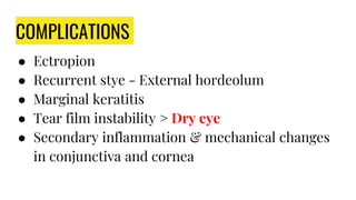
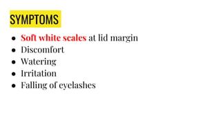
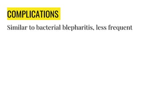
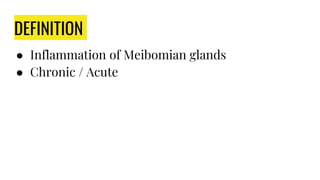
Blepharitis is a chronic inflammation of the lid margins that can be anterior (squamous or ulcerative) or posterior (meibomitis) in nature. Hordeolum (stye) is an acute suppurative infection of the eyelash follicle or meibomian gland that presents as a tender, red swelling and is usually caused by Staphylococcus aureus. Chalazion is a chronic non-infectious cyst of the meibomian gland that appears as a painless, firm nodule.
Dry eye occurs when there is inadequate tear production or function, resulting in an unstable tear film and ocular surface disorder. It can be caused by conditions that reduce tear production such as Sjogren's syndrome, vitamin A deficiency, Stevens-Johnson syndrome, or medications. Other causes affect the tear film layers, like meibomian gland dysfunction reducing the outer lipid layer. Symptoms include dryness, burning, and blurred vision. Treatment focuses on replacing tears, improving ocular surface health, addressing underlying causes, and escalating care based on severity through the DEWS treatment guidelines.
Blepharitis is inflammation of the eyelid margin that can be anterior (involving the front of the eyelid) or posterior (involving the glands of the eyelid). Anterior blepharitis includes bacterial, seborrheic, and parasitic types. Bacterial blepharitis is common and caused by staphylococci and streptococci, causing chronic irritation, crusting, and redness. Treatment involves lid hygiene, antibiotics, and anti-inflammatory drugs. Seborrheic blepharitis is associated with dandruff and treated with lid cleaning and antibiotic/steroid ointment. Parasitic blepharitis involves lice inf
This document discusses various bacterial skin infections including impetigo contagiosa, bullous impetigo, staphylococcal scalded skin syndrome, ecthyma, folliculitis, furunculosis, carbuncle, erysipelas, and cellulitis. It covers the etiology, clinical features, morphology, sites of involvement, investigations, differential diagnosis, and treatment of each condition. The main causative organisms are Staphylococcus aureus and Streptococcus pyogenes. Conditions vary in depth and spread, from superficial to deep infections, and treatment involves antibiotics, incision and drainage, and supportive measures.
A stye, also known as a hordeolum, is an inflammation of the sebaceous gland of the eyelid that results in a red, tender bump on the edge of the eyelid. Common causes of a stye include bacterial infection by Staphylococcus bacteria, excessive use of cosmetics, poor nutrition, lack of hygiene, rubbing of the eyes, and lack of sleep. Symptoms include a red lump on the eyelid that looks like a boil or pimple, eyelid pain, and eyelid swelling. Treatment involves warm compresses, analgesics like acetaminophen, and antibiotics like erythromycin ophthalmic ointment or amoxicillin.
Bacterial eyelid infections and blepharitis.SristiThakur
This document discusses bacterial infections and blepharitis that can affect the eyelids. It begins by covering the anatomy of the eyelid and then describes several specific bacterial infections including external hordeolum, impetigo, erysipelas, necrotizing fasciitis, anthrax, and syphilis. It then discusses blepharitis in detail, describing the different types (staphylococcal, seborrheic, posterior), associated conditions like meibomian gland dysfunction, symptoms, signs, treatment, and complications.
Blepharitis is an inflammation of the eyelid margins that can be caused by bacteria, seborrhea, or meibomian gland dysfunction. It commonly presents with irritation, itching, crusting or scaling of the eyelids. Treatment involves warm compresses, lid scrubs, antibiotic ointments or drops, and oral antibiotics if needed. More severe forms include hordeolum (styes) which are infections of the eyelid glands, and chalazion which are non-infected cysts of the meibomian glands that may require incision and drainage. Regular lid hygiene is important to prevent complications and recurrence.
Simple eye education for EHW, Ophthalmic eye student, school eye education & first - second year optometry students only .
common eye lid inflammatory conditions .
stye or hordeolum ( external / internal hordeolum ), lid abscess , chalazion or mebomian retention cyst, accessory lacrimal glands , lacrimal gland etc...
A corneal ulcer is a loss of corneal epithelium with inflammation caused by bacterial or fungal infection that can lead to blindness if not treated properly. Common causes include infections from Staphylococcus or Pseudomonas bacteria. Treatment involves topical antibiotics like fortified cephazolin or tobramycin drops every few hours to control the infection along with cycloplegic drops for pain relief and prevention of complications like perforation or secondary glaucoma.
5-hydroxytryptamine or 5-HT or Serotonin is a neurotransmitter that serves a range of roles in the human body. It is sometimes referred to as the happy chemical since it promotes overall well-being and happiness.
It is mostly found in the brain, intestines, and blood platelets.
5-HT is utilised to transport messages between nerve cells, is known to be involved in smooth muscle contraction, and adds to overall well-being and pleasure, among other benefits. 5-HT regulates the body's sleep-wake cycles and internal clock by acting as a precursor to melatonin.
It is hypothesised to regulate hunger, emotions, motor, cognitive, and autonomic processes.
These lecture slides, by Dr Sidra Arshad, offer a simplified look into the mechanisms involved in the regulation of respiration:
Learning objectives:
1. Describe the organisation of respiratory center
2. Describe the nervous control of inspiration and respiratory rhythm
3. Describe the functions of the dorsal and respiratory groups of neurons
4. Describe the influences of the Pneumotaxic and Apneustic centers
5. Explain the role of Hering-Breur inflation reflex in regulation of inspiration
6. Explain the role of central chemoreceptors in regulation of respiration
7. Explain the role of peripheral chemoreceptors in regulation of respiration
8. Explain the regulation of respiration during exercise
9. Integrate the respiratory regulatory mechanisms
10. Describe the Cheyne-Stokes breathing
Study Resources:
1. Chapter 42, Guyton and Hall Textbook of Medical Physiology, 14th edition
2. Chapter 36, Ganong’s Review of Medical Physiology, 26th edition
3. Chapter 13, Human Physiology by Lauralee Sherwood, 9th edition
Nano-gold for Cancer Therapy chemistry investigatory projectSIVAVINAYAKPK
chemistry investigatory project
The development of nanogold-based cancer therapy could revolutionize oncology by providing a more targeted, less invasive treatment option. This project contributes to the growing body of research aimed at harnessing nanotechnology for medical applications, paving the way for future clinical trials and potential commercial applications.
Cancer remains one of the leading causes of death worldwide, prompting the need for innovative treatment methods. Nanotechnology offers promising new approaches, including the use of gold nanoparticles (nanogold) for targeted cancer therapy. Nanogold particles possess unique physical and chemical properties that make them suitable for drug delivery, imaging, and photothermal therapy.
DECLARATION OF HELSINKI - History and principlesanaghabharat01
This SlideShare presentation provides a comprehensive overview of the Declaration of Helsinki, a foundational document outlining ethical guidelines for conducting medical research involving human subjects.
low birth weight presentation. Low birth weight (LBW) infant is defined as the one whose birth weight is less than 2500g irrespective of their gestational age. Premature birth and low birth weight(LBW) is still a serious problem in newborn. Causing high morbidity and mortality rate worldwide. The nursing care provide to low birth weight babies is crucial in promoting their overall health and development. Through careful assessment, diagnosis,, planning, and evaluation plays a vital role in ensuring these vulnerable infants receive the specialize care they need. In India every third of the infant weight less than 2500g.
Birth period, socioeconomical status, nutritional and intrauterine environment are the factors influencing low birth weight
Kosmoderma Academy, a leading institution in the field of dermatology and aesthetics, offers comprehensive courses in cosmetology and trichology. Our specialized courses on PRP (Hair), DR+Growth Factor, GFC, and Qr678 are designed to equip practitioners with advanced skills and knowledge to excel in hair restoration and growth treatments.
Osteoporosis - Definition , Evaluation and Management .pdfJim Jacob Roy
Osteoporosis is an increasing cause of morbidity among the elderly.
In this document , a brief outline of osteoporosis is given , including the risk factors of osteoporosis fractures , the indications for testing bone mineral density and the management of osteoporosis
Lecture 6 -- Memory 2015.pptlearning occurs when a stimulus (unconditioned st...AyushGadhvi1
learning occurs when a stimulus (unconditioned stimulus) eliciting a response (unconditioned response) • is paired with another stimulus (conditioned stimulus)
How to Control Your Asthma Tips by gokuldas hospital.Gokuldas Hospital
Respiratory issues like asthma are the most sensitive issue that is affecting millions worldwide. It hampers the daily activities leaving the body tired and breathless.
The key to a good grip on asthma is proper knowledge and management strategies. Understanding the patient-specific symptoms and carving out an effective treatment likewise is the best way to keep asthma under control.
Test bank for karp s cell and molecular biology 9th edition by gerald karp.pdfrightmanforbloodline
Test bank for karp s cell and molecular biology 9th edition by gerald karp.pdf
Promoting Wellbeing - Applied Social Psychology - Psychology SuperNotesPsychoTech Services
A proprietary approach developed by bringing together the best of learning theories from Psychology, design principles from the world of visualization, and pedagogical methods from over a decade of training experience, that enables you to: Learn better, faster!
8. SIGNS
● Yellow crusts at the root of cilia
● On removal of crusts - Small ulcers that bleed
easily
● Red, thickened lid margins
● Mild papillary conjunctivitis
● Conjunctival hyperemia
17. ETIOLOGY
Glands of Zeis → Excessive, abnormal Neutral lipids
→ Split by Corynebacterium acne into FFA
A/w seborrhoea of scalp
18. SYMPTOMS
● Soft white scales at lid margin
● Discomfort
● Watering
● Irritation
● Falling of eyelashes
19. SIGNS
● White scales on the lid margin along the lashes
● Removal of scales - Hyperemic & Greasy surface
● Lashes fall out easily
● Thickened lid margin
● Rounded posterior border → Epiphora
21. TREATMENT
● Lukewarm soln of 3% soda bicarb for scale
removal & cleaning of lid margin
● Antibiotic & Steroid eye drops
● Oral antibiotics are used if mixed with bacterial
blepharitis
26. SIGNS - CHRONIC
● White frothy secretions at lid margins & canthi
● Opening of meibomian glands clogged with thick secretions >
May show pouting, recession
● On pressure - secretions can be expressed
● Vertical yellowish streaks seen through conjunctiva -
Meibomian ducts
● Hyperemia & telangiectasia of post lid margin
● Oily & foamy tear film
33. ETIOLOGY
Lice infestation - poor hygiene
● Phthiriasis palpebrum - infestation by phthirus
pubis (crab louse), an STI
● Pediculosis - infestation by pediculus humanus
corporis or capitis (head louse)
35. SIGNS
● Red, inflamed lid margins
● Lice visible clawing on to lashes on SLE
● Nits at the base of cilia
● Conjunctival congestion & follicles in long-
standing cases
36. TREATMENT
● Removal of lice & nits with forceps
● Apply antibiotic ointment to lid margin
● Apply Yellow Mercuric Oxide 1% to lid margin &
lashes
● Delousing of pt, pt belongings (bed, clothes),
family members