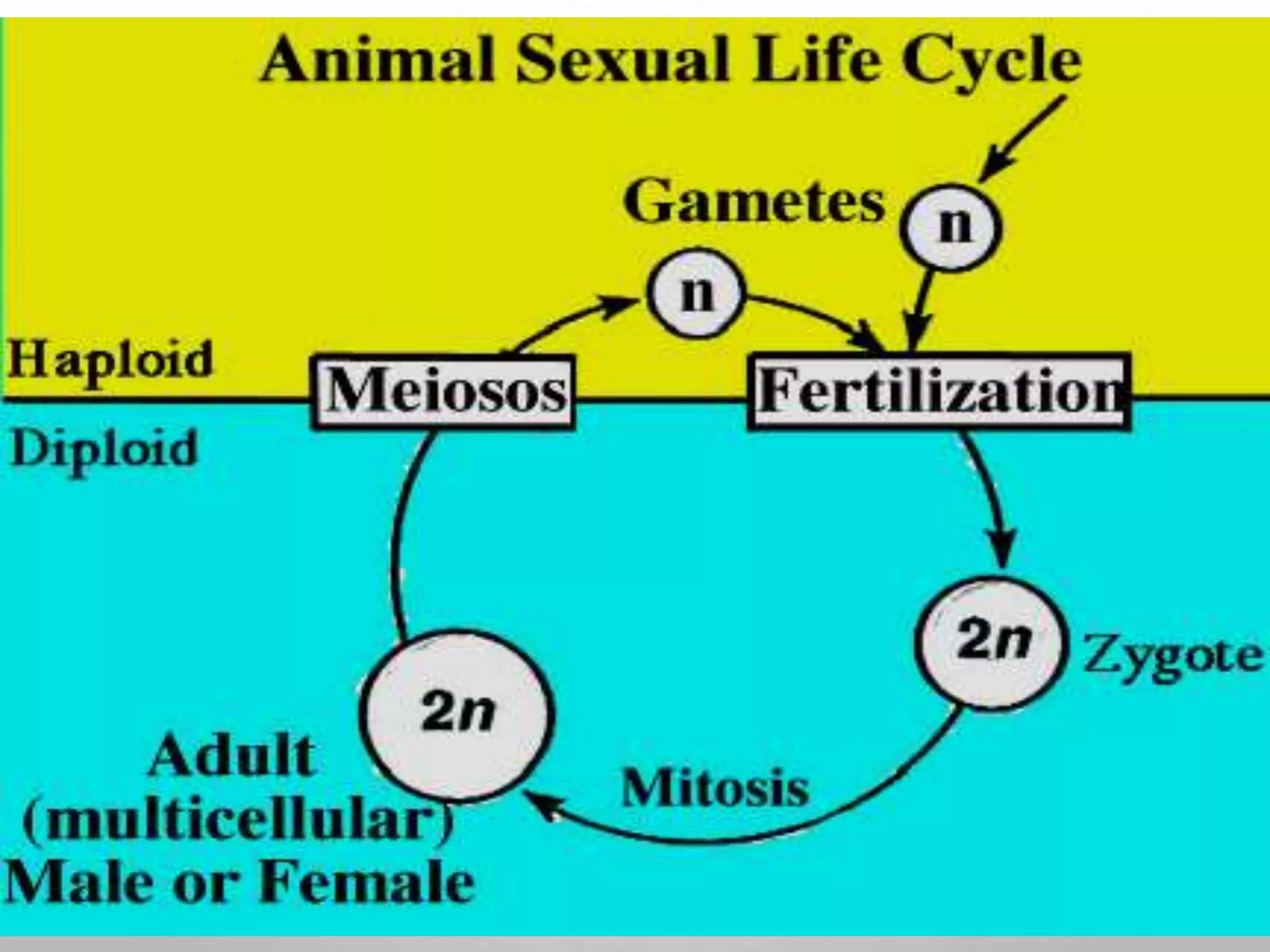
1. Fertilization involves the fusion of a sperm and egg nuclei to form a zygote.
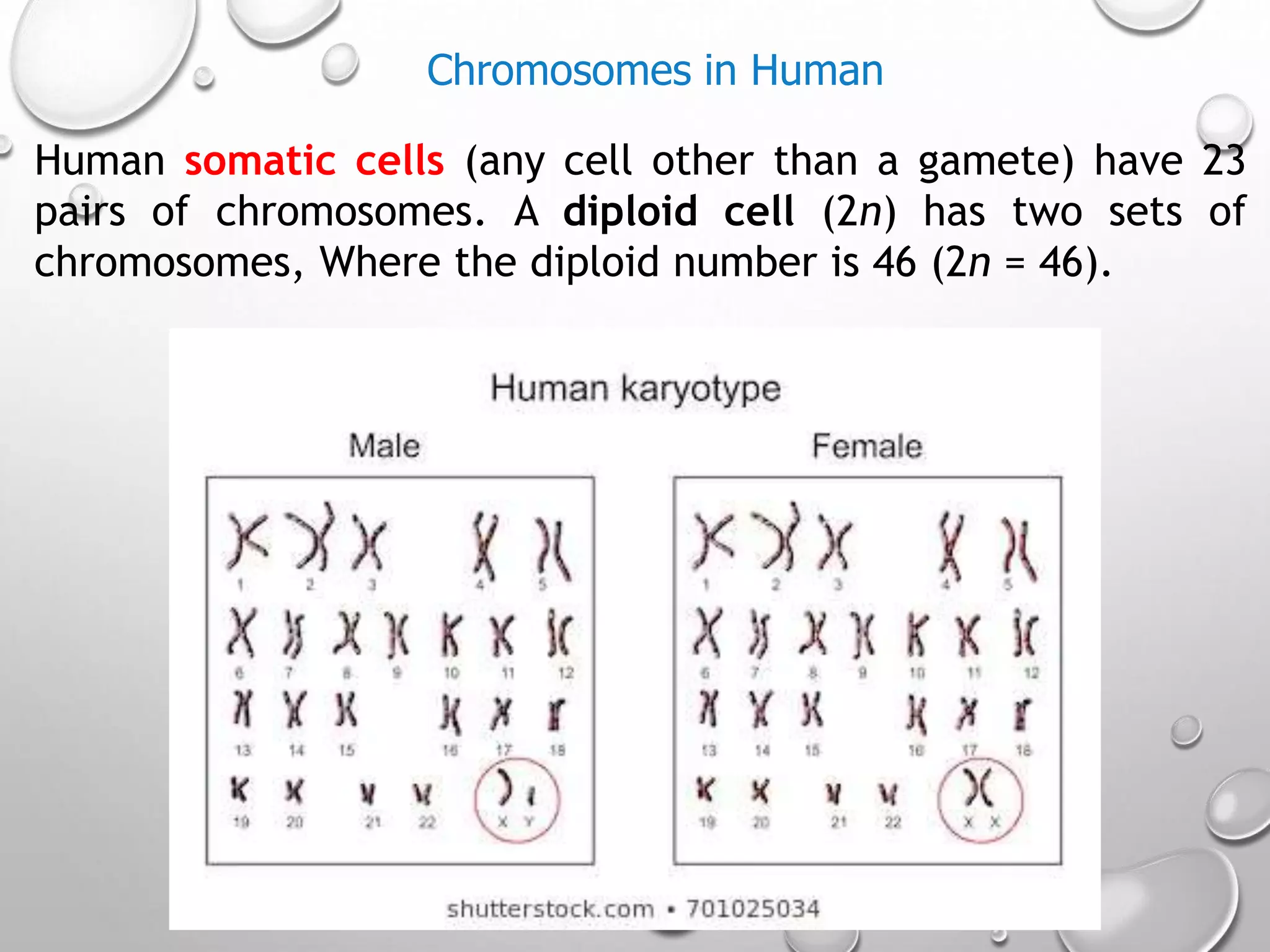
2. It restores the diploid number of chromosomes and determines the sex of the embryo.
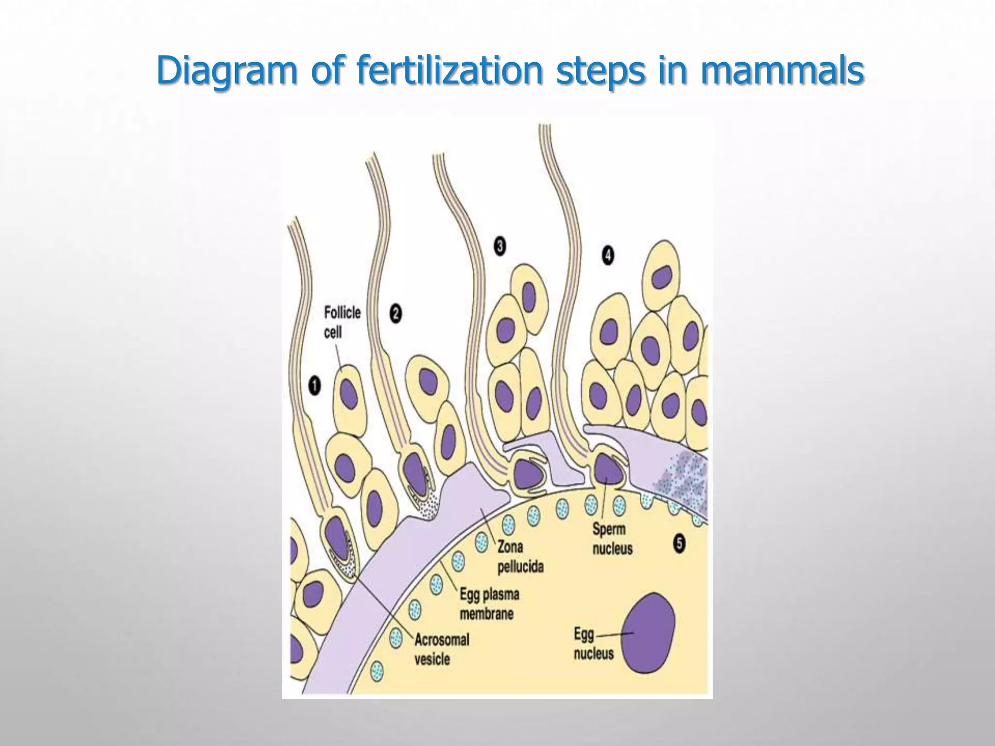
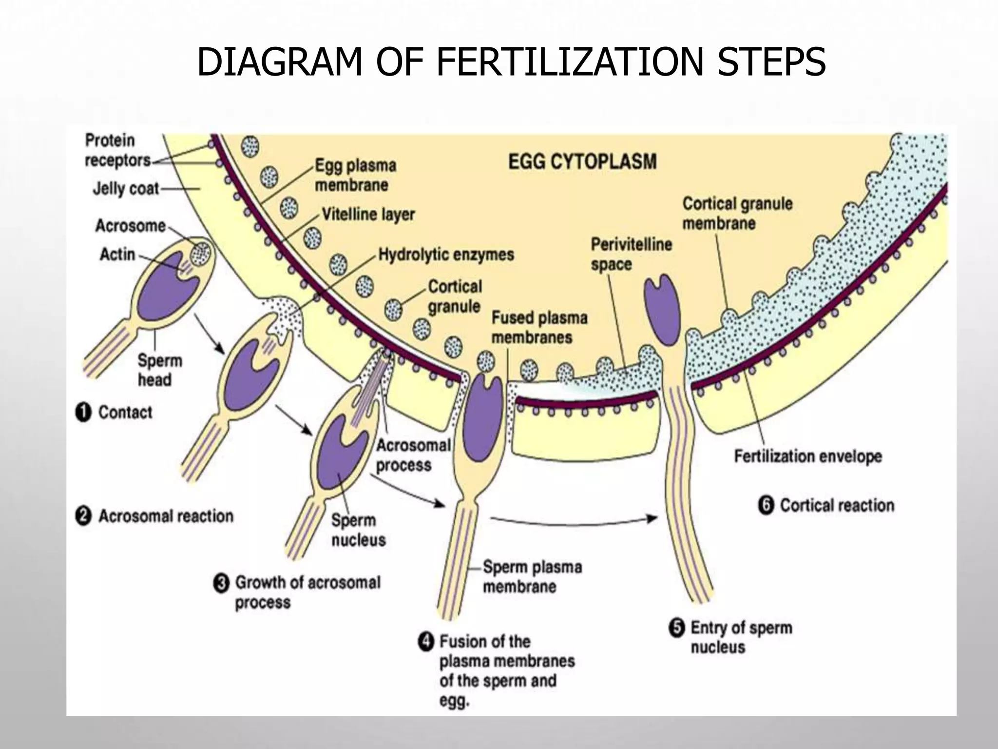
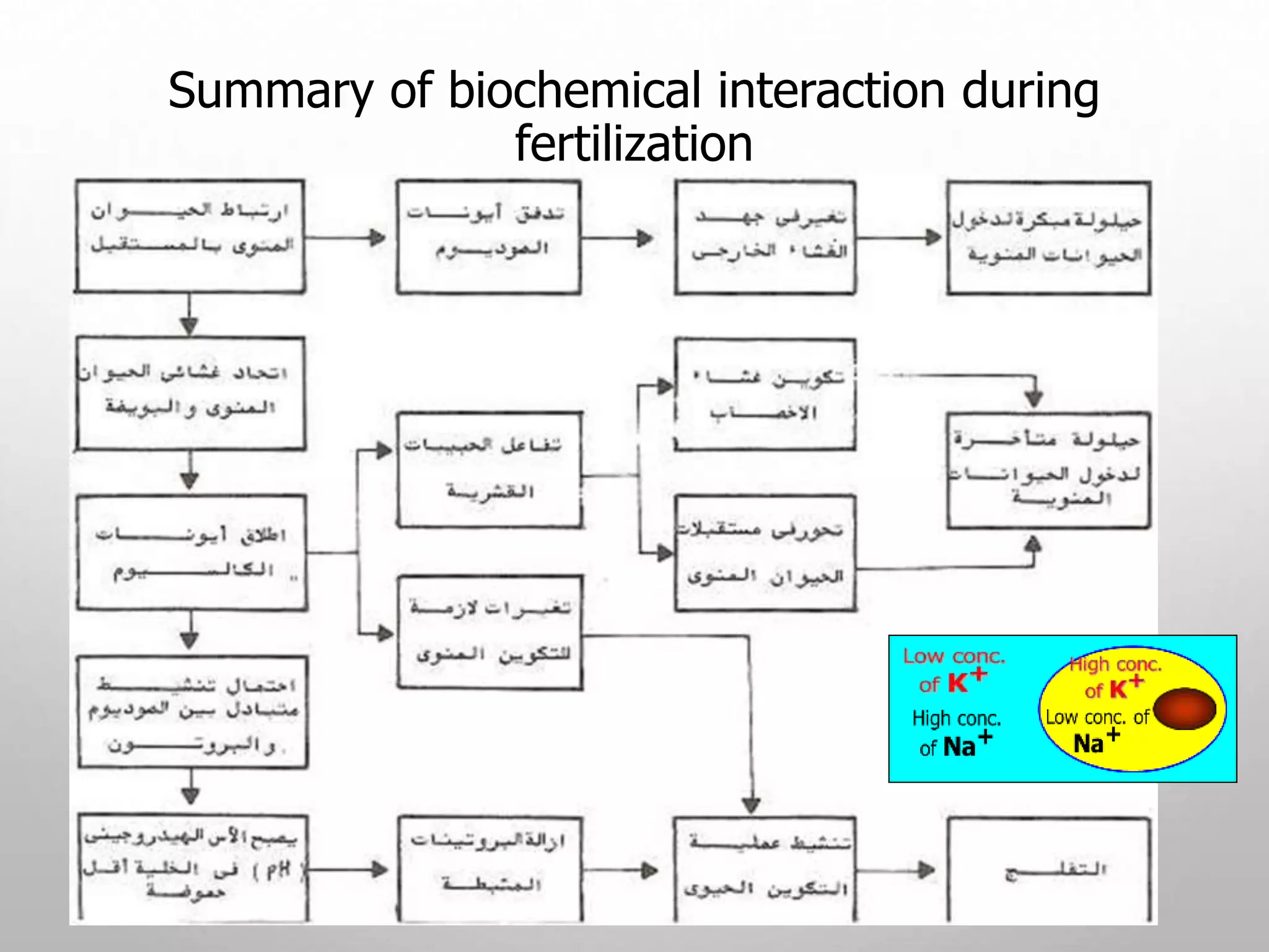
3. A series of events must occur for fertilization to be successful, including capacitation of sperm, penetration of the zona pellucida, fusion of membranes, and formation of male and female pronuclei.
Implantation and placentation , and overviewPranjal Gupta
Implantation and formation of placenta is an essential developmental process during human embryogenesis as it marks the connection between maternal and fetal blood, a condition specific to mammals more precisely eutherians. It works as a passage of required nutrients to the growing embryo and collection of its waste. It also discusses various types of placenta that are seen in mammals.
A brief account of different parts of sperm and its constitutions and,ovum parts and different envelops.all things are explained by a simple attractive diagram.
Polyspermy describes an egg that has been fertilized by more than one sperm. Diploid organisms normally contain two copies of each chromosome, one from each parent. The cell resulting from polyspermy
The first issue that an egg and a sperm of any organism type face in successfully producing an embryo is the possibility of polyspermy. Polyspermy is the fertilization of an egg by multiple sperm, and the results of such unions are lethal.
If multiple sperm fertilize an egg, the embryo inherits multiple paternal centrioles. This causes competition for extra chromosomes and results in the disruption of the creation of the cleavage furrow, thus causing the zygote to die. As an important model organism in the study of fertilization and embryonic development, polyspermy in sea urchins has been studied in detail. The sea urchin’s methods of polyspermy prevention have been broken down into two main pathways. These two primary pathways are known as the fast block and the slow block to polyspermy
After the sperm’s receptors come into contact with the egg’s jelly layer and the acrosomal enzymes are released and break down the jelly layer, the sperm head comes into contact with the vitelline and plasma membranes of the egg. When the two plasma membranes contact one another, signals in the egg are initiated.
First, Na+ channels in the egg open, allowing Na+ to flood into the egg. This causes a depolarization of the egg from it’s normal resting potential of -70 mV.
While depolarization is occurring, the remainder of the jelly layer is dissolving. With the dissolution of the jelly layer and the depolarization of the plasma membrane, the first block to preventing fertilization by multiple sperm is put into place.
These two simple changes are part of the first block to polyspermy, known as the fast block. Within 1/10th of a second of contact, the fast block t
How 3 germ layers are formed in Chick that are endoderm, mesoderm and ectoderm.As Chick are polylecithal so cell movements are somewhat restricted and gastrulation is modified as compared to frog.
The term implantation is used to describe the attachment of the developing embryo to the endometrium.
After fertilization, the embryo reaches the uterus in the blastocyst stage. Then attached to the wall of the uterus. Though the implantation may occur at any period between the sixth to the tenth day after the fertilization generally it occurs on the seventh day after fertilization.
INTRODUCTION
DEFINATION
GAMETES
STRUCTURE OF GAMETES
SPERM
OVUM
RECOGNITION OF EGG AND SPERM
CAPACITATION
ACROSOME REACTION
SPECIES-SPECIFIC RECOGNITION
GAMETE BINDING AND RECOGNITION
GAMETE FUSION
PREVENTION OF POLYSPERMY
ACTIVATION OF GAMETE METABOLISM
FUSION OF THE GENETIC MATERIAL
SIGNIFICANCE OF FERTILIZATION
CONCLUSIONS
REFERENCES
USMLE GENERAL EMBRYOLOGY 008 First week of development A embryo .pdfAHMED ASHOUR
The initial week of embryonic development is a vital period commencing with fertilization, leading to the creation of the zygote and early cell divisions. It's noteworthy that, throughout this week, the developing embryo remains in the pre-implantation stage, journeying from the fallopian tube toward the uterus. Key events such as fertilization, cleavage, and the formation of the blastocyst are crucial for the embryo's early development.
These events lay the foundation for subsequent processes in the following weeks. The successful implantation of the blastocyst into the uterus marks the transition from the first week to the second week of embryonic development.
Here provided contents for learning that what are the male and female gametes. How to they form? And described how to both gametes fused (fertilization)on the molecular basis.
Multi-source connectivity as the driver of solar wind variability in the heli...Sérgio Sacani
The ambient solar wind that flls the heliosphere originates from multiple
sources in the solar corona and is highly structured. It is often described
as high-speed, relatively homogeneous, plasma streams from coronal
holes and slow-speed, highly variable, streams whose source regions are
under debate. A key goal of ESA/NASA’s Solar Orbiter mission is to identify
solar wind sources and understand what drives the complexity seen in the
heliosphere. By combining magnetic feld modelling and spectroscopic
techniques with high-resolution observations and measurements, we show
that the solar wind variability detected in situ by Solar Orbiter in March
2022 is driven by spatio-temporal changes in the magnetic connectivity to
multiple sources in the solar atmosphere. The magnetic feld footpoints
connected to the spacecraft moved from the boundaries of a coronal hole
to one active region (12961) and then across to another region (12957). This
is refected in the in situ measurements, which show the transition from fast
to highly Alfvénic then to slow solar wind that is disrupted by the arrival of
a coronal mass ejection. Our results describe solar wind variability at 0.5 au
but are applicable to near-Earth observatories.
Seminar of U.V. Spectroscopy by SAMIR PANDASAMIR PANDA
Spectroscopy is a branch of science dealing the study of interaction of electromagnetic radiation with matter.
Ultraviolet-visible spectroscopy refers to absorption spectroscopy or reflect spectroscopy in the UV-VIS spectral region.
Ultraviolet-visible spectroscopy is an analytical method that can measure the amount of light received by the analyte.
Introduction:
RNA interference (RNAi) or Post-Transcriptional Gene Silencing (PTGS) is an important biological process for modulating eukaryotic gene expression.
It is highly conserved process of posttranscriptional gene silencing by which double stranded RNA (dsRNA) causes sequence-specific degradation of mRNA sequences.
dsRNA-induced gene silencing (RNAi) is reported in a wide range of eukaryotes ranging from worms, insects, mammals and plants.
This process mediates resistance to both endogenous parasitic and exogenous pathogenic nucleic acids, and regulates the expression of protein-coding genes.
What are small ncRNAs?
micro RNA (miRNA)
short interfering RNA (siRNA)
Properties of small non-coding RNA:
Involved in silencing mRNA transcripts.
Called “small” because they are usually only about 21-24 nucleotides long.
Synthesized by first cutting up longer precursor sequences (like the 61nt one that Lee discovered).
Silence an mRNA by base pairing with some sequence on the mRNA.
Discovery of siRNA?
The first small RNA:
In 1993 Rosalind Lee (Victor Ambros lab) was studying a non- coding gene in C. elegans, lin-4, that was involved in silencing of another gene, lin-14, at the appropriate time in the
development of the worm C. elegans.
Two small transcripts of lin-4 (22nt and 61nt) were found to be complementary to a sequence in the 3' UTR of lin-14.
Because lin-4 encoded no protein, she deduced that it must be these transcripts that are causing the silencing by RNA-RNA interactions.
Types of RNAi ( non coding RNA)
MiRNA
Length (23-25 nt)
Trans acting
Binds with target MRNA in mismatch
Translation inhibition
Si RNA
Length 21 nt.
Cis acting
Bind with target Mrna in perfect complementary sequence
Piwi-RNA
Length ; 25 to 36 nt.
Expressed in Germ Cells
Regulates trnasposomes activity
MECHANISM OF RNAI:
First the double-stranded RNA teams up with a protein complex named Dicer, which cuts the long RNA into short pieces.
Then another protein complex called RISC (RNA-induced silencing complex) discards one of the two RNA strands.
The RISC-docked, single-stranded RNA then pairs with the homologous mRNA and destroys it.
THE RISC COMPLEX:
RISC is large(>500kD) RNA multi- protein Binding complex which triggers MRNA degradation in response to MRNA
Unwinding of double stranded Si RNA by ATP independent Helicase
Active component of RISC is Ago proteins( ENDONUCLEASE) which cleave target MRNA.
DICER: endonuclease (RNase Family III)
Argonaute: Central Component of the RNA-Induced Silencing Complex (RISC)
One strand of the dsRNA produced by Dicer is retained in the RISC complex in association with Argonaute
ARGONAUTE PROTEIN :
1.PAZ(PIWI/Argonaute/ Zwille)- Recognition of target MRNA
2.PIWI (p-element induced wimpy Testis)- breaks Phosphodiester bond of mRNA.)RNAse H activity.
MiRNA:
The Double-stranded RNAs are naturally produced in eukaryotic cells during development, and they have a key role in regulating gene expression .
This pdf is about the Schizophrenia.
For more details visit on YouTube; @SELF-EXPLANATORY;
https://www.youtube.com/channel/UCAiarMZDNhe1A3Rnpr_WkzA/videos
Thanks...!
Richard's aventures in two entangled wonderlandsRichard Gill
Since the loophole-free Bell experiments of 2020 and the Nobel prizes in physics of 2022, critics of Bell's work have retreated to the fortress of super-determinism. Now, super-determinism is a derogatory word - it just means "determinism". Palmer, Hance and Hossenfelder argue that quantum mechanics and determinism are not incompatible, using a sophisticated mathematical construction based on a subtle thinning of allowed states and measurements in quantum mechanics, such that what is left appears to make Bell's argument fail, without altering the empirical predictions of quantum mechanics. I think however that it is a smoke screen, and the slogan "lost in math" comes to my mind. I will discuss some other recent disproofs of Bell's theorem using the language of causality based on causal graphs. Causal thinking is also central to law and justice. I will mention surprising connections to my work on serial killer nurse cases, in particular the Dutch case of Lucia de Berk and the current UK case of Lucy Letby.
2. Fertilization
Fertilization is part of mating or copulation between male
and female.
Fertilization is the fusion of the sperm nucleus with the ova
nucleus to produce a zygote (fertilized egg).
Fertilization brings the haploid nuclei of sperm and egg
together, forming a diploid zygote.
The sperm’s contact with the egg’s surface initiates
metabolic reactions in the egg that trigger the onset of
embryonic development.
3.
4. Human somatic cells (any cell other than a gamete) have 23
pairs of chromosomes. A diploid cell (2n) has two sets of
chromosomes, Where the diploid number is 46 (2n = 46).
Chromosomes in Human
5. •The sex chromosomes in mammals are called X and Y.
•Human females have a homologous pair of X chromosomes
(XX)
•Human males have one X and one Y chromosome.
•A gamete (sperm or egg) contains a single set of
chromosomes, and is haploid (n).
•For humans, the haploid number is 23 (n = 23)
•Each set of 23 consists of 22 autosomes and a single sex
chromosome
•In an unfertilized egg (ovum), the sex chromosome is X
•In a sperm cell, the sex chromosome may be either X or Y
Sex Determination
6. Fertilization in animal kingdom
There are two types of fertilization (external and
internal).
External fertilization
Occurs mostly in wet
environments .
It’s occur outside the body of
female ,where requires both
the male and the female to
release their gametes into
their surroundings (usually
water)
It’s produce a large number
of offspring.
Ex) fish and amphibians
External fertilization in amphibians
and fish
7. Internal fertilization
It’s occur in most of terrestrial
animals .
It’s occur inside the body of
female, to
protect the developing egg.
It’s produce less number of
offspring.
Ex) mammals, reptiles, birds
Fertilization in animal kingdom
8. Internal fertilization in mammals
• Fertilization in mammals occurs in the oviduct.
• The ova is viable for approximately 24 hours after ovulation.
9. What are the factors that affect the sperm’s potential
for successfully fertilizing the egg?
1- Capacitation of sperm.
2- Structures surrounding the egg – barriers.
10. 1- Capacitation of sperm
• It is the process that allows the acrosome reaction to occur,
so the sperm can penetrate the zona pellucida of the oocyte.
• The physiological changes that confer on the sperm the
ability to fertilize are called capacitation.
• Capacitation includes multiple physiological and biochemical
modifications.
• occurs in the female’s vagina. Vaginal secretions cause a
molecular change in the sperm plasma lemma (removal of
decapacitating factor - semen proteins, results in increased
membrane fluidity,).
• Takes 4-5 hr. in humans, 1 hr. in mice, 6 hr. in rabbits.
11. 1- Capacitation of sperm
• The biochemical changes associated with the capacitation
process include:
1. an efflux of cholesterol from the plasma membrane leading
to an increase in membrane fluidity and permeability to
bicarbonate and calcium ions.
2. hyperpolarization of the plasma membrane, changes in
protein phosphorylation and protein kinase activity
3. increases in bicarbonate (HCO3) concentration and
intracellular pH, Calcium and cyclic adenosine
monophosphate (cAMP) levels.
4. Galactosyltransferase enzyme is a protein in the sperm
plasma lemma that binds to the zona pellucida 3 protein
(ZP3) receptor around the ova and initiates the acrosome
reaction.
13. Increased rate of metabolism.
Flagellum beats more rapidly; Result: Sperm are more
motile.
Changes in sperm plasma lemma proteins allow sperm-
egg binding and occurrence of the acrosome reaction.
Pro-Acrosin (inactive) is converted to acrosin (active).
Sperm become capable of chemotaxis.
Effects of Capacitation on Sperm
14. 2- Structures surrounding the egg - barriers
1. Follicle cells.
2. Zona pellucida (ZP)in
mammals.
3. (vitelline membrane in non-
mammals).
4. Oolemma (plasma membrane
of ova).
15. Ovum is a larger nutritive cell.
Like an animal cell it also contains
all cell organelles.
Its size varies in different animals.
Ovum is generally round/spherical
in shape.
It is covered by a thin membrane
known as plasma membrane.
Beside this, it is also covered by
other covering membranes known
as egg membranes.
2- Structure of Ovum (egg)
16. These are divided into three-
1. Primary egg membrane- Secreted by ovum itself.( viteline .m)
2. Secondary egg membrane- Secreted by ovarian tissue like
follicular membrane.
3. Tertiary egg membrane- Secreted or generated by various
regions of uterus or oviduct.
Importance of the egg membranes:-
These membranes provide nutrition and protection for developing
embryo.
Egg membranes
17. Fertilization Stage
1. The acrosome reaction.
2. The Penetration of the zona pellucida.
3. Fusion of plasma membranes of oocyte and sperm.
4. 2nd meiotic division of oocyte is completed
5. Formation of male and female pronuclei.
6. Membranes of the pronuclei break down, chromosomes
condense and arrange themselves for mitotic cell division.
18. Fertilization
1- Acrosome reaction
• The acrosome reaction must be completed before the sperm
can fuse with the secondary oocyte.
• Occurs when sperms come into contact with the corona
radiata of the oocyte
• Perforations develop in the acrosome
• Point fusions of the sperm plasma membrane and the
external acrosomal membrane occur
• The acrosome reaction is associated with the release of
acrosome enzymes that facilitate fertilization
• Passage of sperm through the corona radiata depends on
enzyme action:
• Hyaluronidase released from sperm acrosome
• Tubal mucosal enzymes
• Flagella action also aids corona radiata penetration
20. Ovum and Sperms: (in vitro)
Advanced Fertility Center of Chicago
http://www.advancedfertility.com/
From this photograph, it should be
clear that the heads of human
sperm are less than 1/20 the
diameter of human eggs.
Arrows point to sperm heads
21. Fertilization
2- penetration of the zona pellucida
• Penetration of the zona pellucida around the oocyte:
• Acrosomal enzymes: esterases, acrosin, and neuraminidase
cause lysis of the zona pellucida.
• Once sperm penetrates zona pellucida, the zona reaction
occurs: fertilization membrane will form.
• This reaction makes the zona pellucida impermeable to
other sperms. Block to poly spermy.
• When more than one sperm manages to enter the ovum
(dispermy = 2; triploidy = 3 the called poly spermy), the
fetus nearly always aborts.
23. Fertilization
3- fusion of plasma membranes of oocyte and
sperm
• Fusion of plasma membranes of oocyte and sperm.
• Head of a sperm enter the cytoplasm of the oocyte, but the
sperm plasma membrane remains behind.
24. Ova cortical granules reaction
• Once the sperm cell membrane fused the ova membrane,
the ova cortical granules fused with ova membrane and
released its content to the out side of the ova in the
perivitelline space.
• The released materials from the cortical granule will form a
hyaline layer between the ova membrane and the vitelline
membrane.
• This layer will prevent the entry of any other sperm to the
ova and considered as the second bloke to polyspermy.
26. Fertilization
4- second meiotic division of oocyte is completed
• 2nd meiotic division of oocyte is completed.
• The secondary oocyte was previously arrested in
metaphase of the 2nd meiotic division, and now forms
the mature ovum and another polar body.
27. Fertilization
5- formation of male and female pronuclei
• Formation of male and female pronuclei:
• Chromosomal material of the sperm decondensates
and enlarges
• Chromosomal material of the ovum decondensates
following the completion of meiosis
• At this stage, the male and female pronuclei are
indistinguishable.
• As they grow, the pronuclei replicate their dna still 1n
(haploid)-( 23 chromosomes, each in chromatid pairs in
human).
28. • The male and female pronuclei
are indistinguishable from one
another.
• The second polar body can be
seen (blue arrow).
• The plasma membranes of the
two pronuclei are dissolving
and one diploid nucleus will
remain.
Fusion of the pronuclei
(in vitro)
Advanced Fertility Center of Chicago
http://www.advancedfertility.com/
29. Fertilization
6- membranes of the pronuclei break down
• Membranes of the pronuclei break down, chromosomes
condense and arrange themselves for mitotic cell division
• On membrane dissolution, there is 1 cell with 46
chromosomes = diploid (2N)
• The first cleavage follows shortly, leaving 2 cells, each
with 46 chromosomes in human.
• Mitosis in the new zygote uses centrioles derived
from the sperm.
• The oocyte has no centrioles.
30.
31. Fertilization facts
• Completed within 24 hours of ovulation in human
• Approximately 400 to 600 MILLION sperms are deposited at
cervical opening during ejaculation.
• Some sperm are held up by the folds of the cervix and
are gradually released into the cervical canal; this
gradual release increases the chances of fertilization.
• Most human sperms do not survive longer than 48 hours
in the female genital tract.
• Only about 200 sperms reach the fertilization site; most
degenerate and are absorbed by the female genital tract.
32. The results of fertilization
• Stimulates the secondary oocyte to complete meiosis.
• Restores the normal diploid number of chromosomes (46 in
human).
• Results in variation of human species as maternal and
paternal chromosomes intermingle.
• The embryo contains only maternal mitochondria because the
sperm mitochondria are dispersed into the egg cytoplasm and
discarded.
• Determines the sex of the embryo.
• The sex chromosome (y or x) carried by the successful sperm
determines embryonic sex.
34. After fertilization, embryonic development proceeds
through cleavage, gastrulation, and organogenesis
• Important events regulating development occur during
fertilization and the three stages that build the animal’s
body
• Cleavage: cell division creates a hollow ball of cells
called a blastula
• Gastrulation: cells are rearranged into a three-layered
gastrula
• Organogenesis: the three layers interact and move to
give rise to organs