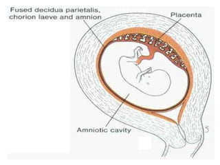
Formation and Structure of
Placenta
Presenter:
Dr. Anju B. Uppin
Assistant Professor
Department of Rachana Sharira
KAHER’s Shri BMK
Ayurveda Mahavidyalaya
Belagavi, Karnataka
Definition of Placenta
The placenta is the organ that facilitates
nutrient and exchange of gases between the
maternal and foetal compartments.
Implantation :
It is a process by which the embryo is embedded and fixed with
the endometrium of the uterus.
Certain Changes takes place:
 At the time of implantation, the embryo is in the form of
blastocyst. It usually occurs during 6–10 days after ovulation.
 The intimate contact of fetal and maternal tissues occurs by
implantation of the embryo.
 The blastocyst surrounded by zona pellucida enters the uterus
on the sixth day. The zona pellucida pre-vents it from sticking
to the wall of the uterus.
 As the blastocyst enlarges, the zona pellucida cover-ing it
becomes stretched and ultimately disappears. Consequently the
trophoblast is exposed.
 The trophoblast has the property of attaching itself to
any tissue with which it comes in contact with.
 The trophoblast sticks to the uterine endometrium.
The cells of trophoblast divide mitotically and form
new cells that lose their cell membranes and form a
mass of cells called syncytiotrophoblast.
 The syncy-tiotrophoblast invades the endometrium
with the help of proteolytic enzymes secreted by its
cells.
 The blastocyst goes deeper and deeper until it com-
pletely lies within the endometrium (interstitial
implantation).
Normal Site of Implantation
Normally the blastocyst is implanted in the upper
part of the posterior wall of the uterine cavity.
8-12 DAYS
6TH DAY
Decidua basalis
Amnion
Chorion
Decidua parietalis
Extra embryonic coelom
(Chorionic cavity)
Uterine cavity
Myometrium
Decidua capsularis
Amniotic cavity INTERSTITIAL
IMPLANTATION
11th day
Decidua
 After implantation of the blastocyst, the functional layer
of the endometrium of the uterus is termed Decidua.
 The endometrium of the uterus is in the secretory phase
of menstruation at the time of implantation.
 As the embryo is implanted the syncytiotrophoblast
starts secreting human chorionic gonadotrophin (HCG)
hormone, which intensifies the changes that occur in the
secretary phase, viz., stromal cells enlarge, vacuo-late,
and get filled to the brim with glycogen and lipids.
 This change in the stromal cells is called Decidual
Reaction and changed character of the endometrium
after implantation of the ovum is now called Decidua.
Parts of Decidua
1. Decidua Basalis: It is that part of decidua which lies
deep to the embryo (developing blastocyst).This
contributes to the development of the placenta.
2. Decidua Capsularis: It is that part of decidua which
forms a capsule around the embryo and separates it
from the uterine cavity.
3. Decidua Parietalis: Rest of the decidua (excluding
decidua basalis and decidua capsularis) is termed
decidua parietalis.
Formation of Placenta
The placenta develops from two entirely different
sources.
 The fetal portion develops from chorion frondosum
and the maternal portion from the decidua basalis.
 By the end of third week, the anatomical
arrangements necessary for physiological exchange
between the mother and embryo are established.
 By the end of fourth week, a complex vascular
network is formed within the placenta, which
facilitates the exchange of gases, nutrients, and
metabolic waste products between the mother and
embryo.
1. Formation of chorion frondosum and chorion laeve
(a) During the development of placenta small finger-like
projections arise from chorion (tro-phoblast and underlying
mesoderm) into the decidua.
(b) Initially chorionic villi are formed all around the chorionic sac.
(c) As the chorionic sac enlarges the chorionic villi in relation to
decidua capsularis get com-pressed and degenerate. As a result,
this part of chorion becomes smooth and is now called chorion
laeve.
(d) The chorionic villi in relation to decidua basa-lis grow
extensively into the decidua basalis. This part of the chorion is
called chorion frondosum
2. Formation of trabeculae and intervillous spaces
(a) The cells of trophoblast (made up of single layer of
cells) proliferate, move on the surface, and losetheir
cell membranes to form a continuous sheetof
cytoplasm containing many nuclei. This sheet/layer
is called syncytiotrophoblast. Thus, trophoblast is
differentiated into two layers:
 A deep layer called cytotrophoblast and
 Asuperficial layer called syncytiotrophoblast.
(b) The syncytiotrophoblast grows rapidly and erodes
the decidua basalis, and becomes thick. The small
cavities (lacunae) appear in thickened
syncytiotrophoblast.
The syncytio-trophoblast continues to erode the decidua
basalis. The lacunae increase in size. They lie radially
around the developing embryo (blas-tocyst). They are
separated from each other by portions of
syncytiotrophoblast termed trabeculae (villous
primordia).
(c) As the syncytiotrophoblast further grows it erodes the
blood vessels of the endometrium and the blood enters
into the lacunae along with the secretions of the
endometrial glands. Later the lacunae communicate
with each other around the trabeculae and form the
intervillous spaces. Thus, each trabeculus gets
surrounded all around by the blood-filled space in the
endometrium.
Development of Villi
1. Primary villi: The cytotrophoblast forms finger-like projections
that invade the trabeculae in its center. This finger-like
projection of cytotropho-blast surrounded by a layer of
syncytiotrophoblast is called the primary villus.
2. Secondary villi: The extraembryonic somatopleu-ric mesoderm
lying deep to the cytotrophoblast now invades the center of each
villus.As a result, now each villus consists of three layers. From
inside to outside these are mesoderm, cytotrophoblast, and
syncytiotrophoblast. This villus is now termed secondary villus.
3. Tertiary villi: The blood vessels develop in the mesoderm of the
secondary villi. The secondary villus with blood vessels in its
mesoderm is called tertiary villus.
Decidua basalis
Trophoblast
Extraembryonic mesoderm
STAGES IN FORMATION OF CHORIONIC VILLI
Cytotrophoblast
Syncytiotrophoblast
Decidua basalis
Lacunae in
Syncytiotrophoblast
8th Day
9th Day
Trabeculae
Decidua basalis
Lacunar space
Lacunar space
Y
X
Transverse section along XY
Trabeculae Maternal blood
in Lacunar space
Uterine blood vessel
Transverse section along XY
X Y
Decidua basalis
Syncytiotrophoblast
Maternal blood in Lacunar space
Cytotrophoblast
X Y
Cytotrophoblast
Syncytiotrophoblast
PRIMA RY VILLUS
Maternal blood in Lacunar space
Transverse section along XY
13th Day
Decidua basalis
SECONDARY VILLUS
Syncytiotrophoblast
Cytotrophoblast
Extraembryonic mesoderm
Maternal blood in Lacunar space
X Y
Cytotrophoblast
Syncytiotrophoblast
Extraembryonic mesoderm
Transverse section along XY
16th Day
Decidua basalis
TERTIARY VILLUS
Maternal blood in Lacunar space
Capillary in extraembryonic mesoderm
Cytotrophoblast
Syncytiotrophoblast
Cytotrophoblast
Intervillous space
Syncytiotrophoblast
Capillary in extraembryonic mesoderm
X Y
Transverse section along XY
16th Day
Uterine blood vessel
Cytotrophoblastic shell
Syncytiotrophoblast
Maternal blood in Lacunar space
Capillary in villus
Fetal blood vessels in
extraembryonic mesoderm
Decidua basalis
Chorionic villi
Decidua capsularis
Uterine lumen
2nd month
Chorion frondosum
Chorion laevae
(Decidual plate)
2nd MONTH
Definitive Placenta
4th month
3rd MONTH
PLACENTA
MATERNALASPECT
PLACENTA FETAL
ASPECT
Extraembryonic
mesoderm
PLACENTAL MEMBRANE
ANOMALIES IN FORM OF PLACENTA
Bidiscoidal Placenta Lobed placenta
Diffuse placenta Placenta succenturiata
Fenestrated Placenta Circumvallate placenta
Thank you

PLACENTA FORMATION,STRUCTURE, APPLIED IMPORTANCE.ppt

  • 1.
    Formation and Structureof Placenta Presenter: Dr. Anju B. Uppin Assistant Professor Department of Rachana Sharira KAHER’s Shri BMK Ayurveda Mahavidyalaya Belagavi, Karnataka
  • 2.
    Definition of Placenta Theplacenta is the organ that facilitates nutrient and exchange of gases between the maternal and foetal compartments.
  • 3.
    Implantation : It isa process by which the embryo is embedded and fixed with the endometrium of the uterus. Certain Changes takes place:  At the time of implantation, the embryo is in the form of blastocyst. It usually occurs during 6–10 days after ovulation.  The intimate contact of fetal and maternal tissues occurs by implantation of the embryo.  The blastocyst surrounded by zona pellucida enters the uterus on the sixth day. The zona pellucida pre-vents it from sticking to the wall of the uterus.  As the blastocyst enlarges, the zona pellucida cover-ing it becomes stretched and ultimately disappears. Consequently the trophoblast is exposed.
  • 4.
     The trophoblasthas the property of attaching itself to any tissue with which it comes in contact with.  The trophoblast sticks to the uterine endometrium. The cells of trophoblast divide mitotically and form new cells that lose their cell membranes and form a mass of cells called syncytiotrophoblast.  The syncy-tiotrophoblast invades the endometrium with the help of proteolytic enzymes secreted by its cells.  The blastocyst goes deeper and deeper until it com- pletely lies within the endometrium (interstitial implantation).
  • 5.
    Normal Site ofImplantation Normally the blastocyst is implanted in the upper part of the posterior wall of the uterine cavity.
  • 6.
  • 7.
    Decidua basalis Amnion Chorion Decidua parietalis Extraembryonic coelom (Chorionic cavity) Uterine cavity Myometrium Decidua capsularis Amniotic cavity INTERSTITIAL IMPLANTATION 11th day
  • 8.
    Decidua  After implantationof the blastocyst, the functional layer of the endometrium of the uterus is termed Decidua.  The endometrium of the uterus is in the secretory phase of menstruation at the time of implantation.  As the embryo is implanted the syncytiotrophoblast starts secreting human chorionic gonadotrophin (HCG) hormone, which intensifies the changes that occur in the secretary phase, viz., stromal cells enlarge, vacuo-late, and get filled to the brim with glycogen and lipids.  This change in the stromal cells is called Decidual Reaction and changed character of the endometrium after implantation of the ovum is now called Decidua.
  • 9.
    Parts of Decidua 1.Decidua Basalis: It is that part of decidua which lies deep to the embryo (developing blastocyst).This contributes to the development of the placenta. 2. Decidua Capsularis: It is that part of decidua which forms a capsule around the embryo and separates it from the uterine cavity. 3. Decidua Parietalis: Rest of the decidua (excluding decidua basalis and decidua capsularis) is termed decidua parietalis.
  • 10.
  • 11.
    The placenta developsfrom two entirely different sources.  The fetal portion develops from chorion frondosum and the maternal portion from the decidua basalis.  By the end of third week, the anatomical arrangements necessary for physiological exchange between the mother and embryo are established.  By the end of fourth week, a complex vascular network is formed within the placenta, which facilitates the exchange of gases, nutrients, and metabolic waste products between the mother and embryo.
  • 17.
    1. Formation ofchorion frondosum and chorion laeve (a) During the development of placenta small finger-like projections arise from chorion (tro-phoblast and underlying mesoderm) into the decidua. (b) Initially chorionic villi are formed all around the chorionic sac. (c) As the chorionic sac enlarges the chorionic villi in relation to decidua capsularis get com-pressed and degenerate. As a result, this part of chorion becomes smooth and is now called chorion laeve. (d) The chorionic villi in relation to decidua basa-lis grow extensively into the decidua basalis. This part of the chorion is called chorion frondosum
  • 18.
    2. Formation oftrabeculae and intervillous spaces (a) The cells of trophoblast (made up of single layer of cells) proliferate, move on the surface, and losetheir cell membranes to form a continuous sheetof cytoplasm containing many nuclei. This sheet/layer is called syncytiotrophoblast. Thus, trophoblast is differentiated into two layers:  A deep layer called cytotrophoblast and  Asuperficial layer called syncytiotrophoblast. (b) The syncytiotrophoblast grows rapidly and erodes the decidua basalis, and becomes thick. The small cavities (lacunae) appear in thickened syncytiotrophoblast.
  • 19.
    The syncytio-trophoblast continuesto erode the decidua basalis. The lacunae increase in size. They lie radially around the developing embryo (blas-tocyst). They are separated from each other by portions of syncytiotrophoblast termed trabeculae (villous primordia). (c) As the syncytiotrophoblast further grows it erodes the blood vessels of the endometrium and the blood enters into the lacunae along with the secretions of the endometrial glands. Later the lacunae communicate with each other around the trabeculae and form the intervillous spaces. Thus, each trabeculus gets surrounded all around by the blood-filled space in the endometrium.
  • 23.
    Development of Villi 1.Primary villi: The cytotrophoblast forms finger-like projections that invade the trabeculae in its center. This finger-like projection of cytotropho-blast surrounded by a layer of syncytiotrophoblast is called the primary villus. 2. Secondary villi: The extraembryonic somatopleu-ric mesoderm lying deep to the cytotrophoblast now invades the center of each villus.As a result, now each villus consists of three layers. From inside to outside these are mesoderm, cytotrophoblast, and syncytiotrophoblast. This villus is now termed secondary villus. 3. Tertiary villi: The blood vessels develop in the mesoderm of the secondary villi. The secondary villus with blood vessels in its mesoderm is called tertiary villus.
  • 24.
    Decidua basalis Trophoblast Extraembryonic mesoderm STAGESIN FORMATION OF CHORIONIC VILLI Cytotrophoblast Syncytiotrophoblast Decidua basalis Lacunae in Syncytiotrophoblast 8th Day 9th Day
  • 25.
    Trabeculae Decidua basalis Lacunar space Lacunarspace Y X Transverse section along XY
  • 27.
    Trabeculae Maternal blood inLacunar space Uterine blood vessel Transverse section along XY X Y
  • 28.
    Decidua basalis Syncytiotrophoblast Maternal bloodin Lacunar space Cytotrophoblast X Y Cytotrophoblast Syncytiotrophoblast PRIMA RY VILLUS Maternal blood in Lacunar space Transverse section along XY 13th Day
  • 29.
    Decidua basalis SECONDARY VILLUS Syncytiotrophoblast Cytotrophoblast Extraembryonicmesoderm Maternal blood in Lacunar space X Y Cytotrophoblast Syncytiotrophoblast Extraembryonic mesoderm Transverse section along XY 16th Day
  • 30.
    Decidua basalis TERTIARY VILLUS Maternalblood in Lacunar space Capillary in extraembryonic mesoderm Cytotrophoblast Syncytiotrophoblast Cytotrophoblast Intervillous space Syncytiotrophoblast Capillary in extraembryonic mesoderm X Y Transverse section along XY 16th Day
  • 31.
    Uterine blood vessel Cytotrophoblasticshell Syncytiotrophoblast Maternal blood in Lacunar space Capillary in villus Fetal blood vessels in extraembryonic mesoderm
  • 32.
    Decidua basalis Chorionic villi Deciduacapsularis Uterine lumen 2nd month
  • 33.
  • 34.
  • 35.
  • 39.
  • 40.
  • 41.
    ANOMALIES IN FORMOF PLACENTA Bidiscoidal Placenta Lobed placenta Diffuse placenta Placenta succenturiata Fenestrated Placenta Circumvallate placenta
  • 42.