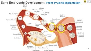
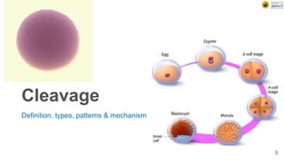
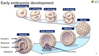
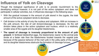
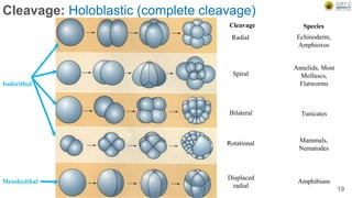
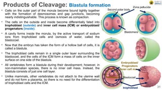
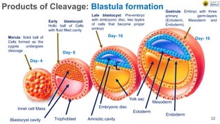
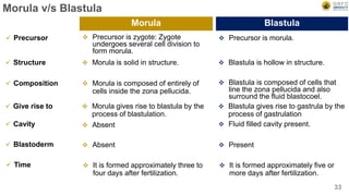
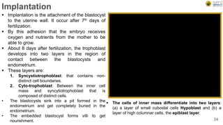
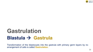
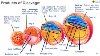
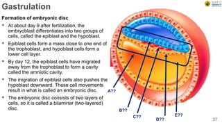
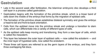
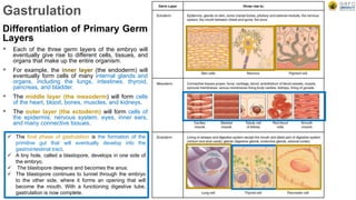
By – Dr. Mafatlal M. Kher
Developmental biology: Early embryonic development
Date & Time : Saturday, 11 September 2021
Semester : III
Program : B.Sc. Biotechnology
School : School of Science
Subject code : BSBO303 (Unit II)
1
Early embryonic development
 Cleavage: Definition, types, patterns & mechanism
 Morula to Blastulation:
 Gastrulation: Morphogenetic movements–epiboly, emboly, extension,
invagination, convergence, de-lamination. Formation & differentiation of
primary germ layers, Fate Maps in early embryos.
2
Early embryonic development
Most animals proceeds through these stages during development:
 Zygote
 Early cleavage stages
Morula
Blastula: Establishment of polarity and body axis
 Gastrulation: Establishment of germ layers
 Body plan (segmentation): In vertebrates, this involves neurulation
 Morphogenesis: Organogenesis
3
Early Embryonic Development: From ovule to implantation
4
Cleavage
Definition, types, patterns & mechanism
5
Early embryonic development
6
Fertilized egg
2- cell stage 4- cell stage 8- cell stage
Morula
Blastula
Early Gastrula
Gastrula
Ectoderm
Endoderm
Blastocoel
Blastopore
Gastrulation
Gut cavity
Influence of Yolk on Cleavage
Though the biological significance of yolk is to provide nourishment to the
developing embryo however, it is not part of the active cytoplasm and does not
participate in cellular activities, but, it influences cleavage in the following ways:
 With the gradual increase in the amount of yolk stored in the zygote, the total
amount of the active cytoplasm tends to decrease.
 Cell division is the activity of only the nucleus and cytoplasm. With an increase in
the yolk amount, the rate of cell division/cleavage is decreases. As the cell
division takes place in the active cytoplasm and nucleus, which is a relatively
smaller area of the zygote and its daughter blastomeres.
 The speed of cleavage is inversely proportional to the amount of yolk
present. In fertilized telolecithal eggs, the blastomeres nearer to the animal pole
divide at a faster rate than the blastomeres located towards the vegetal pole
because of the passive behavior of the inert yolk in the yolky parts of the zygote
and its daughter blastomeres obstructs the formation of cleavage furrows.
Animal
Pole
(Nucleus)
Vegital Pole
(Yolk)
Therefore, the nature of various
metabolic activities of the egg
and the blastomeres derived
from it depend upon the amount
and placement of the yolk mass
in the zygote.
7
Animal
Pole
(Nucleus)
Vegital Pole
(Yolk)
Cleavage: Definition and introduction
 After fertilization successfully activates the egg, the egg begins a series of rapid
cell divisions called cleavage. Or Cleavage is a series of cell divisions of the
fertilised egg through which it is converted into a multicellular structure, called
blastula. The developing embryo is called a blastula following completion of
cleavage.
 Rapid, multiple rounds of mitotic cell division where the overall size of the embryo
does not increase. Practically no growth takes place during cleavage only the
large volume of the zygote’s cytoplasm is divided into numerous smaller cells
called blastomeres.
 The pattern and types of embryonic cleavage particular to a species is determined
by two major parameters:
 The amount and distribution of yolk protein within the cytoplasm, and
 Factors in the egg cytoplasm that influence the angle of the mitotic spindle and
the timing of its formation. 8
Characteristic features of cleavage
 All the divisions of the zygote are mitotic and occur in quick succession.
 Synchronisation of cell divisions of blastomeres: The early blastomeres divide
simultaneously (synchronously) producing two blastomeres from zygote followed
by 4,8,16.32,64 blastomeres and so on, in most cases. However, such
synchronisation is lost, during later cleavage divisions.
 There is no interphase between two successive cleavage divisions in
blastomeres, or the Interphase period in the cleavage divisions is very short
and does not involve growth so that the resulting blastomeres becomes smaller in
size as their number increases i.e. there is no growth in the amount of cytoplasm
in the derived blastomeres with the result that the size of daughter blastomeres
continues to decrease during successive cleavages.
 The size of the nucleus remains practically unchanged. Therefore, the nucleus:
cytoplasm ratio, which is very small in the fertilized egg cell or zygote, continues to
increase in the blastomeres, derived from successive cleavage divisions. 9
Characteristic features of cleavage
 The rate of cell divisions is very rapid and a very large number of cells are
produced during cleavage.
For example in fast cleaving embryos, such as embryos of frog Xenopus laevis, the
cell division results in 37000 cells in 43 hours and
In Drosophila melanogaster about 50,000 cells or blastomeres are produced in just 12
hours.
 This is possible due to absence of G1 and G2 phases in the cell cycle in their
blastomeres. Which just have a mitotic phase M and a synthesis Phase S, where
only the nuclear material is replicated and so blastomeres go directly from M to S
without the intervening G1 or G2 stages as present in the somatic cell cycles
(normal mitotic cell division).
 The cell division rate slows down later on before gastrulation starts.
10
Cleavage: Humans
 About 30 hrs after fertilization, the newly
formed zygote divide into two cells, the
blastomers, in the upper portion of the
Fallopian tube.
 This is the first cleavage. The next division
occurs within 40 hours after fertilization
(Four Cell).
 The third division occurs about 2.5 days
after fertilization. During these early
cleavages, the embryo is slowly moving
down the Fallopian tube towards uterus.
 At the end of 3-4 day, the embryo reaches
uterus. It has 8-16 blastomers and this solid
mass of cell is known as morula (little
mulberry) as it looks like a mulberry.
 When the blastomers divide completely the
cleavage is called holoblastic. 11
Cleavage: Key points
 Cleavage is the division of cells in the early embryo.
 The process follows fertilization, with the transfer being triggered by the activation of a cyclin-
dependent kinase complex.
 The zygotes of many species undergo rapid cell cycles with no significant overall growth,
producing a cluster of cells the same size as the original zygote.
 The different cells derived from cleavage are called blastomeres and form a compact mass called
the morula.
 Cleavage ends with the formation of the blastula.
 Depending mostly on the amount of yolk in the egg, the cleavage can be holoblastic (total or entire
cleavage) or meroblastic (partial cleavage).
 The pole of the egg with the highest concentration of yolk is referred to as the vegetal pole while
the opposite is referred to as the animal pole.
 Cleavage differs from other forms of cell division in that it increases the number of cells and
nuclear mass without increasing the cytoplasmic mass.
 This means that with each successive subdivision, there is roughly half the cytoplasm in each
daughter cell than before that division, and thus the ratio of nuclear to cytoplasmic material
increases.
12
Cleavage: Patterns
The planes (patterns) of division are: Meridional, Equatorial, Vertical and
Latitudinal
13
Cleavage
 The ova of most of the animal groups (except some specific cases like insects) are
spherical or nearly spherical having their own actual centre which is comparable to
the earth shapes.
 Similar to north and south poles on earth, the egg has animal and vegetal poles.
 The yolk platelets have more density than the active cytoplasm which also
contains the nucleus.
 The yolk platelets are concentrated more towards vegetal hemisphere.
 Therefore, when the egg lies in any fluid medium (the fundamental feature of most
of the eggs even in the apparently terrestrial eggs like those of birds etc.), the
vegetal pole tends to face the centre of gravity and animal pole is away from it.
14
Cleavage: The planes (patterns) of division are:
 Meridional plane of cleavage: When a furrow bisect both
the poles of the egg passing through the median axis or
centre of egg it is called meridional plane of cleavage. The
median axis runs between the centre of animal pole and
vegetal pole.
 Equatorial plane of cleavage: This type of cleavage plane
divides the egg halfway between the animal and vegetal
poles and the line of division runs at right angle to the
median axis.
 Vertical plane of cleavage: When a furrow passes in any
direction (does not pass through the median axis) from the
animal pole towards the opposite pole.
 Latitudinal plane of cleavage: This is almost similar to the
equatorial plane of cleavage, but the furrow runs through
the cytoplasm on either side of the equatorial plane.
Equatorial
Meridional
Latitudinal
Vertical
15
Cleavage types
Holoblastic or Meroblastic
16
Cleavage: Types
Depending mostly on the amount of yolk in the egg, the cleavage can be:-
 Holoblastic (total or entire cleavage) or
 Meroblastic (partial cleavage).
The pole of the egg with the yolk is referred to as the vegetal pole while the portion
with nucleus is referred to as the animal pole.
17
Cleavage: Meroblastic (incomplete cleavage)
Telolecithal
Centrolecithal
Cleavage Species
Bilateral Cephalopod,
Molluscs
Discoidal Fish, Reptiles,
Birds
Superficial Most insects
18
Cleavage: Holoblastic (complete cleavage)
Isolecithal
Mesolecithal
Cleavage Species
Radial Echinoderm,
Amphioxus
Spiral
Annelids, Most
Molluscs,
Flatworms
Bilateral Tunicates
Rotational Mammals,
Nematodes
Displaced
radial
Amphibians
19
Cleavage
 In meroblastic cleavage the cleavage of egg remains
incomplete that means a small portion of cytoplasm is
cleaved because of yolk rich cytoplasm.
 The cleavage furrow does not go through the whole
egg.
 Occurs in large eggs that contains a high amount of
yolk.
 It occurs in Macro and Meso (Telolecithal and
Centrolecithal) eggs.
 It gives rise to Telolecithal (bilateral, discoidal),
Centrolecithal (superficial) cleavage.
 In holoblastic cleavage complete cleavage of the egg
occurs due to the less yolk and more cytoplasm.
 The cleavage furrow goes all the way through the
fertilized egg.
 It occurs in small eggs that contains moderate to
sparse yolk.
 It occurs in Isolacithal and micro and alecithal
eggs.
 It gives rise to Isolacithal (radial, spiral, bilateral and
rotational), Mesolecithal (Displaced radial) cleavage.
20
Meroblastic Cleavage Holoblastic Cleavage
Cleavage: Mechanism
Like mitotic divisions that takes place in any cell, cleavage is the result of
two events: mitotic nuclear division (karyokinesis) followed by
cytoplasmic division (cytokinesis).
21
Cleavage: Mechanism
 Fertilization by sperm activates the
metabolic processes in the egg cytoplasm
and initiates cleavage according to the
programme already set by the maternal
genes during oogenesis.
 There is much evidence to prove that
cleavage is guided by the genetic
information received by the egg cytoplasm
from the mother during oogenesis. There is
little or no transcriptional activity in the
zygotic nucleus during early cleavage.
Therefore, the effects of paternal genes that
come into the egg with sperm nucleus are
transcribed only later. The regulative factors
for such biphasic cleavage are said to lie in
the egg cytoplasm itself.
22
Cleavage: Mechanism
 Karyokinesis and cytokinesis are co-ordinated processes with the latter following the former.
 However, the exact mechanism which brings about this coordination is not known so far.
 The available evidence suggests that mitotic spindle dictates the location of cleavage furrows.
 The furrow always forms perpendicular to the long axis of the spindle.
 Although karyokinesis and cytokinesis are coordinated they are however, independent processes.
 Nuclear divisions can take place without being followed by cytoplasmic division.
 Similarly cleavage of cytoplasm can take place even if karyokinesis is blocked, e.g. if the zygotic
nucleus of a fertilized egg is removed the enucleate egg cytoplasm undergoes cleavage divisions up
to about the blastula stage.
23
Cleavage: Mechanism
Karyokinesis
 The mitotic division of nucleus depends upon the formation of the mitotic spindle.
 The mitotic spindle is constituted by microtubules of which the tubulin protein is the structural unit.
 If the egg is treated with the drug colchicine the microtubules are disrupted and karyokinesis is arrested at
metaphase.
Cytokinesis
 Division of the cell depends upon the contractile microfilaments of which protein actin is the structural unit.
 A ring of microfilament appears in the cortex around the cell where the cleavage furrow is formed.
 Contraction of the microfilament ring in a purse-string manner deepens the furrow, ultimately cutting the cell
into two.
 Treatment of the egg with cytochalasin B inhibits the organisation of contractile ring of microfilaments so
that cleavage furrow is not formed and cytokinesis does not take place
24
25
Cleavage:
Mechanism
(Mitosis)
Cleavage: Mechanism
Formation of New Membrane
Division of the egg or a blastomere increase the total surface area of the two daughter cells
that are required to be covered by the membrane at each cleavage.
The existing membrane of the parent cell is insufficient. From the evidence available so far, it
is indicated that this insufficiency of membranes for daughter blastomeres during cleavage is
made up from two sources:
A portion of the membranes covering the daughter cells is provided by stretching and
extension of the original plasma membrane of the zygote or the blastomeres.
A portion of the cell membrane is newly synthesized by the daughter cells.
26
Difference between typical cleavage and mitosis
1. It occurs in zygote.
2. Interphase is short.
3. Growth does not occur.
4. Oxygen consumption is high as it is
very rapid process.
5. End products size is very small as
compare to parental cell
6. Size of blastomere (endproduct)
decreases.
7. DNA synthesis is faster.
8. Nucleoplasmic-cytoplasmic ratio is
increases.
1. It occurs in most of the body cells.
2. Long duration interphase.
3. Growth occurs during interphase.
4. Oxygen consumption is low as it is a
slow process.
5. End products size is same to parental
cell
6. Size of daughter cell remains same
after growth (endproduct).
7. DNA synthesis is slow.
8. Nucleoplasmic-cytoplasmic ratio
remains same.
27
Cleavage Mitosis
Significance of Cleavage
Cleavage brings about:-
 The distribution of the cytoplasm of the zygote, amongst the blastomers.
 Increased the mobility of the protoplasm, which facilitates morphogenetic
movements necessary for cell differentiation, germ layer formation and formation
of tissue and organ.
 The restoration of the cell size and nucleo-cytoplasmic ratio characteristic of the
species.
 Unicellular zygote is converted into multicellular embryo.
28
Products of Cleavage:-
Morula  Blastula
29
Products of Cleavage: Morula
 Soon after fertilization, the mammalian zygote begins the process of
cleavage, which is the process by which the zygote rapidly divides without
growing to become multicellular.
 Cleavage uses the process of mitosis to replicate the genome and then
divide the cells in half; however, unlike normal mitosis, there is no growth
phase between divisions during cleavage.
 The first division occurs about a day after fertilization, and subsequent
divisions occur every 12 to 24 hours after that. the zygote divides several
times to form a mass of cells called a morula, which is an embryonic stage
consisting of a solid, compact mass of 16 or more cells.
 The morula is the first embryonic stage where mammalian cells can be
categorized as being either internal or external.
 The cells continue to divide, and when the mammalian morula reaches
the 64 cell stage, the internal and external cells become separate lineages.
 The internal cells are called the inner cell mass or ICM for short. The ICM will
eventually become the embryo itself and its surrounding membranes.
 The external cells are called trophoblast cells. The trophoblast cells will play
a key role in the process of implantation in the uterine wall and will eventually
become the chorion, which is the embryonic portion of the placenta.
30
A morula (Latin, morus: mulberry)
is an early-stage embryo
consisting of 16 cells (called
blastomeres) in a solid ball
contained within the zona
pellucida.
As a result, after some cleavage
divisions have taken place the
embryo has a shape resembling
mulberry.
Because of this superficial
resemblance this stage of
embryonic development of many
animals has been referred to as
morula (Latin for mulberry)
Second polar body
Blastomere
Zona pellucida
Embryoblast/
Progenators
Inner cell mass
Blastocoel
trophoblast
Products of Cleavage: Blastula formation
• Cells on the outer part of the morula become bound tightly together
with the formation of desmosomes and gap junctions, becoming
nearly indistinguishable. This process is known as compaction.
• The cells on the outside and inside become differentially fated into
trophoblast (outside) and inner cell mass (ICM) or embryoblast/
progenators (inside).
• A cavity forms inside the morula, by the active transport of sodium
ions from trophoblast cells and osmosis of water, called the
blastocoel.
• Now that the embryo has taken the form of a hollow ball of cells, it is
called a blastula.
• The trophoblast cells remain in a single outer layer surrounding the
blastocoel, and the cells of the ICM form a mass of cells on the inner
surface on one side of the blastula.
• All vertebrates form a blastula during their development; however, in
non-mammalian species, there is no inner cell mass. Instead, the
blastula consists of just one cell layer.
• Unlike mammals, other vertebrates do not attach to the uterine wall
and do not form a placenta, so there is no need for the differentiation
of trophoblast cells and the ICM.
31
Products of Cleavage: Blastula formation
Day- 4
Day- 6
Day- 10
Day- 16
Inner cell Mass
Blastocyst cavity Trophoblast
Embryonic disc
Amniotic cavity
Yolk sac
Ectoderm
Mesoderm
Endoderm
Morula: Solid ball of
Cells formed as the
zygote undergoes
cleavage
Early blastocyst:
Hollo ball of Cells
with fluid filled cavity
Late blastocyst: Pre-embryo
with embryonic disc, two layers
of cells that become proper
embryo
Gastrula: Embryo with three
primary germ-layers
(Ectoderm, Mesoderm and
Endoderm).
32
Morula v/s Blastula
 Precursor is zygote: Zygote
undergoes several cell division to
form morula.
 Precursor is morula.
33
Morula Blastula
 Precursor
 Morula is solid in structure.  Blastula is hollow in structure.
 Structure
 Morula is composed of entirely of
cells inside the zona pellucida.
 Blastula is composed of cells that
line the zona pellucida and also
surround the fluid blastocoel.
 Composition
 Morula gives rise to blastula by the
process of blastulation.
 Blastula gives rise to gastrula by the
process of gastrulation
 Give rise to
 Absent  Fluid filled cavity present.
 Cavity
 Absent  Present
 Blastoderm
 It is formed approximately three to
four days after fertilization.
 It is formed approximately five or
more days after fertilization.
 Time
Implantation
 Implantation is the attachment of the blastocyst
to the uterine wall. It occur after 7th days of
fertilization.
 By this adhesion that the embryo receives
oxygen and nutrients from the mother to be
able to grow.
 About 8 days after fertilization, the trophoblast
develops into two layers in the region of
contact between the blastocysts and
endometrium.
 These layers are:
1. Syncystiotrophoblast: that contains non-
distinct cell boundaries.
2. Cyto-trophoblast: Between the inner cell
mass and syncytiotrophoblast that is
composed of distinct cells.
• The blastocysts sink into a pit formed in the
endometrium and get completely buried in the
endometrium.
• The embedded blastocyst forms villi to get
nourishment. 34
 The cells of inner mass differentiate into two layers:
(a) a layer of small cuboidal cells Hypoblast and (b) a
layer of high columnar cells, the epiblast layer.
Gastrulation
Transformation of the blastocysts into the gastrula with primary germ layers by re-
arrangement of cells is called Gastrulation.
35
Blastula  Gastrula
Products of Cleavage:
Day- 4
Day- 6
Day- 10
Day- 16
Inner cell Mass
Blastocyst cavity Trophoblast
Embryonic disc
Amniotic cavity
Yolk sac
Ectoderm
Mesoderm
Endoderm
Morula: Solid ball of
Cells formed as the
zygote undergoes
cleavage
Early blastocyst:
Hollo ball of Cells
with fluid filled cavity
Late blastocyst: Pre-embryo
with embryonic disc, two layers
of cells that become proper
embryo
Gastrula: Embryo with three
primary germ-layers
(Ectoderm, Mesoderm and
Endoderm).
36
Gastrulation
Formation of embryonic disc
 At about day 9 after fertilization, the
embryoblast differentiates into two groups of
cells, called the epiblast and the hypoblast.
 Epiblast cells form a mass close to one end of
the trophoblast, and hypoblast cells form a
lower cell layer.
 By day 12, the epiblast cells have migrated
away from the trophoblast to form a cavity
called the amniotic cavity.
 The migration of epiblast cells also pushes the
hypoblast downward. These cell movements
result in what is called an embryonic disc.
 The embryonic disc consists of two layers of
cells, so it is called a bilaminar (two-layered)
disc.
37
A??
B??
C??
D??
E??
Gastrulation
 Late in the second week after fertilization, the bilaminar embryonic disc develops a third
cell layer in a process called gastrulation.
 Gastrulation begins with the formation of the primitive streak, which is a linear band of
cells down the middle of the embryo that forms by the migration of epiblast cells.
 The formation of the primitive streak establishes bilateral symmetry and gives the embryo
a head-to-tail and front-to-back orientation.
 Cells from the epiblast move into the primitive streak and undergo a transition to stem
cells, which can differentiate into a variety of different types of cells.
 As the epiblast cells keep moving and transitioning, they form a new layer of cells, which
is called the mesoderm.
 This layer lies between the outer layer of epiblast cells — now called the ectoderm — and
the inner layer of hypoblast cells, now called the endoderm.
 These three cell layers are referred to as the germ layers of the embryo, and they form
three overlapping flat discs.
38
Gastrulation
Differentiation of Primary Germ
Layers
 Each of the three germ layers of the embryo will
eventually give rise to different cells, tissues, and
organs that make up the entire organism.
 For example, the inner layer (the endoderm) will
eventually form cells of many internal glands and
organs, including the lungs, intestines, thyroid,
pancreas, and bladder.
 The middle layer (the mesoderm) will form cells
of the heart, blood, bones, muscles, and kidneys.
 The outer layer (the ectoderm) will form cells of
the epidermis, nervous system, eyes, inner ears,
and many connective tissues.
 The final phase of gastrulation is the formation of the
primitive gut that will eventually develop into the
gastrointestinal tract.
 A tiny hole, called a blastopore, develops in one side of
the embryo.
 The blastopore deepens and becomes the anus.
 The blastopore continues to tunnel through the embryo
to the other side, where it forms an opening that will
become the mouth. With a functioning digestive tube,
gastrulation is now complete.
Gastrulation
 Transformation of the blastocysts into the gastrula
with primary germ layers by re-arrangement of cells
is called Gastrulation.
 Gastrulation involves cell movements that helps to
attain new shape and morphology of the embryo.
These cell movements are called morphogenetic
movements.
 In all the triploblastic animals, three germ layers
namely ectoderm, mesoderm and endoderm are
formed by morphogenetic movements.
40
Gastrulation outcomes:-
 The formation of the three germ layers namely ectoderm, mesoderm and
endoderm.
 The formation of the embryonic gut or archenteron
 The appearance of the major body axes. Though in some animals the information
specifying the body axes is already present in egg in the form of cytoplasmic
determinants and or the polarity of the yolk. However, the polarity becomes
actually visible during gastrulation.
 Rearrangement of cells of the embryo by means of morphogenetic movements.
 The nuclei become more active in controlling the activities of the embryonic cells.
The influence of paternal genes becomes evident during gastrulation.
 Proteins of many new types that were not present in the egg or blastula begin to
be synthesized.
41
Gastrulation: Morphogenetic Movements
 The movement of cells in the embryo from one place to another to form particular
structures is known as morphogenetic movements.
 In the embryo the morphogenetic movements of cells from one place to another in
order to establish a particular form or structural arrangement, occurs during
embryonic development from the beginning of gastrulation onwards as well as in
the adult body.
42
Gastrulation: Morphogenetic Movements
43
Epiboly
 Epiboly means to throw on or to extend
upon.
 It is the movement of epidermal cell
sheets spreading over as a unit to
cover the deeper layers of the embryo.
 It occurs only in the presumptive
ectodermal layers (epidermal and
neural areas). The cells of this area
have an inherent property of flattening,
expanding and stretching.
 The cells of the presumptive
ectodermal areas remain on the
surface, eventually forming the outer
layer covering the entire embryo and
enveloping the inwardly migrating
presumptive mesodermal and
endodermal layers
44
Epiboly
 During epiboly, a sheet of cells spreads by
thinning while its overall surface area
increases in the other two directions.
 Epiboly can involve a monolayer (i.e. a
sheet of cells which is one cell layer thick),
in which case the individual cells must
undergo a change in shape.
 In other cases, however, a sheet that has
several cell layers can become thin by
changes in position of its cells. In this
case, epiboly occurs via intercalation.
 The analogy with epiboly in which a viscous
liquid is poured over a sphere. The liquid
slowly spreads covering the surface of the
sphere.
45
Emboly
 Emboly means to throw in or to thrust in.
 Such movements bring about the migration of presumptive mesodermal and
endodermal cells from the external surface of the embryo into its interior.
 Emboly includes several different types of cell movements which are as follows:
1. Invagination
2. Involution
3. Intercalation
4. Delamination
5. Ingression
46
Emboly: Invagination
 Invagination specifically includes the process
of infolding or rolling in of the presumptive
endodermal areas and is the most widely
observed embolic movement during
gastrulation in most animals, e.g.
echinoderms, Amphioxus and amphibians
etc.
 Invagination may be passive, occurring as a
result of the activity of other cells, or active,
resulting from the inherent forces within the
invaginating cells.
47
Invagination
Basal
Apical
Animal Pole
Blastocoel
Ectoderm
Archenteron
Invaginating
Endoderm
Blastopore
Vegetal pole
Emboly: Invagination
 It is the local inward movement of cells towards cavity
Emboly: Involution
 Involution: Involution denotes inward movement of an expanding outer layer so
that it spreads over the internal surface of the remaining external cells. Involution
of mesodermal blastomeres has been observed in Amphioxus, amphibians, birds
and reptiles.
49
During involution, a tissue sheet rolls inward to form an underlying layer via bulk movement of tissue.
Emboly: Involution
 It is similar to invagination, but more dramatic. It is an inward expansion of
epithelial cells around an edge such as the blastophore.
Emboly: Intercalation
 Intercalation is another form of
morphogenetic movement.
 During intercalation, two or more rows of
cells move between one another, creating an
array of cells that is longer (in one or more
dimensions) but thinner.
 The overall change in shape of the tissue
results from cell rearrangement.
 Intercalation can be a powerful means of
expanding a tissue sheet.
 A specialized form of intercalation is
convergent extension
51
Intercalation involves two or more rows of cells
that move between one another creating an
array of cells.
Emboly: Delamination
 Delamination, denotes the separation of
groups of cells from other cell groups to form
separate cell layers.
 It includes splitting of a pre-existing sheet
(layer) of cells into two more or less parallel
sheets, usually with a space separating
them.
52
Delamination results in the formation of the
hypoblast from epiblast in amniotes.
Emboly: Ingression
 In this process, migration of individual cells
from the surface blastoderm or blastodisc
into the embryo’s interior takes place.
Individual cells become mesenchymal (i.e.,
separate from one another) and migrate
independently into the cavity or spaces
developed within the embryo.
 The primary mesodermal cells of sea urchin
embryo become internal by this process.
 Neural crest cell are an example of a
mesenchymal cell type that emigrates out of
an epithelium
53
Ingression:
(A) During ingression, cells leave an epithelial sheet
by transforming from typical epithelial cells into freely
migrating mesenchyme cells. To do so, they must
presumably alter their cellular architecture and their
adhesive relationship to the surrounding cells.
(B) Diagrammatic representation of ingression of
primary mesenchymal cells in sea urchin embryo.
Apical
Basal
(B)
(A)
Fate Map in early embryo
To understand how a particular cell develops into a final cell type, known as
fate determination or fate map.
54
Fate Maps in early embryo:
 Within an embryo, several processes play out at the cellular and tissue level to create
an organism.
 These processes include cell proliferation, differentiation, cellular movement and
programmed cell death.
 Each cell in an embryo receives molecular signals from neighbouring cells in the form of
proteins, RNAs and even surface interactions.
 Almost all animals undergo a similar sequence of events during very early development,
a conserved process known as embryogenesis.
 During embryogenesis, cells exist in three germ layers, and undergo gastrulation.
 A fate map is a diagram of an egg or blastula, indicating the fate of each cell or region,
at a later stage of development.
 Fate maps are essential tool in most embryological experiments.
 It provide researchers with information on which portions of the embryo will normally
become which larval or adult structure.
 The analysis of the fate of each blastomere after first and second cleavage is called
cytogeny or cell lineage study.
55
Fate Maps in early embryo:
 The development of new molecular tools including Green Fluorescent Protein (GFP),
and major advances in imaging technology including fluorescence microscopy, have
made possible the mapping of the cell lineage of Caenorhabditis elegans including its
embryo.
 The technique of fate mapping is used to study cells as they differentiate and gain
specified function.
 Merely observing a cell as it becomes differentiated during embryogenesis provides no
indication of the mechanisms that drive the specification.
 The use of molecular techniques, including gene and protein knock downs, knock outs
and overexpression allows investigation into the mechanisms of fate determination.
 Improvements in imaging tools including live confocal microscopy and super resolution
microscopy allow visualization of molecular changes in experimentally manipulated cells
as compared to controls.
 Transplantation experiments can also be used in conjunction with the genetic
manipulation and lineage tracing.
 Newer cell fate determination techniques include lineage tracing performed using Cre-
lox transgenic mice, where specific cell populations can be experimentally mapped
using reporters like brainbow, a colourful reporter that is useful in the brain and other
tissues to follow the differentiation path of a cell.
56
Fate Maps in early embryo: How?
 Fate mapping is a method used in developmental biology to study the embryonic
origin of various adult tissues and structures.
 The "fate" of each cell or group of cells is mapped onto the embryo, showing which
parts of the embryo will develop into which tissue.
 The process is carried out at single-cell resolution, this process is called cell
lineage tracing.
 Fate mapping is accomplished by inserting a heritable genetic mark into a cell.
Typically, this is a fluorescent protein.
 Therefore, any progeny of the cell will have this genetic mark. It can also be done
through the use of molecular barcodes, which are introduced to the cell by
retroviruses
57
Fate Maps in early embryo: some useful methods
Observing Living Embryos
 In some invertebrates, the embryos being transparent and having relatively few
daughter cells that remain close to one another, it has been possible to look
through the microscope and trace the descendants of a particular cell to the organ
they subsequently formed.
 This type of study was performed by Edwin G. Conklin (1905) in the tunicate,
Styela partita, where the different cells contain different pigments. As for example,
the muscle-forming cells always have a yellow colour.
58
Fate Maps in early embryo: some useful methods
Vital dye marking
 Most embryos, however, do not have the facilities (transparent, few cells, different
colours etc.) as in Styela partita.
 It was in 1929 that Vogt was able to trace the fate of different areas of amphibian
eggs by applying vital dyes. These vital dyes stain the cells without killing them.
59
Fate Maps in early embryo: some useful methods
Radioactive Labelling and Fluorescent Dyes
 A variation of the dye marking technique is to make one area of the embryo
radioactive.
 A donor embryo is taken and grown in a solution containing radioactive thymidine.
 This thymidine base is subsequently incorporated into the DNA of the dividing
embryo.
 A second embryo, acting as the host embryo, is grown under normal conditions.
 The region of interest is cut off from the host embryo and is replaced by a radioac-
tive graft from the donor embryo.
 The cells that are radioactive will be the descendants of the cells of the graft, and
are distinguished by autoradiography.
60
Fate Maps in early embryo: some useful methods
CRISPR/Cas9 system
 More recently, researchers have begun using synthetic biology approaches and
the CRISPR/Cas9 system to engineer new genetic systems that enable cells to
autonomously record lineage information in their own genome.
 These systems are based on engineered, targeted mutation of defined genetic
elements.
 By generating new, random genomic alterations in each cell generation these
approaches facilitate reconstruction of lineage trees.
 These approaches promise to provide more comprehensive analysis of lineage
relationships in model organisms.
 Computational tree reconstruction methods are also being developed for datasets
generated by such approaches.
61
Fate Maps in early embryo: Modes of specification
Autonomous specification:
 This type of specification results from cell-intrinsic properties; it gives rise to
mosaic development.
 The cell-intrinsic properties arise from a cleavage of a cell with asymmetrically
expressed maternal cytoplasmic determinants (proteins, small regulatory RNAs
and mRNA). Thus, the fate of the cell depends on factors secreted into its
cytoplasm during cleavage.
62
Fate Maps in early embryo: Modes of specification
Conditional specification:
 Specification is a cell-extrinsic process that relies on cues and interactions
between cells or from concentration-gradients of morphogens.
 Inductive interactions between neighbouring cells is the most common mode of
tissue patterning.
 In this mechanism, one or two cells from a group of cells with the same
developmental potential are exposed to a signal (morphogen) from outside the
group.
 Only the cells exposed to the signal are induced to follow a different
developmental pathway, leaving the rest of the equivalence group unchanged.
 Another mechanism that determines the cell fate is regional determination. As
implied by the name, this specification occurs based on where within the embryo
the cell is positioned, it is also known as positional value.
63
Fate Maps in early embryo: Modes of specification
Syncytial specification:
 This type of a specification is a hybrid of the autonomous and conditional that
occurs in insects.
 This method involves the action of morphogen gradients within the syncytium.
 As there are no cell boundaries in the syncytium, these morphogens can influence
nuclei in a concentration-dependent manner.
64

Cleavage: Definition, types, and mechanism

  • 1.
    By – Dr.Mafatlal M. Kher Developmental biology: Early embryonic development Date & Time : Saturday, 11 September 2021 Semester : III Program : B.Sc. Biotechnology School : School of Science Subject code : BSBO303 (Unit II) 1
  • 2.
    Early embryonic development Cleavage: Definition, types, patterns & mechanism  Morula to Blastulation:  Gastrulation: Morphogenetic movements–epiboly, emboly, extension, invagination, convergence, de-lamination. Formation & differentiation of primary germ layers, Fate Maps in early embryos. 2
  • 3.
    Early embryonic development Mostanimals proceeds through these stages during development:  Zygote  Early cleavage stages Morula Blastula: Establishment of polarity and body axis  Gastrulation: Establishment of germ layers  Body plan (segmentation): In vertebrates, this involves neurulation  Morphogenesis: Organogenesis 3
  • 4.
    Early Embryonic Development:From ovule to implantation 4
  • 5.
  • 6.
    Early embryonic development 6 Fertilizedegg 2- cell stage 4- cell stage 8- cell stage Morula Blastula Early Gastrula Gastrula Ectoderm Endoderm Blastocoel Blastopore Gastrulation Gut cavity
  • 7.
    Influence of Yolkon Cleavage Though the biological significance of yolk is to provide nourishment to the developing embryo however, it is not part of the active cytoplasm and does not participate in cellular activities, but, it influences cleavage in the following ways:  With the gradual increase in the amount of yolk stored in the zygote, the total amount of the active cytoplasm tends to decrease.  Cell division is the activity of only the nucleus and cytoplasm. With an increase in the yolk amount, the rate of cell division/cleavage is decreases. As the cell division takes place in the active cytoplasm and nucleus, which is a relatively smaller area of the zygote and its daughter blastomeres.  The speed of cleavage is inversely proportional to the amount of yolk present. In fertilized telolecithal eggs, the blastomeres nearer to the animal pole divide at a faster rate than the blastomeres located towards the vegetal pole because of the passive behavior of the inert yolk in the yolky parts of the zygote and its daughter blastomeres obstructs the formation of cleavage furrows. Animal Pole (Nucleus) Vegital Pole (Yolk) Therefore, the nature of various metabolic activities of the egg and the blastomeres derived from it depend upon the amount and placement of the yolk mass in the zygote. 7 Animal Pole (Nucleus) Vegital Pole (Yolk)
  • 8.
    Cleavage: Definition andintroduction  After fertilization successfully activates the egg, the egg begins a series of rapid cell divisions called cleavage. Or Cleavage is a series of cell divisions of the fertilised egg through which it is converted into a multicellular structure, called blastula. The developing embryo is called a blastula following completion of cleavage.  Rapid, multiple rounds of mitotic cell division where the overall size of the embryo does not increase. Practically no growth takes place during cleavage only the large volume of the zygote’s cytoplasm is divided into numerous smaller cells called blastomeres.  The pattern and types of embryonic cleavage particular to a species is determined by two major parameters:  The amount and distribution of yolk protein within the cytoplasm, and  Factors in the egg cytoplasm that influence the angle of the mitotic spindle and the timing of its formation. 8
  • 9.
    Characteristic features ofcleavage  All the divisions of the zygote are mitotic and occur in quick succession.  Synchronisation of cell divisions of blastomeres: The early blastomeres divide simultaneously (synchronously) producing two blastomeres from zygote followed by 4,8,16.32,64 blastomeres and so on, in most cases. However, such synchronisation is lost, during later cleavage divisions.  There is no interphase between two successive cleavage divisions in blastomeres, or the Interphase period in the cleavage divisions is very short and does not involve growth so that the resulting blastomeres becomes smaller in size as their number increases i.e. there is no growth in the amount of cytoplasm in the derived blastomeres with the result that the size of daughter blastomeres continues to decrease during successive cleavages.  The size of the nucleus remains practically unchanged. Therefore, the nucleus: cytoplasm ratio, which is very small in the fertilized egg cell or zygote, continues to increase in the blastomeres, derived from successive cleavage divisions. 9
  • 10.
    Characteristic features ofcleavage  The rate of cell divisions is very rapid and a very large number of cells are produced during cleavage. For example in fast cleaving embryos, such as embryos of frog Xenopus laevis, the cell division results in 37000 cells in 43 hours and In Drosophila melanogaster about 50,000 cells or blastomeres are produced in just 12 hours.  This is possible due to absence of G1 and G2 phases in the cell cycle in their blastomeres. Which just have a mitotic phase M and a synthesis Phase S, where only the nuclear material is replicated and so blastomeres go directly from M to S without the intervening G1 or G2 stages as present in the somatic cell cycles (normal mitotic cell division).  The cell division rate slows down later on before gastrulation starts. 10
  • 11.
    Cleavage: Humans  About30 hrs after fertilization, the newly formed zygote divide into two cells, the blastomers, in the upper portion of the Fallopian tube.  This is the first cleavage. The next division occurs within 40 hours after fertilization (Four Cell).  The third division occurs about 2.5 days after fertilization. During these early cleavages, the embryo is slowly moving down the Fallopian tube towards uterus.  At the end of 3-4 day, the embryo reaches uterus. It has 8-16 blastomers and this solid mass of cell is known as morula (little mulberry) as it looks like a mulberry.  When the blastomers divide completely the cleavage is called holoblastic. 11
  • 12.
    Cleavage: Key points Cleavage is the division of cells in the early embryo.  The process follows fertilization, with the transfer being triggered by the activation of a cyclin- dependent kinase complex.  The zygotes of many species undergo rapid cell cycles with no significant overall growth, producing a cluster of cells the same size as the original zygote.  The different cells derived from cleavage are called blastomeres and form a compact mass called the morula.  Cleavage ends with the formation of the blastula.  Depending mostly on the amount of yolk in the egg, the cleavage can be holoblastic (total or entire cleavage) or meroblastic (partial cleavage).  The pole of the egg with the highest concentration of yolk is referred to as the vegetal pole while the opposite is referred to as the animal pole.  Cleavage differs from other forms of cell division in that it increases the number of cells and nuclear mass without increasing the cytoplasmic mass.  This means that with each successive subdivision, there is roughly half the cytoplasm in each daughter cell than before that division, and thus the ratio of nuclear to cytoplasmic material increases. 12
  • 13.
    Cleavage: Patterns The planes(patterns) of division are: Meridional, Equatorial, Vertical and Latitudinal 13
  • 14.
    Cleavage  The ovaof most of the animal groups (except some specific cases like insects) are spherical or nearly spherical having their own actual centre which is comparable to the earth shapes.  Similar to north and south poles on earth, the egg has animal and vegetal poles.  The yolk platelets have more density than the active cytoplasm which also contains the nucleus.  The yolk platelets are concentrated more towards vegetal hemisphere.  Therefore, when the egg lies in any fluid medium (the fundamental feature of most of the eggs even in the apparently terrestrial eggs like those of birds etc.), the vegetal pole tends to face the centre of gravity and animal pole is away from it. 14
  • 15.
    Cleavage: The planes(patterns) of division are:  Meridional plane of cleavage: When a furrow bisect both the poles of the egg passing through the median axis or centre of egg it is called meridional plane of cleavage. The median axis runs between the centre of animal pole and vegetal pole.  Equatorial plane of cleavage: This type of cleavage plane divides the egg halfway between the animal and vegetal poles and the line of division runs at right angle to the median axis.  Vertical plane of cleavage: When a furrow passes in any direction (does not pass through the median axis) from the animal pole towards the opposite pole.  Latitudinal plane of cleavage: This is almost similar to the equatorial plane of cleavage, but the furrow runs through the cytoplasm on either side of the equatorial plane. Equatorial Meridional Latitudinal Vertical 15
  • 16.
  • 17.
    Cleavage: Types Depending mostlyon the amount of yolk in the egg, the cleavage can be:-  Holoblastic (total or entire cleavage) or  Meroblastic (partial cleavage). The pole of the egg with the yolk is referred to as the vegetal pole while the portion with nucleus is referred to as the animal pole. 17
  • 18.
    Cleavage: Meroblastic (incompletecleavage) Telolecithal Centrolecithal Cleavage Species Bilateral Cephalopod, Molluscs Discoidal Fish, Reptiles, Birds Superficial Most insects 18
  • 19.
    Cleavage: Holoblastic (completecleavage) Isolecithal Mesolecithal Cleavage Species Radial Echinoderm, Amphioxus Spiral Annelids, Most Molluscs, Flatworms Bilateral Tunicates Rotational Mammals, Nematodes Displaced radial Amphibians 19
  • 20.
    Cleavage  In meroblasticcleavage the cleavage of egg remains incomplete that means a small portion of cytoplasm is cleaved because of yolk rich cytoplasm.  The cleavage furrow does not go through the whole egg.  Occurs in large eggs that contains a high amount of yolk.  It occurs in Macro and Meso (Telolecithal and Centrolecithal) eggs.  It gives rise to Telolecithal (bilateral, discoidal), Centrolecithal (superficial) cleavage.  In holoblastic cleavage complete cleavage of the egg occurs due to the less yolk and more cytoplasm.  The cleavage furrow goes all the way through the fertilized egg.  It occurs in small eggs that contains moderate to sparse yolk.  It occurs in Isolacithal and micro and alecithal eggs.  It gives rise to Isolacithal (radial, spiral, bilateral and rotational), Mesolecithal (Displaced radial) cleavage. 20 Meroblastic Cleavage Holoblastic Cleavage
  • 21.
    Cleavage: Mechanism Like mitoticdivisions that takes place in any cell, cleavage is the result of two events: mitotic nuclear division (karyokinesis) followed by cytoplasmic division (cytokinesis). 21
  • 22.
    Cleavage: Mechanism  Fertilizationby sperm activates the metabolic processes in the egg cytoplasm and initiates cleavage according to the programme already set by the maternal genes during oogenesis.  There is much evidence to prove that cleavage is guided by the genetic information received by the egg cytoplasm from the mother during oogenesis. There is little or no transcriptional activity in the zygotic nucleus during early cleavage. Therefore, the effects of paternal genes that come into the egg with sperm nucleus are transcribed only later. The regulative factors for such biphasic cleavage are said to lie in the egg cytoplasm itself. 22
  • 23.
    Cleavage: Mechanism  Karyokinesisand cytokinesis are co-ordinated processes with the latter following the former.  However, the exact mechanism which brings about this coordination is not known so far.  The available evidence suggests that mitotic spindle dictates the location of cleavage furrows.  The furrow always forms perpendicular to the long axis of the spindle.  Although karyokinesis and cytokinesis are coordinated they are however, independent processes.  Nuclear divisions can take place without being followed by cytoplasmic division.  Similarly cleavage of cytoplasm can take place even if karyokinesis is blocked, e.g. if the zygotic nucleus of a fertilized egg is removed the enucleate egg cytoplasm undergoes cleavage divisions up to about the blastula stage. 23
  • 24.
    Cleavage: Mechanism Karyokinesis  Themitotic division of nucleus depends upon the formation of the mitotic spindle.  The mitotic spindle is constituted by microtubules of which the tubulin protein is the structural unit.  If the egg is treated with the drug colchicine the microtubules are disrupted and karyokinesis is arrested at metaphase. Cytokinesis  Division of the cell depends upon the contractile microfilaments of which protein actin is the structural unit.  A ring of microfilament appears in the cortex around the cell where the cleavage furrow is formed.  Contraction of the microfilament ring in a purse-string manner deepens the furrow, ultimately cutting the cell into two.  Treatment of the egg with cytochalasin B inhibits the organisation of contractile ring of microfilaments so that cleavage furrow is not formed and cytokinesis does not take place 24
  • 25.
  • 26.
    Cleavage: Mechanism Formation ofNew Membrane Division of the egg or a blastomere increase the total surface area of the two daughter cells that are required to be covered by the membrane at each cleavage. The existing membrane of the parent cell is insufficient. From the evidence available so far, it is indicated that this insufficiency of membranes for daughter blastomeres during cleavage is made up from two sources: A portion of the membranes covering the daughter cells is provided by stretching and extension of the original plasma membrane of the zygote or the blastomeres. A portion of the cell membrane is newly synthesized by the daughter cells. 26
  • 27.
    Difference between typicalcleavage and mitosis 1. It occurs in zygote. 2. Interphase is short. 3. Growth does not occur. 4. Oxygen consumption is high as it is very rapid process. 5. End products size is very small as compare to parental cell 6. Size of blastomere (endproduct) decreases. 7. DNA synthesis is faster. 8. Nucleoplasmic-cytoplasmic ratio is increases. 1. It occurs in most of the body cells. 2. Long duration interphase. 3. Growth occurs during interphase. 4. Oxygen consumption is low as it is a slow process. 5. End products size is same to parental cell 6. Size of daughter cell remains same after growth (endproduct). 7. DNA synthesis is slow. 8. Nucleoplasmic-cytoplasmic ratio remains same. 27 Cleavage Mitosis
  • 28.
    Significance of Cleavage Cleavagebrings about:-  The distribution of the cytoplasm of the zygote, amongst the blastomers.  Increased the mobility of the protoplasm, which facilitates morphogenetic movements necessary for cell differentiation, germ layer formation and formation of tissue and organ.  The restoration of the cell size and nucleo-cytoplasmic ratio characteristic of the species.  Unicellular zygote is converted into multicellular embryo. 28
  • 29.
  • 30.
    Products of Cleavage:Morula  Soon after fertilization, the mammalian zygote begins the process of cleavage, which is the process by which the zygote rapidly divides without growing to become multicellular.  Cleavage uses the process of mitosis to replicate the genome and then divide the cells in half; however, unlike normal mitosis, there is no growth phase between divisions during cleavage.  The first division occurs about a day after fertilization, and subsequent divisions occur every 12 to 24 hours after that. the zygote divides several times to form a mass of cells called a morula, which is an embryonic stage consisting of a solid, compact mass of 16 or more cells.  The morula is the first embryonic stage where mammalian cells can be categorized as being either internal or external.  The cells continue to divide, and when the mammalian morula reaches the 64 cell stage, the internal and external cells become separate lineages.  The internal cells are called the inner cell mass or ICM for short. The ICM will eventually become the embryo itself and its surrounding membranes.  The external cells are called trophoblast cells. The trophoblast cells will play a key role in the process of implantation in the uterine wall and will eventually become the chorion, which is the embryonic portion of the placenta. 30 A morula (Latin, morus: mulberry) is an early-stage embryo consisting of 16 cells (called blastomeres) in a solid ball contained within the zona pellucida. As a result, after some cleavage divisions have taken place the embryo has a shape resembling mulberry. Because of this superficial resemblance this stage of embryonic development of many animals has been referred to as morula (Latin for mulberry)
  • 31.
    Second polar body Blastomere Zonapellucida Embryoblast/ Progenators Inner cell mass Blastocoel trophoblast Products of Cleavage: Blastula formation • Cells on the outer part of the morula become bound tightly together with the formation of desmosomes and gap junctions, becoming nearly indistinguishable. This process is known as compaction. • The cells on the outside and inside become differentially fated into trophoblast (outside) and inner cell mass (ICM) or embryoblast/ progenators (inside). • A cavity forms inside the morula, by the active transport of sodium ions from trophoblast cells and osmosis of water, called the blastocoel. • Now that the embryo has taken the form of a hollow ball of cells, it is called a blastula. • The trophoblast cells remain in a single outer layer surrounding the blastocoel, and the cells of the ICM form a mass of cells on the inner surface on one side of the blastula. • All vertebrates form a blastula during their development; however, in non-mammalian species, there is no inner cell mass. Instead, the blastula consists of just one cell layer. • Unlike mammals, other vertebrates do not attach to the uterine wall and do not form a placenta, so there is no need for the differentiation of trophoblast cells and the ICM. 31
  • 32.
    Products of Cleavage:Blastula formation Day- 4 Day- 6 Day- 10 Day- 16 Inner cell Mass Blastocyst cavity Trophoblast Embryonic disc Amniotic cavity Yolk sac Ectoderm Mesoderm Endoderm Morula: Solid ball of Cells formed as the zygote undergoes cleavage Early blastocyst: Hollo ball of Cells with fluid filled cavity Late blastocyst: Pre-embryo with embryonic disc, two layers of cells that become proper embryo Gastrula: Embryo with three primary germ-layers (Ectoderm, Mesoderm and Endoderm). 32
  • 33.
    Morula v/s Blastula Precursor is zygote: Zygote undergoes several cell division to form morula.  Precursor is morula. 33 Morula Blastula  Precursor  Morula is solid in structure.  Blastula is hollow in structure.  Structure  Morula is composed of entirely of cells inside the zona pellucida.  Blastula is composed of cells that line the zona pellucida and also surround the fluid blastocoel.  Composition  Morula gives rise to blastula by the process of blastulation.  Blastula gives rise to gastrula by the process of gastrulation  Give rise to  Absent  Fluid filled cavity present.  Cavity  Absent  Present  Blastoderm  It is formed approximately three to four days after fertilization.  It is formed approximately five or more days after fertilization.  Time
  • 34.
    Implantation  Implantation isthe attachment of the blastocyst to the uterine wall. It occur after 7th days of fertilization.  By this adhesion that the embryo receives oxygen and nutrients from the mother to be able to grow.  About 8 days after fertilization, the trophoblast develops into two layers in the region of contact between the blastocysts and endometrium.  These layers are: 1. Syncystiotrophoblast: that contains non- distinct cell boundaries. 2. Cyto-trophoblast: Between the inner cell mass and syncytiotrophoblast that is composed of distinct cells. • The blastocysts sink into a pit formed in the endometrium and get completely buried in the endometrium. • The embedded blastocyst forms villi to get nourishment. 34  The cells of inner mass differentiate into two layers: (a) a layer of small cuboidal cells Hypoblast and (b) a layer of high columnar cells, the epiblast layer.
  • 35.
    Gastrulation Transformation of theblastocysts into the gastrula with primary germ layers by re- arrangement of cells is called Gastrulation. 35 Blastula  Gastrula
  • 36.
    Products of Cleavage: Day-4 Day- 6 Day- 10 Day- 16 Inner cell Mass Blastocyst cavity Trophoblast Embryonic disc Amniotic cavity Yolk sac Ectoderm Mesoderm Endoderm Morula: Solid ball of Cells formed as the zygote undergoes cleavage Early blastocyst: Hollo ball of Cells with fluid filled cavity Late blastocyst: Pre-embryo with embryonic disc, two layers of cells that become proper embryo Gastrula: Embryo with three primary germ-layers (Ectoderm, Mesoderm and Endoderm). 36
  • 37.
    Gastrulation Formation of embryonicdisc  At about day 9 after fertilization, the embryoblast differentiates into two groups of cells, called the epiblast and the hypoblast.  Epiblast cells form a mass close to one end of the trophoblast, and hypoblast cells form a lower cell layer.  By day 12, the epiblast cells have migrated away from the trophoblast to form a cavity called the amniotic cavity.  The migration of epiblast cells also pushes the hypoblast downward. These cell movements result in what is called an embryonic disc.  The embryonic disc consists of two layers of cells, so it is called a bilaminar (two-layered) disc. 37 A?? B?? C?? D?? E??
  • 38.
    Gastrulation  Late inthe second week after fertilization, the bilaminar embryonic disc develops a third cell layer in a process called gastrulation.  Gastrulation begins with the formation of the primitive streak, which is a linear band of cells down the middle of the embryo that forms by the migration of epiblast cells.  The formation of the primitive streak establishes bilateral symmetry and gives the embryo a head-to-tail and front-to-back orientation.  Cells from the epiblast move into the primitive streak and undergo a transition to stem cells, which can differentiate into a variety of different types of cells.  As the epiblast cells keep moving and transitioning, they form a new layer of cells, which is called the mesoderm.  This layer lies between the outer layer of epiblast cells — now called the ectoderm — and the inner layer of hypoblast cells, now called the endoderm.  These three cell layers are referred to as the germ layers of the embryo, and they form three overlapping flat discs. 38
  • 39.
    Gastrulation Differentiation of PrimaryGerm Layers  Each of the three germ layers of the embryo will eventually give rise to different cells, tissues, and organs that make up the entire organism.  For example, the inner layer (the endoderm) will eventually form cells of many internal glands and organs, including the lungs, intestines, thyroid, pancreas, and bladder.  The middle layer (the mesoderm) will form cells of the heart, blood, bones, muscles, and kidneys.  The outer layer (the ectoderm) will form cells of the epidermis, nervous system, eyes, inner ears, and many connective tissues.  The final phase of gastrulation is the formation of the primitive gut that will eventually develop into the gastrointestinal tract.  A tiny hole, called a blastopore, develops in one side of the embryo.  The blastopore deepens and becomes the anus.  The blastopore continues to tunnel through the embryo to the other side, where it forms an opening that will become the mouth. With a functioning digestive tube, gastrulation is now complete.
  • 40.
    Gastrulation  Transformation ofthe blastocysts into the gastrula with primary germ layers by re-arrangement of cells is called Gastrulation.  Gastrulation involves cell movements that helps to attain new shape and morphology of the embryo. These cell movements are called morphogenetic movements.  In all the triploblastic animals, three germ layers namely ectoderm, mesoderm and endoderm are formed by morphogenetic movements. 40
  • 41.
    Gastrulation outcomes:-  Theformation of the three germ layers namely ectoderm, mesoderm and endoderm.  The formation of the embryonic gut or archenteron  The appearance of the major body axes. Though in some animals the information specifying the body axes is already present in egg in the form of cytoplasmic determinants and or the polarity of the yolk. However, the polarity becomes actually visible during gastrulation.  Rearrangement of cells of the embryo by means of morphogenetic movements.  The nuclei become more active in controlling the activities of the embryonic cells. The influence of paternal genes becomes evident during gastrulation.  Proteins of many new types that were not present in the egg or blastula begin to be synthesized. 41
  • 42.
    Gastrulation: Morphogenetic Movements The movement of cells in the embryo from one place to another to form particular structures is known as morphogenetic movements.  In the embryo the morphogenetic movements of cells from one place to another in order to establish a particular form or structural arrangement, occurs during embryonic development from the beginning of gastrulation onwards as well as in the adult body. 42
  • 43.
  • 44.
    Epiboly  Epiboly meansto throw on or to extend upon.  It is the movement of epidermal cell sheets spreading over as a unit to cover the deeper layers of the embryo.  It occurs only in the presumptive ectodermal layers (epidermal and neural areas). The cells of this area have an inherent property of flattening, expanding and stretching.  The cells of the presumptive ectodermal areas remain on the surface, eventually forming the outer layer covering the entire embryo and enveloping the inwardly migrating presumptive mesodermal and endodermal layers 44
  • 45.
    Epiboly  During epiboly,a sheet of cells spreads by thinning while its overall surface area increases in the other two directions.  Epiboly can involve a monolayer (i.e. a sheet of cells which is one cell layer thick), in which case the individual cells must undergo a change in shape.  In other cases, however, a sheet that has several cell layers can become thin by changes in position of its cells. In this case, epiboly occurs via intercalation.  The analogy with epiboly in which a viscous liquid is poured over a sphere. The liquid slowly spreads covering the surface of the sphere. 45
  • 46.
    Emboly  Emboly meansto throw in or to thrust in.  Such movements bring about the migration of presumptive mesodermal and endodermal cells from the external surface of the embryo into its interior.  Emboly includes several different types of cell movements which are as follows: 1. Invagination 2. Involution 3. Intercalation 4. Delamination 5. Ingression 46
  • 47.
    Emboly: Invagination  Invaginationspecifically includes the process of infolding or rolling in of the presumptive endodermal areas and is the most widely observed embolic movement during gastrulation in most animals, e.g. echinoderms, Amphioxus and amphibians etc.  Invagination may be passive, occurring as a result of the activity of other cells, or active, resulting from the inherent forces within the invaginating cells. 47 Invagination Basal Apical Animal Pole Blastocoel Ectoderm Archenteron Invaginating Endoderm Blastopore Vegetal pole
  • 48.
    Emboly: Invagination  Itis the local inward movement of cells towards cavity
  • 49.
    Emboly: Involution  Involution:Involution denotes inward movement of an expanding outer layer so that it spreads over the internal surface of the remaining external cells. Involution of mesodermal blastomeres has been observed in Amphioxus, amphibians, birds and reptiles. 49 During involution, a tissue sheet rolls inward to form an underlying layer via bulk movement of tissue.
  • 50.
    Emboly: Involution  Itis similar to invagination, but more dramatic. It is an inward expansion of epithelial cells around an edge such as the blastophore.
  • 51.
    Emboly: Intercalation  Intercalationis another form of morphogenetic movement.  During intercalation, two or more rows of cells move between one another, creating an array of cells that is longer (in one or more dimensions) but thinner.  The overall change in shape of the tissue results from cell rearrangement.  Intercalation can be a powerful means of expanding a tissue sheet.  A specialized form of intercalation is convergent extension 51 Intercalation involves two or more rows of cells that move between one another creating an array of cells.
  • 52.
    Emboly: Delamination  Delamination,denotes the separation of groups of cells from other cell groups to form separate cell layers.  It includes splitting of a pre-existing sheet (layer) of cells into two more or less parallel sheets, usually with a space separating them. 52 Delamination results in the formation of the hypoblast from epiblast in amniotes.
  • 53.
    Emboly: Ingression  Inthis process, migration of individual cells from the surface blastoderm or blastodisc into the embryo’s interior takes place. Individual cells become mesenchymal (i.e., separate from one another) and migrate independently into the cavity or spaces developed within the embryo.  The primary mesodermal cells of sea urchin embryo become internal by this process.  Neural crest cell are an example of a mesenchymal cell type that emigrates out of an epithelium 53 Ingression: (A) During ingression, cells leave an epithelial sheet by transforming from typical epithelial cells into freely migrating mesenchyme cells. To do so, they must presumably alter their cellular architecture and their adhesive relationship to the surrounding cells. (B) Diagrammatic representation of ingression of primary mesenchymal cells in sea urchin embryo. Apical Basal (B) (A)
  • 54.
    Fate Map inearly embryo To understand how a particular cell develops into a final cell type, known as fate determination or fate map. 54
  • 55.
    Fate Maps inearly embryo:  Within an embryo, several processes play out at the cellular and tissue level to create an organism.  These processes include cell proliferation, differentiation, cellular movement and programmed cell death.  Each cell in an embryo receives molecular signals from neighbouring cells in the form of proteins, RNAs and even surface interactions.  Almost all animals undergo a similar sequence of events during very early development, a conserved process known as embryogenesis.  During embryogenesis, cells exist in three germ layers, and undergo gastrulation.  A fate map is a diagram of an egg or blastula, indicating the fate of each cell or region, at a later stage of development.  Fate maps are essential tool in most embryological experiments.  It provide researchers with information on which portions of the embryo will normally become which larval or adult structure.  The analysis of the fate of each blastomere after first and second cleavage is called cytogeny or cell lineage study. 55
  • 56.
    Fate Maps inearly embryo:  The development of new molecular tools including Green Fluorescent Protein (GFP), and major advances in imaging technology including fluorescence microscopy, have made possible the mapping of the cell lineage of Caenorhabditis elegans including its embryo.  The technique of fate mapping is used to study cells as they differentiate and gain specified function.  Merely observing a cell as it becomes differentiated during embryogenesis provides no indication of the mechanisms that drive the specification.  The use of molecular techniques, including gene and protein knock downs, knock outs and overexpression allows investigation into the mechanisms of fate determination.  Improvements in imaging tools including live confocal microscopy and super resolution microscopy allow visualization of molecular changes in experimentally manipulated cells as compared to controls.  Transplantation experiments can also be used in conjunction with the genetic manipulation and lineage tracing.  Newer cell fate determination techniques include lineage tracing performed using Cre- lox transgenic mice, where specific cell populations can be experimentally mapped using reporters like brainbow, a colourful reporter that is useful in the brain and other tissues to follow the differentiation path of a cell. 56
  • 57.
    Fate Maps inearly embryo: How?  Fate mapping is a method used in developmental biology to study the embryonic origin of various adult tissues and structures.  The "fate" of each cell or group of cells is mapped onto the embryo, showing which parts of the embryo will develop into which tissue.  The process is carried out at single-cell resolution, this process is called cell lineage tracing.  Fate mapping is accomplished by inserting a heritable genetic mark into a cell. Typically, this is a fluorescent protein.  Therefore, any progeny of the cell will have this genetic mark. It can also be done through the use of molecular barcodes, which are introduced to the cell by retroviruses 57
  • 58.
    Fate Maps inearly embryo: some useful methods Observing Living Embryos  In some invertebrates, the embryos being transparent and having relatively few daughter cells that remain close to one another, it has been possible to look through the microscope and trace the descendants of a particular cell to the organ they subsequently formed.  This type of study was performed by Edwin G. Conklin (1905) in the tunicate, Styela partita, where the different cells contain different pigments. As for example, the muscle-forming cells always have a yellow colour. 58
  • 59.
    Fate Maps inearly embryo: some useful methods Vital dye marking  Most embryos, however, do not have the facilities (transparent, few cells, different colours etc.) as in Styela partita.  It was in 1929 that Vogt was able to trace the fate of different areas of amphibian eggs by applying vital dyes. These vital dyes stain the cells without killing them. 59
  • 60.
    Fate Maps inearly embryo: some useful methods Radioactive Labelling and Fluorescent Dyes  A variation of the dye marking technique is to make one area of the embryo radioactive.  A donor embryo is taken and grown in a solution containing radioactive thymidine.  This thymidine base is subsequently incorporated into the DNA of the dividing embryo.  A second embryo, acting as the host embryo, is grown under normal conditions.  The region of interest is cut off from the host embryo and is replaced by a radioac- tive graft from the donor embryo.  The cells that are radioactive will be the descendants of the cells of the graft, and are distinguished by autoradiography. 60
  • 61.
    Fate Maps inearly embryo: some useful methods CRISPR/Cas9 system  More recently, researchers have begun using synthetic biology approaches and the CRISPR/Cas9 system to engineer new genetic systems that enable cells to autonomously record lineage information in their own genome.  These systems are based on engineered, targeted mutation of defined genetic elements.  By generating new, random genomic alterations in each cell generation these approaches facilitate reconstruction of lineage trees.  These approaches promise to provide more comprehensive analysis of lineage relationships in model organisms.  Computational tree reconstruction methods are also being developed for datasets generated by such approaches. 61
  • 62.
    Fate Maps inearly embryo: Modes of specification Autonomous specification:  This type of specification results from cell-intrinsic properties; it gives rise to mosaic development.  The cell-intrinsic properties arise from a cleavage of a cell with asymmetrically expressed maternal cytoplasmic determinants (proteins, small regulatory RNAs and mRNA). Thus, the fate of the cell depends on factors secreted into its cytoplasm during cleavage. 62
  • 63.
    Fate Maps inearly embryo: Modes of specification Conditional specification:  Specification is a cell-extrinsic process that relies on cues and interactions between cells or from concentration-gradients of morphogens.  Inductive interactions between neighbouring cells is the most common mode of tissue patterning.  In this mechanism, one or two cells from a group of cells with the same developmental potential are exposed to a signal (morphogen) from outside the group.  Only the cells exposed to the signal are induced to follow a different developmental pathway, leaving the rest of the equivalence group unchanged.  Another mechanism that determines the cell fate is regional determination. As implied by the name, this specification occurs based on where within the embryo the cell is positioned, it is also known as positional value. 63
  • 64.
    Fate Maps inearly embryo: Modes of specification Syncytial specification:  This type of a specification is a hybrid of the autonomous and conditional that occurs in insects.  This method involves the action of morphogen gradients within the syncytium.  As there are no cell boundaries in the syncytium, these morphogens can influence nuclei in a concentration-dependent manner. 64

Editor's Notes

  • #47 It should be kept in mind that not any one factor causes invagination but a combination of different factors may be involved in various animals.
  • #50 It should be kept in mind that not any one factor causes invagination but a combination of different factors may be involved in various animals.