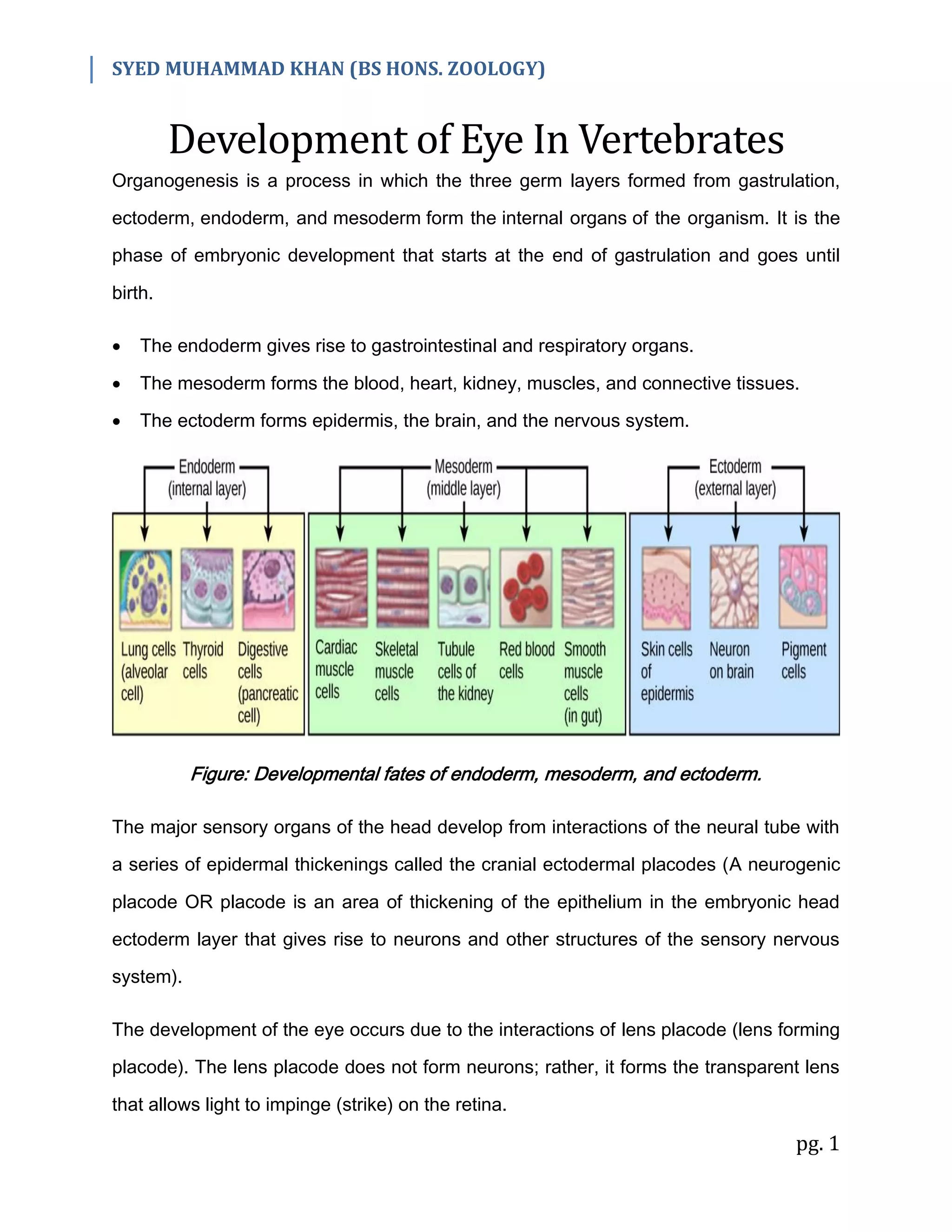
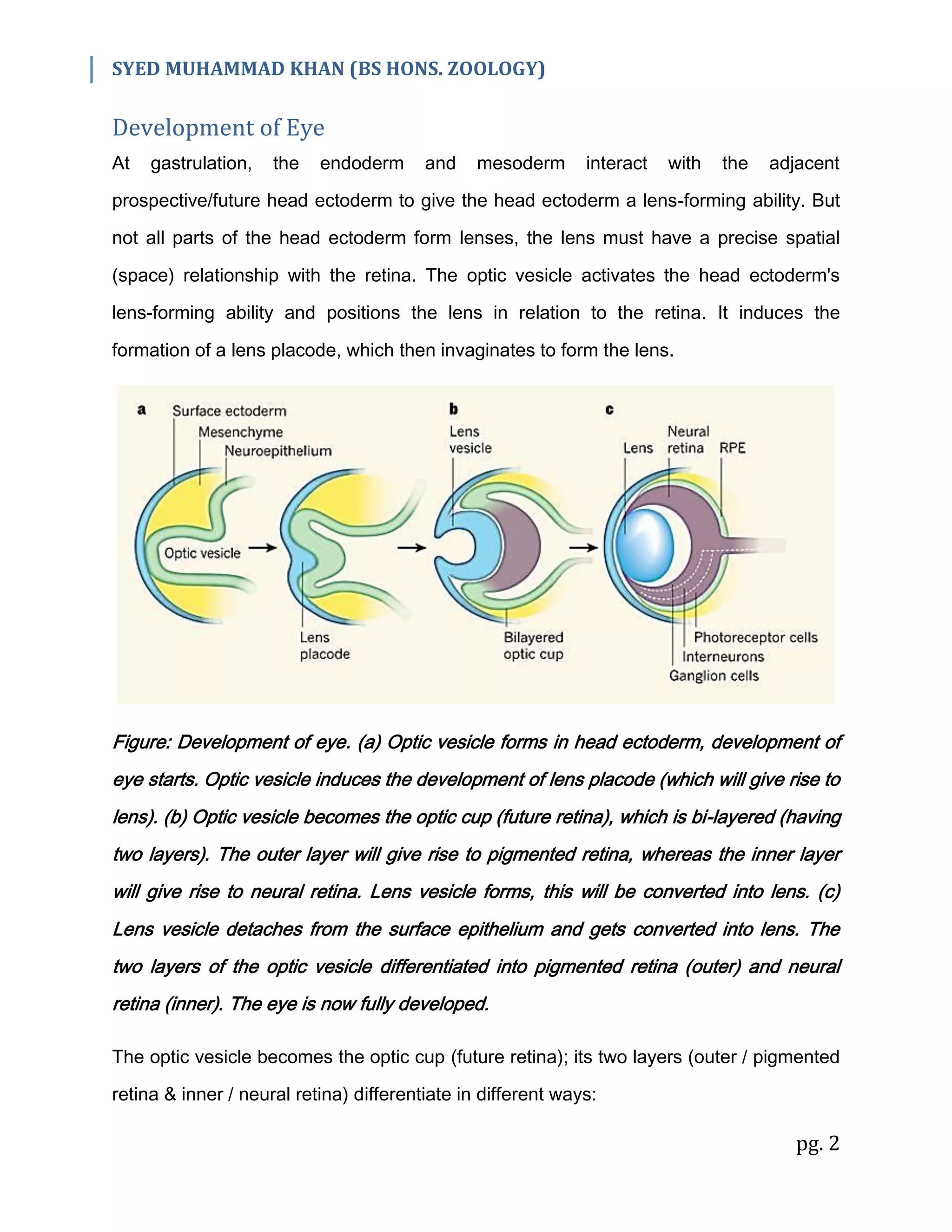
SYED MUHAMMAD KHAN (BS HONS. ZOOLOGY)
pg. 1
Development of Eye In Vertebrates
Organogenesis is a process in which the three germ layers formed from gastrulation,
ectoderm, endoderm, and mesoderm form the internal organs of the organism. It is the
phase of embryonic development that starts at the end of gastrulation and goes until
birth.
 The endoderm gives rise to gastrointestinal and respiratory organs.
 The mesoderm forms the blood, heart, kidney, muscles, and connective tissues.
 The ectoderm forms epidermis, the brain, and the nervous system.
Figure: Developmental fates of endoderm, mesoderm, and ectoderm.
The major sensory organs of the head develop from interactions of the neural tube with
a series of epidermal thickenings called the cranial ectodermal placodes (A neurogenic
placode OR placode is an area of thickening of the epithelium in the embryonic head
ectoderm layer that gives rise to neurons and other structures of the sensory nervous
system).
The development of the eye occurs due to the interactions of lens placode (lens forming
placode). The lens placode does not form neurons; rather, it forms the transparent lens
that allows light to impinge (strike) on the retina.
SYED MUHAMMAD KHAN (BS HONS. ZOOLOGY)
pg. 2
Development of Eye
At gastrulation, the endoderm and mesoderm interact with the adjacent
prospective/future head ectoderm to give the head ectoderm a lens-forming ability. But
not all parts of the head ectoderm form lenses, the lens must have a precise spatial
(space) relationship with the retina. The optic vesicle activates the head ectoderm's
lens-forming ability and positions the lens in relation to the retina. It induces the
formation of a lens placode, which then invaginates to form the lens.
Figure: Development of eye. (a) Optic vesicle forms in head ectoderm, development of
eye starts. Optic vesicle induces the development of lens placode (which will give rise to
lens). (b) Optic vesicle becomes the optic cup (future retina), which is bi-layered (having
two layers). The outer layer will give rise to pigmented retina, whereas the inner layer
will give rise to neural retina. Lens vesicle forms, this will be converted into lens. (c)
Lens vesicle detaches from the surface epithelium and gets converted into lens. The
two layers of the optic vesicle differentiated into pigmented retina (outer) and neural
retina (inner). The eye is now fully developed.
The optic vesicle becomes the optic cup (future retina); its two layers (outer / pigmented
retina & inner / neural retina) differentiate in different ways:
SYED MUHAMMAD KHAN (BS HONS. ZOOLOGY)
pg. 3
Outer Layer / Pigmented Retina: The cells of the outer layer produce melanin pigment
(brown) and ultimately become the pigmented retina.
Inner Layer / Neural Retina: The cells of the inner layer proliferate rapidly (rapid cell
divisions) and generate a variety of glia (Glia/Glial cells/Neuroglia – non-neuronal cells
in the central nervous system), ganglion cells (a type of neuron located near the inner
surface of the retina), interneurons, and light-sensitive photoreceptor neurons.
Collectively, these cells constitute the neural retina.
The retinal ganglion cells are neurons whose axons send electric impulses to the brain.
Their axons meet at the base of the eye and travel down the optic stalk, which is then
called the optic nerve.
Cell Differentiation in Eye
The differentiation of cells of various parts of the eye takes place in the following ways:
(1) Neural Retina Differentiation:
The retinal precursor cells divide before they differentiate. The neural retina develops
into a layered array/arrangement of different neuronal types, these layers include:
1. Rods – Light-sensitive photoreceptor cells
2. Cones – Color-sensitive photoreceptor cells
3. Cell bodies of the ganglion cells
4. Bipolar interneurons that transmit electric stimuli from the rods and cones to the
ganglion cells
5. Numerous Muller glial cells that maintain the retina's integrity (keep it together)
6. Amacrine neurons (which lack large axons)
7. Horizontal neurons that transmit electric impulses in the plane of the retina (same
axis/direction)
SYED MUHAMMAD KHAN (BS HONS. ZOOLOGY)
pg. 4
The neuroblasts (neuron/nerve forming cells) of the retina are competent to generate all
seven retinal cell types (that have been mentioned above).
(2) Lens & Cornea Differentiation:
The eye can't focus on the retina unless it has a lens and a cornea.
Cornea Differentiation: Shortly after the lens vesicle has detached from the surface
ectoderm, mesenchymal cells from the neural crest (embryonic structure that gives rise
to the peripheral nervous system) migrate into the space between the lens and the
surface epithelium. These cells condense to form several flat layers of cells, which
become the corneal precursor cells. As these cells mature, they dehydrate and form
tight junctions among the cells, forming the cornea. Fluid pressure from the aqueous
humor (transparent fluid between lens and cornea) is necessary for the correct
curvature of the cornea.
Lens Differentiation: The differentiation of the lens tissue into a transparent membrane
capable of directing light onto the retina involves changes in cell structure and shape as
well as the synthesis of transparent, lens-specific proteins called crystallins. The cells at
the inner portion of the lens vesicle elongate and, under the influence of the neural
retina, become the lens fibers. As these fibers continue to grow, they synthesize
crystallins (transparent and lens-specific proteins), which eventually fill up the cell and
cause the extrusion of the nucleus (it is thrown out of the cell). The lens contains three
regions: an anterior zone of dividing epithelial cells, an equatorial zone (middle) of
cellular elongation, and a posterior and central zone of crystallin-containing fiber cells.
This arrangement persists throughout the lifetime of the animal.

Development of Eye In Vertebrates

  • 1.
    SYED MUHAMMAD KHAN(BS HONS. ZOOLOGY) pg. 1 Development of Eye In Vertebrates Organogenesis is a process in which the three germ layers formed from gastrulation, ectoderm, endoderm, and mesoderm form the internal organs of the organism. It is the phase of embryonic development that starts at the end of gastrulation and goes until birth.  The endoderm gives rise to gastrointestinal and respiratory organs.  The mesoderm forms the blood, heart, kidney, muscles, and connective tissues.  The ectoderm forms epidermis, the brain, and the nervous system. Figure: Developmental fates of endoderm, mesoderm, and ectoderm. The major sensory organs of the head develop from interactions of the neural tube with a series of epidermal thickenings called the cranial ectodermal placodes (A neurogenic placode OR placode is an area of thickening of the epithelium in the embryonic head ectoderm layer that gives rise to neurons and other structures of the sensory nervous system). The development of the eye occurs due to the interactions of lens placode (lens forming placode). The lens placode does not form neurons; rather, it forms the transparent lens that allows light to impinge (strike) on the retina.
  • 2.
    SYED MUHAMMAD KHAN(BS HONS. ZOOLOGY) pg. 2 Development of Eye At gastrulation, the endoderm and mesoderm interact with the adjacent prospective/future head ectoderm to give the head ectoderm a lens-forming ability. But not all parts of the head ectoderm form lenses, the lens must have a precise spatial (space) relationship with the retina. The optic vesicle activates the head ectoderm's lens-forming ability and positions the lens in relation to the retina. It induces the formation of a lens placode, which then invaginates to form the lens. Figure: Development of eye. (a) Optic vesicle forms in head ectoderm, development of eye starts. Optic vesicle induces the development of lens placode (which will give rise to lens). (b) Optic vesicle becomes the optic cup (future retina), which is bi-layered (having two layers). The outer layer will give rise to pigmented retina, whereas the inner layer will give rise to neural retina. Lens vesicle forms, this will be converted into lens. (c) Lens vesicle detaches from the surface epithelium and gets converted into lens. The two layers of the optic vesicle differentiated into pigmented retina (outer) and neural retina (inner). The eye is now fully developed. The optic vesicle becomes the optic cup (future retina); its two layers (outer / pigmented retina & inner / neural retina) differentiate in different ways:
  • 3.
    SYED MUHAMMAD KHAN(BS HONS. ZOOLOGY) pg. 3 Outer Layer / Pigmented Retina: The cells of the outer layer produce melanin pigment (brown) and ultimately become the pigmented retina. Inner Layer / Neural Retina: The cells of the inner layer proliferate rapidly (rapid cell divisions) and generate a variety of glia (Glia/Glial cells/Neuroglia – non-neuronal cells in the central nervous system), ganglion cells (a type of neuron located near the inner surface of the retina), interneurons, and light-sensitive photoreceptor neurons. Collectively, these cells constitute the neural retina. The retinal ganglion cells are neurons whose axons send electric impulses to the brain. Their axons meet at the base of the eye and travel down the optic stalk, which is then called the optic nerve. Cell Differentiation in Eye The differentiation of cells of various parts of the eye takes place in the following ways: (1) Neural Retina Differentiation: The retinal precursor cells divide before they differentiate. The neural retina develops into a layered array/arrangement of different neuronal types, these layers include: 1. Rods – Light-sensitive photoreceptor cells 2. Cones – Color-sensitive photoreceptor cells 3. Cell bodies of the ganglion cells 4. Bipolar interneurons that transmit electric stimuli from the rods and cones to the ganglion cells 5. Numerous Muller glial cells that maintain the retina's integrity (keep it together) 6. Amacrine neurons (which lack large axons) 7. Horizontal neurons that transmit electric impulses in the plane of the retina (same axis/direction)
  • 4.
    SYED MUHAMMAD KHAN(BS HONS. ZOOLOGY) pg. 4 The neuroblasts (neuron/nerve forming cells) of the retina are competent to generate all seven retinal cell types (that have been mentioned above). (2) Lens & Cornea Differentiation: The eye can't focus on the retina unless it has a lens and a cornea. Cornea Differentiation: Shortly after the lens vesicle has detached from the surface ectoderm, mesenchymal cells from the neural crest (embryonic structure that gives rise to the peripheral nervous system) migrate into the space between the lens and the surface epithelium. These cells condense to form several flat layers of cells, which become the corneal precursor cells. As these cells mature, they dehydrate and form tight junctions among the cells, forming the cornea. Fluid pressure from the aqueous humor (transparent fluid between lens and cornea) is necessary for the correct curvature of the cornea. Lens Differentiation: The differentiation of the lens tissue into a transparent membrane capable of directing light onto the retina involves changes in cell structure and shape as well as the synthesis of transparent, lens-specific proteins called crystallins. The cells at the inner portion of the lens vesicle elongate and, under the influence of the neural retina, become the lens fibers. As these fibers continue to grow, they synthesize crystallins (transparent and lens-specific proteins), which eventually fill up the cell and cause the extrusion of the nucleus (it is thrown out of the cell). The lens contains three regions: an anterior zone of dividing epithelial cells, an equatorial zone (middle) of cellular elongation, and a posterior and central zone of crystallin-containing fiber cells. This arrangement persists throughout the lifetime of the animal.