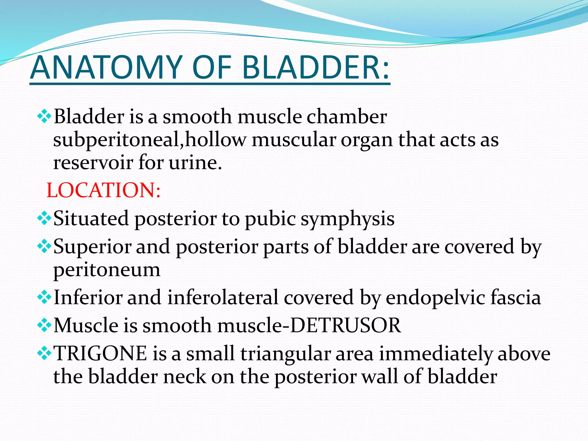
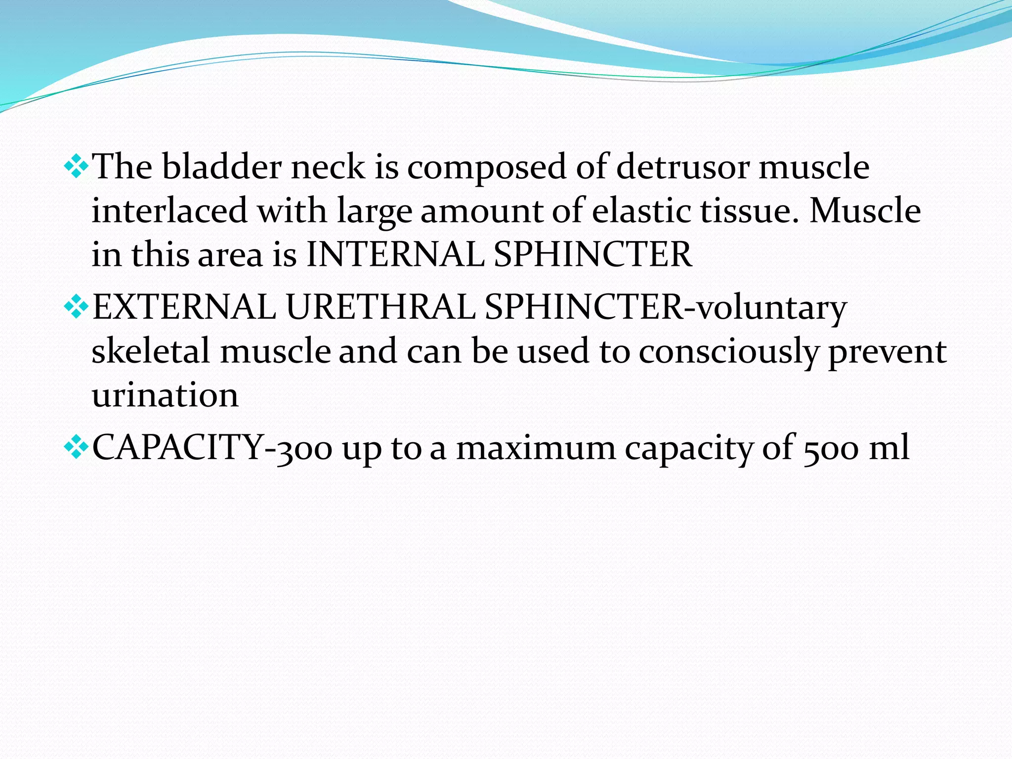
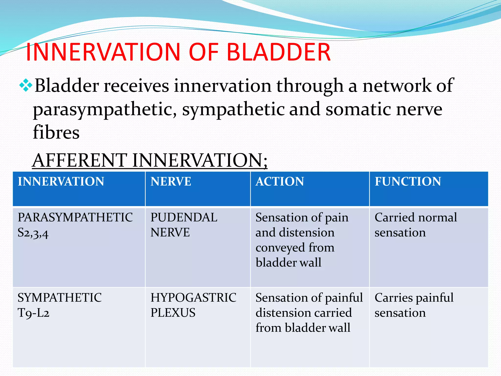
MODERATOR:- Dr.BALASUBRAMANYAM SIR
PRESENTER:- Dr.MOUNICA.K
ANATOMY OF BLADDER:
Bladder is a smooth muscle chamber
subperitoneal,hollow muscular organ that acts as
reservoir for urine.
LOCATION:
Situated posterior to pubic symphysis
Superior and posterior parts of bladder are covered by
peritoneum
Inferior and inferolateral covered by endopelvic fascia
Muscle is smooth muscle-DETRUSOR
TRIGONE is a small triangular area immediately above
the bladder neck on the posterior wall of bladder
Parts of bladder
The bladder neck is composed of detrusor muscle
interlaced with large amount of elastic tissue. Muscle
in this area is INTERNAL SPHINCTER
EXTERNAL URETHRAL SPHINCTER-voluntary
skeletal muscle and can be used to consciously prevent
urination
CAPACITY-300 up to a maximum capacity of 500 ml
INNERVATION OF BLADDER
Bladder receives innervation through a network of
parasympathetic, sympathetic and somatic nerve
fibres
AFFERENT INNERVATION;
INNERVATION NERVE ACTION FUNCTION
PARASYMPATHETIC
S2,3,4
PUDENDAL
NERVE
Sensation of pain
and distension
conveyed from
bladder wall
Carried normal
sensation
SYMPATHETIC
T9-L2
HYPOGASTRIC
PLEXUS
Sensation of painful
distension carried
from bladder wall
Carries painful
sensation
EFFERENT INNERVATION;
INNERVATION NERVE ACTION FUNCTION
PARASYMPATHETIC
S2,3,4
PELVIC NERVE
(NERVI
ERIGENTES)-
HYPOGASTRIC
PLEXUS
Detrusor muscle
contraction and
internal sphincter
relaxation
voiding
SYMPATHETIC
T11-L2
HYPOGASTRIC
NERVES
Detrusor muscle
relaxation and
internal sphincter
contraction
storage
SOMATIC-FROM
ANTERIOR HORN
CELLS S2,3,4
PUDENDAL
NERVE
Voluntary
innervations
initiate or inhibit
micturition
through cortical
control
Voluntary control
INNERVATION OF BLADDER:
RECEPTORS AND INNERVATION;
 DETRUSOR-
intermediolateral gray
column of s2,3,4-
parasympathetic pelvic
nerve (M2 receptors)
 EUS-Somatomotor S2,3,4
nucleus(onuf’s nucleus)-
pudendal nerve (nicotinic
receptors)
 TRIGONE and IUS-
Innervated by sympathetic
nervous system acts
through beta2 and alpha1
receptors
SPINAL REFLEX ARC
•
 Afferrent arc-Sensation of
stretch arising from bladder
wall travels through
parasympathetic nerves to
centres for micturition
 Detrusor centre or sacral
parasympathetic nucleus-
sacral segments s2,3,4 of
spinal cord
 Efferent arc-travels through
pelvic nerves to pelvis plexus
and short postganglionic
fibres travel from plexus to
detrusor muscle
HIGHER CENTRES
1.CORTICAL CENTRES
a.Medial frontal lobe
b.Cingulate gyrus
c.Corpus callosum
2.SUBCORTICAL CENTRES
a.Thalamic nucleus
b.Limbic system
c.Red nucleus
d.Substantia nigra
e.Hypothalamus
f.Subthalamic nucleus
3.CEREBELLUM
a.Anterior vermis
b.Fastigial nucleus
FRONTAL LOBE OF BRAIN
 Contains PRIMARY MICTURITION CENTRE
 It helps in voluntary urination
 It sends inhibitory impulses to detrusor muscle via
pons to prevent bladder emptying until socially
acceptable place is available
 Lesion results in loss of voluntary control to
normal micturition
 PONS
 Pons contains pontine mictutrition centre –
BARRINGTON NUCLEUS
• PONTOMESENCEPHALIC RETICULAR
FORMATION MICTURITION CENTER-Located in
locus ceruleus,pontomesencephalic gray matter and
nucleus tegmentolateralis dorsalis
• Collection of cell bodies located in rostral pons in
brainstem involved in supraspinal regulation of
micturition
• Connections with medial frontal cortex,insular
cortex,hypothalamus,periaqueductal gray
 PERIAQUEDUCTAL GRAY
 It is the relay station for ascending information from
spinal cord and incoming information from higher
brain areas
 SACRAL SPINAL CORD
 Terminal portion of spinalcord
 It has sacral spinal reflex centre
 It is a primitive micturition centre
 Upto 3-4 yrs of age micturition is controlled by
SRC
PHYSIOLOGY OF MICTURITION
Micturition is a spinobulbospinal reflex
 In response to stretch impulses afferent impulses are
carried to spinal cord
 From spinal cord information is carried to pontine
micturition centre(Barringtons nucleus)
 PMC sends descending fibres to preganglionic
parasympathetic motor neurons in spinal cord
innervating bladder
 PMC is under control of centres in forebrain
Neural regulation of storage phase and
voiding phase
STORAGE PHASE
 Storage reflexes. During filling,
there is low-level activity from
bladder afferent fiberssignaling
distension via the pelvic nerve,
which in turn stimulates
sympathetic outflow to the bladder
neck and wall via the hypogastric
nerve. This sympathetic
stimulation relaxes the detrusor
and contracts the bladder neck at
the internal sphincter. Afferent
pelvic nerve impulses also
stimulate the pudendal (somatic)
outflow to the external sphincter
causing contraction and
maintenance of continence.
VOIDING PHASE
Voiding reflexes. Upon initiation of
micturition, there is high-intensity
afferent activity signaling wall
tension, which activates the
brainstem pontine micturition
center. Spinobulbospinal reflex can
be seen as an ascending signal from
afferent pelvic nerve stimulation
(left side), which passes through the
periaqueductal gray matter before
reaching the pontine micturition
center and descending (right side)
to elicit parasympathetic
contraction of the detrusor, and
somatic relaxation via the pudendal
nerve.
 In infants bladder function is purely reflex but with
cortical maturation and completion of myelination
inhibitory control over this reflex develops as well as
voluntary regulation of external sphincter
 Normal micturition requires intact autonomic and
spinal pathways ,cerebral inhibition and control of
EUS must be normal
NEUROGENIC BLADDER
 Neurogenic bladder refers to dysfunction of urinary
bladder caused due to disease of central nervous
system or peripheral nerves involved in control of
micturition
 NEWER CLASSIFICATION OF BLADDER
DYSFUNCTION
1. SUPRAPONTINE
2. INFRAPONTINE/SUPRASACRAL
3. INFRASACRAL
SUPRAAPONTINE SUPRASACRAL/
INFRAPONTINE
INFRASACRAL
SYMPTOMS STORAGE PROBLEM
FREQUENCY
,URGENCY AND URGE
INCONTINENCE
MIXED OVERLAP
OF HESITANCY
,INTERRUPTED
STREAM,FREQUEN
CY ,URGENCY
VOIDING
PROBLEM,HESIT
ANCY,OVERFLO
W
INCONTINENCE
POSTVOIDAL
RESIDUAL URIE
NORMAL HIGH VERY HIGH
UROFLOWMETRY NORMAL INTERRUPTED
FLOW
POOR OR
ABSENT FLOW
URODYNAMICS DETRUSOR
OVERACTIVITY
DETRUSOR
SPHINCTER
DYSSYNERGIA
DETRUSOR
UNDERACTIVITY
,SPHINCTER
INSUFFICIENCY
NEUROGENIC BLADDER
 OLDER CLASSIFICATION-LIPIDES
CLASSIFICATION
 1.LOSS OF SUPRASPINAL CONTROL-UNINHIBITED
BLADDER
 2.SPINAL CORD LESION ABOVE SACRAL LEVEL-
REFLEX NEUROGENIC BLADDER –AUTOMATIC
 3.SPINAL CORD LESION INVOLVING SACRAL
LEVEL-AUTONOMOUS BLADDER
 4.LESIONS INVOLVING AFFERENT SENSORY
NEURONS-SENSORY NEUROGENIC BLADDER
 5.LESIONS INVOLVING EFFERENT MOTOR
NEURONS-MOTOR PARALYTIC BLADDER
 6.OTHERS-STROKE/DEMENTIA/SPINAL SHOCK
1.UNINHIBITED/CORTICAL BLADDER
 Lesion of CNS involving area above pons.
 Uninhibited impulses to pontine micturition
centre/barrington nucleus leading to s2,3,4 and
micturition
 C/f-frequency,urgency and urge incontinence
 No or absent residual volume so called safe bladder
 Also called as social disinhibition bladder syndrome
 Seen in CVA, frontal tumors, parkinsons
disease,demyelinating disease
2.REFLEX NEUROGENIC BLADDER/SPASTIC
 Spinalcord lesion above sacral level and below pons
 Occurs with severe myelopathy or extensive brain
lesions causing interruption of both the descending
autonomic tracts to the bladder and the ascending
sensory pathways above sacral segments of cord
 UMN CUT OFF BUT LMN IS INTACT
 Detrusor sphincter synergia is lost
 This results in obstructed voiding,interrupted urinary
stream,incomplete emptying and high intravesical
pressures because sphincter fails to relax correctly
 Loss of normal inhibition from higher centres results
in detrusor contraction during filling
 Detrusor becomes overactive so there is urinary
frequency,urgency,urge incontinene
 Inability to initiate micturition voluntarily
 Associated with quadriplegia or paraplegia and in
advanced cases of multiple sclerosis
3.AUTONOMOUS BLADDER
 Spinal cord lesion lesion involving sacral level
 Denervation of both afferent and efferent supply to
bladder
 LMN type of bladder-large painless dribbling bladder
 Bladder tone flaccid ,sensation absent, inability to
initiate micturition
 Increased bladder capacity and residual urine
 Overflow incontinence, no urgency
 No bladder reflex activity, infection risk high
 Causes-neoplastic, traumatic lesions, cauda equina
,conus medullaris syndrome
4.SENSORY NEUROGENIC BLADDER
 Impaired bladder sensation but motor control is intact
 Sensation is absent and there is no desire to void
 Initiation of micturition is possible
 c/o urinary retention or overflow incontinence
 More prone for urinary tract infections
 If bladder not voided at regular intervals over
distention of bladder
 Causes-Tabes dorsalis,Diabetic neuropathy
5.MOTOR PARALYTIC BLADDER
 Motor supply to bladder is interrupted but sensation is
intact
 Bladder tone flaccid
 c/o painful retention of urine or impaired bladder
emptying
 Inability to initiate or maintain micturition
 Bladder capacity and residual urine markedly
increased.HIGH risk of UTI
 Causes-lumbosacral meningomyelocele,tethered cord
syndrome,lumbar spinal stenosis
UMN/LMN/SENSORY LESION
REFLEXIC-
SPASTIC/UNIHIBITED/
UMN
AREFLEXIC –
AUTONOMUS/Flaccid/
LMN
Sensory
Characteristics
No inhibitions influence
time & place of voiding
bladder empties in response
to stretching of bladder wall
Characteristics
Bladder acts as if there were
paralysis of all motor
functions
Characteristics
Lack of sensation of need to
urinate
Clinical manifestations
-Incontinence
-Frequency
-Urgency
-Voiding is unpredictable
and incomplete
Clinical manifestations
Fills without emptying
Retention
Dribbling incontinence
Clinical manifestations
Poor bladder sensation,
infrequent voiding of large
residual volume
Causes;
Corticospinal tract lesion
observed in
SCI/stroke/multiple
sclerosis/brain tumor/brain
trauma
Causes;
Lower motor neuron lesion
caused by trauma involving
S2-S4 lesions of cauda
equina/pelvic nerves
Causes;
Damage to sensory limb of
bladder spinal reflex arc seen
in multiple scierosis/diabetes
mellitus
URINARY INCONTINENCE AND TYPES
 LOSS OF BLADDER CONTROL
1. Stress – urine loss during activities such as
coughing, sneezing, laughing or lifting.
2. Urge – a sudden need to urinate, occasionally with
large volume urine loss. Can also exist without
incontinence(urgency).
3. Overflow – a frequent dribble of urine as a result of
inefficient bladder emptying symptoms are similar to
stress incontinence.
4. Mixed – stress + urge forms.
5. Functional – urine loss not associated with any
pathology or problem in the urine system.
EVALUATION
HISTORY
Urgency/hesistancy-storage or voiding
EXAMINATION
INVESTIGATIONS
1. Screening for urinary tract infections
2. Bladder scan-to measure post residual volume by usg
3. USG for upper urinary tract abnormalities or renal
scarring
4. Urodynamic studies-to study function of lower
urinary tract
Assessment of lower urinary tract
 Urodynamic studies are necessary to document type of
bladder dysfunction
 Measurement of urine flow rate
 Measurement of post void residual urine
 Cystometry during filling and voiding
 Video cystometry
 Urethral pressure profilometry
Complications of neurogenic
bladder dysfunction
 DETRUSOR OVERACTIVITY-raised intravesical
pressure that leads to structural changes in bladder
wall such as trabeculations and diverticulae
 Upper urinary tract also show changes such as
vesicoureteric reflex,hydronephrosis,renal
impairement and even end stage renal failure
 Also more prone to variety of genitourinary tract
infections such as cystitis,pyelonephritis,and
epididymoorchitis and also to bladder stones
MANAGEMENT
 GOALS-
1. Achieve urinary continence
2. Prevent uti
3. Preseve upper urinary tract function
A. GENERAL MEASURES
If symptoms are mild
Fluid resuscitation
Avoidance of caffeine
 B.VOIDING DYSFUNCTION
a) If residual urine is more than 100ml or more than
1/3rd of bladder capacity-STERILE INTERMITTENT
SELF CATHETERISATION is advised
b) Reflex voiding using trigger techniques and crede
manouvre are generally avoided
c) Alpha blockers-relaxes internal urethral sphincter
and this improves bladder emptying and reduce post
voidal residual urine
 C.STORAGE DYSFUNCTION
a) Antimuscarinic medications are used for detrusor
overactivity
b) DESMOPRESSIN –Synthetic analogue of arginine
vasopressin that decreases urine production and
increases reabsorption of water from DCT and CT
c) BOTULINUM TOXIN-blocks synaptic release of
acetylcholine from the parasympathetic nerve
endings and produces paralysis of detrusor muscle
d) PERIPHERAL NERVE STIMULATION
DRUGS USED
MUSCARINIC
ANTAGONIST
BETA 2 AGONIST ALPHA 1 ANTAGONIST
OXYBUTININ
DOREFENACIN
SOLEFANACIN
TOLTERODINE
MIRABEGRON TERAZOSOIN
DOXAZOSIN
USED IN TREATMENT
OF OVERACTIVE
BLADDE AND URGE
INCONTINEN CE
TRETMENT OF
OVERACTIVE BLADDER
USED IN TREATMENT
OF STRESS
INCONTINENCE AND
BPH
 UROLOGICAL PROCEDURES PERFORMED TO
TREAT VARIOUS CAUSES OF INCONTINENCE
STRESS INCONTINENCE-PELVIC
FLOOR WEAKNESS
BLADDER NECK SUSPENSION
TENSION FREE VAGINAL TAPE
TRANSOBTURATOR TAPE
STRESS INCONTINENCE-SPHINCTER
ABNORMALITY
ARTIFICIAL SPHINCTER
URGENCY INCONTINENCE-
DETRUSOR OVERACTIVITY
BOTULINUM TOXIN
SACRAL NEUROMODULATION
AUGMENTATION CYSTOPLASTY
INTRACTABLE INCONTINENCE URINARY DIVERSION WITH STOMA
 URINARY TRACT INFECTIONS IN
NEUROGENIC BLADDER
 Presence of asymptomatic bacteria alone in a
patient performing cisc should not be a indication
to start antibiotics
 Indication to start antibiotic-presence of
associated symptoms local or systemic features
THANK YOU

NEUROGENIC BLADDER-1.pptx

  • 1.
  • 2.
    ANATOMY OF BLADDER: Bladderis a smooth muscle chamber subperitoneal,hollow muscular organ that acts as reservoir for urine. LOCATION: Situated posterior to pubic symphysis Superior and posterior parts of bladder are covered by peritoneum Inferior and inferolateral covered by endopelvic fascia Muscle is smooth muscle-DETRUSOR TRIGONE is a small triangular area immediately above the bladder neck on the posterior wall of bladder
  • 3.
  • 4.
    The bladder neckis composed of detrusor muscle interlaced with large amount of elastic tissue. Muscle in this area is INTERNAL SPHINCTER EXTERNAL URETHRAL SPHINCTER-voluntary skeletal muscle and can be used to consciously prevent urination CAPACITY-300 up to a maximum capacity of 500 ml
  • 5.
    INNERVATION OF BLADDER Bladderreceives innervation through a network of parasympathetic, sympathetic and somatic nerve fibres AFFERENT INNERVATION; INNERVATION NERVE ACTION FUNCTION PARASYMPATHETIC S2,3,4 PUDENDAL NERVE Sensation of pain and distension conveyed from bladder wall Carried normal sensation SYMPATHETIC T9-L2 HYPOGASTRIC PLEXUS Sensation of painful distension carried from bladder wall Carries painful sensation
  • 6.
    EFFERENT INNERVATION; INNERVATION NERVEACTION FUNCTION PARASYMPATHETIC S2,3,4 PELVIC NERVE (NERVI ERIGENTES)- HYPOGASTRIC PLEXUS Detrusor muscle contraction and internal sphincter relaxation voiding SYMPATHETIC T11-L2 HYPOGASTRIC NERVES Detrusor muscle relaxation and internal sphincter contraction storage SOMATIC-FROM ANTERIOR HORN CELLS S2,3,4 PUDENDAL NERVE Voluntary innervations initiate or inhibit micturition through cortical control Voluntary control
  • 7.
  • 8.
    RECEPTORS AND INNERVATION; DETRUSOR- intermediolateral gray column of s2,3,4- parasympathetic pelvic nerve (M2 receptors)  EUS-Somatomotor S2,3,4 nucleus(onuf’s nucleus)- pudendal nerve (nicotinic receptors)  TRIGONE and IUS- Innervated by sympathetic nervous system acts through beta2 and alpha1 receptors
  • 9.
    SPINAL REFLEX ARC • Afferrent arc-Sensation of stretch arising from bladder wall travels through parasympathetic nerves to centres for micturition  Detrusor centre or sacral parasympathetic nucleus- sacral segments s2,3,4 of spinal cord  Efferent arc-travels through pelvic nerves to pelvis plexus and short postganglionic fibres travel from plexus to detrusor muscle
  • 10.
    HIGHER CENTRES 1.CORTICAL CENTRES a.Medialfrontal lobe b.Cingulate gyrus c.Corpus callosum 2.SUBCORTICAL CENTRES a.Thalamic nucleus b.Limbic system c.Red nucleus d.Substantia nigra e.Hypothalamus f.Subthalamic nucleus 3.CEREBELLUM a.Anterior vermis b.Fastigial nucleus
  • 11.
    FRONTAL LOBE OFBRAIN  Contains PRIMARY MICTURITION CENTRE  It helps in voluntary urination  It sends inhibitory impulses to detrusor muscle via pons to prevent bladder emptying until socially acceptable place is available  Lesion results in loss of voluntary control to normal micturition
  • 12.
     PONS  Ponscontains pontine mictutrition centre – BARRINGTON NUCLEUS • PONTOMESENCEPHALIC RETICULAR FORMATION MICTURITION CENTER-Located in locus ceruleus,pontomesencephalic gray matter and nucleus tegmentolateralis dorsalis • Collection of cell bodies located in rostral pons in brainstem involved in supraspinal regulation of micturition • Connections with medial frontal cortex,insular cortex,hypothalamus,periaqueductal gray
  • 13.
     PERIAQUEDUCTAL GRAY It is the relay station for ascending information from spinal cord and incoming information from higher brain areas
  • 14.
     SACRAL SPINALCORD  Terminal portion of spinalcord  It has sacral spinal reflex centre  It is a primitive micturition centre  Upto 3-4 yrs of age micturition is controlled by SRC
  • 15.
    PHYSIOLOGY OF MICTURITION Micturitionis a spinobulbospinal reflex  In response to stretch impulses afferent impulses are carried to spinal cord  From spinal cord information is carried to pontine micturition centre(Barringtons nucleus)  PMC sends descending fibres to preganglionic parasympathetic motor neurons in spinal cord innervating bladder  PMC is under control of centres in forebrain
  • 17.
    Neural regulation ofstorage phase and voiding phase STORAGE PHASE  Storage reflexes. During filling, there is low-level activity from bladder afferent fiberssignaling distension via the pelvic nerve, which in turn stimulates sympathetic outflow to the bladder neck and wall via the hypogastric nerve. This sympathetic stimulation relaxes the detrusor and contracts the bladder neck at the internal sphincter. Afferent pelvic nerve impulses also stimulate the pudendal (somatic) outflow to the external sphincter causing contraction and maintenance of continence.
  • 18.
    VOIDING PHASE Voiding reflexes.Upon initiation of micturition, there is high-intensity afferent activity signaling wall tension, which activates the brainstem pontine micturition center. Spinobulbospinal reflex can be seen as an ascending signal from afferent pelvic nerve stimulation (left side), which passes through the periaqueductal gray matter before reaching the pontine micturition center and descending (right side) to elicit parasympathetic contraction of the detrusor, and somatic relaxation via the pudendal nerve.
  • 19.
     In infantsbladder function is purely reflex but with cortical maturation and completion of myelination inhibitory control over this reflex develops as well as voluntary regulation of external sphincter  Normal micturition requires intact autonomic and spinal pathways ,cerebral inhibition and control of EUS must be normal
  • 20.
    NEUROGENIC BLADDER  Neurogenicbladder refers to dysfunction of urinary bladder caused due to disease of central nervous system or peripheral nerves involved in control of micturition  NEWER CLASSIFICATION OF BLADDER DYSFUNCTION 1. SUPRAPONTINE 2. INFRAPONTINE/SUPRASACRAL 3. INFRASACRAL
  • 22.
    SUPRAAPONTINE SUPRASACRAL/ INFRAPONTINE INFRASACRAL SYMPTOMS STORAGEPROBLEM FREQUENCY ,URGENCY AND URGE INCONTINENCE MIXED OVERLAP OF HESITANCY ,INTERRUPTED STREAM,FREQUEN CY ,URGENCY VOIDING PROBLEM,HESIT ANCY,OVERFLO W INCONTINENCE POSTVOIDAL RESIDUAL URIE NORMAL HIGH VERY HIGH UROFLOWMETRY NORMAL INTERRUPTED FLOW POOR OR ABSENT FLOW URODYNAMICS DETRUSOR OVERACTIVITY DETRUSOR SPHINCTER DYSSYNERGIA DETRUSOR UNDERACTIVITY ,SPHINCTER INSUFFICIENCY
  • 23.
    NEUROGENIC BLADDER  OLDERCLASSIFICATION-LIPIDES CLASSIFICATION  1.LOSS OF SUPRASPINAL CONTROL-UNINHIBITED BLADDER  2.SPINAL CORD LESION ABOVE SACRAL LEVEL- REFLEX NEUROGENIC BLADDER –AUTOMATIC  3.SPINAL CORD LESION INVOLVING SACRAL LEVEL-AUTONOMOUS BLADDER  4.LESIONS INVOLVING AFFERENT SENSORY NEURONS-SENSORY NEUROGENIC BLADDER  5.LESIONS INVOLVING EFFERENT MOTOR NEURONS-MOTOR PARALYTIC BLADDER  6.OTHERS-STROKE/DEMENTIA/SPINAL SHOCK
  • 24.
    1.UNINHIBITED/CORTICAL BLADDER  Lesionof CNS involving area above pons.  Uninhibited impulses to pontine micturition centre/barrington nucleus leading to s2,3,4 and micturition  C/f-frequency,urgency and urge incontinence  No or absent residual volume so called safe bladder  Also called as social disinhibition bladder syndrome  Seen in CVA, frontal tumors, parkinsons disease,demyelinating disease
  • 25.
    2.REFLEX NEUROGENIC BLADDER/SPASTIC Spinalcord lesion above sacral level and below pons  Occurs with severe myelopathy or extensive brain lesions causing interruption of both the descending autonomic tracts to the bladder and the ascending sensory pathways above sacral segments of cord  UMN CUT OFF BUT LMN IS INTACT  Detrusor sphincter synergia is lost  This results in obstructed voiding,interrupted urinary stream,incomplete emptying and high intravesical pressures because sphincter fails to relax correctly
  • 26.
     Loss ofnormal inhibition from higher centres results in detrusor contraction during filling  Detrusor becomes overactive so there is urinary frequency,urgency,urge incontinene  Inability to initiate micturition voluntarily  Associated with quadriplegia or paraplegia and in advanced cases of multiple sclerosis
  • 27.
    3.AUTONOMOUS BLADDER  Spinalcord lesion lesion involving sacral level  Denervation of both afferent and efferent supply to bladder  LMN type of bladder-large painless dribbling bladder  Bladder tone flaccid ,sensation absent, inability to initiate micturition  Increased bladder capacity and residual urine  Overflow incontinence, no urgency  No bladder reflex activity, infection risk high  Causes-neoplastic, traumatic lesions, cauda equina ,conus medullaris syndrome
  • 28.
    4.SENSORY NEUROGENIC BLADDER Impaired bladder sensation but motor control is intact  Sensation is absent and there is no desire to void  Initiation of micturition is possible  c/o urinary retention or overflow incontinence  More prone for urinary tract infections  If bladder not voided at regular intervals over distention of bladder  Causes-Tabes dorsalis,Diabetic neuropathy
  • 29.
    5.MOTOR PARALYTIC BLADDER Motor supply to bladder is interrupted but sensation is intact  Bladder tone flaccid  c/o painful retention of urine or impaired bladder emptying  Inability to initiate or maintain micturition  Bladder capacity and residual urine markedly increased.HIGH risk of UTI  Causes-lumbosacral meningomyelocele,tethered cord syndrome,lumbar spinal stenosis
  • 30.
    UMN/LMN/SENSORY LESION REFLEXIC- SPASTIC/UNIHIBITED/ UMN AREFLEXIC – AUTONOMUS/Flaccid/ LMN Sensory Characteristics Noinhibitions influence time & place of voiding bladder empties in response to stretching of bladder wall Characteristics Bladder acts as if there were paralysis of all motor functions Characteristics Lack of sensation of need to urinate Clinical manifestations -Incontinence -Frequency -Urgency -Voiding is unpredictable and incomplete Clinical manifestations Fills without emptying Retention Dribbling incontinence Clinical manifestations Poor bladder sensation, infrequent voiding of large residual volume Causes; Corticospinal tract lesion observed in SCI/stroke/multiple sclerosis/brain tumor/brain trauma Causes; Lower motor neuron lesion caused by trauma involving S2-S4 lesions of cauda equina/pelvic nerves Causes; Damage to sensory limb of bladder spinal reflex arc seen in multiple scierosis/diabetes mellitus
  • 31.
    URINARY INCONTINENCE ANDTYPES  LOSS OF BLADDER CONTROL 1. Stress – urine loss during activities such as coughing, sneezing, laughing or lifting. 2. Urge – a sudden need to urinate, occasionally with large volume urine loss. Can also exist without incontinence(urgency). 3. Overflow – a frequent dribble of urine as a result of inefficient bladder emptying symptoms are similar to stress incontinence. 4. Mixed – stress + urge forms. 5. Functional – urine loss not associated with any pathology or problem in the urine system.
  • 33.
    EVALUATION HISTORY Urgency/hesistancy-storage or voiding EXAMINATION INVESTIGATIONS 1.Screening for urinary tract infections 2. Bladder scan-to measure post residual volume by usg 3. USG for upper urinary tract abnormalities or renal scarring 4. Urodynamic studies-to study function of lower urinary tract
  • 34.
    Assessment of lowerurinary tract  Urodynamic studies are necessary to document type of bladder dysfunction  Measurement of urine flow rate  Measurement of post void residual urine  Cystometry during filling and voiding  Video cystometry  Urethral pressure profilometry
  • 35.
    Complications of neurogenic bladderdysfunction  DETRUSOR OVERACTIVITY-raised intravesical pressure that leads to structural changes in bladder wall such as trabeculations and diverticulae  Upper urinary tract also show changes such as vesicoureteric reflex,hydronephrosis,renal impairement and even end stage renal failure  Also more prone to variety of genitourinary tract infections such as cystitis,pyelonephritis,and epididymoorchitis and also to bladder stones
  • 36.
    MANAGEMENT  GOALS- 1. Achieveurinary continence 2. Prevent uti 3. Preseve upper urinary tract function A. GENERAL MEASURES If symptoms are mild Fluid resuscitation Avoidance of caffeine
  • 37.
     B.VOIDING DYSFUNCTION a)If residual urine is more than 100ml or more than 1/3rd of bladder capacity-STERILE INTERMITTENT SELF CATHETERISATION is advised b) Reflex voiding using trigger techniques and crede manouvre are generally avoided c) Alpha blockers-relaxes internal urethral sphincter and this improves bladder emptying and reduce post voidal residual urine
  • 39.
     C.STORAGE DYSFUNCTION a)Antimuscarinic medications are used for detrusor overactivity b) DESMOPRESSIN –Synthetic analogue of arginine vasopressin that decreases urine production and increases reabsorption of water from DCT and CT c) BOTULINUM TOXIN-blocks synaptic release of acetylcholine from the parasympathetic nerve endings and produces paralysis of detrusor muscle d) PERIPHERAL NERVE STIMULATION
  • 40.
    DRUGS USED MUSCARINIC ANTAGONIST BETA 2AGONIST ALPHA 1 ANTAGONIST OXYBUTININ DOREFENACIN SOLEFANACIN TOLTERODINE MIRABEGRON TERAZOSOIN DOXAZOSIN USED IN TREATMENT OF OVERACTIVE BLADDE AND URGE INCONTINEN CE TRETMENT OF OVERACTIVE BLADDER USED IN TREATMENT OF STRESS INCONTINENCE AND BPH
  • 42.
     UROLOGICAL PROCEDURESPERFORMED TO TREAT VARIOUS CAUSES OF INCONTINENCE STRESS INCONTINENCE-PELVIC FLOOR WEAKNESS BLADDER NECK SUSPENSION TENSION FREE VAGINAL TAPE TRANSOBTURATOR TAPE STRESS INCONTINENCE-SPHINCTER ABNORMALITY ARTIFICIAL SPHINCTER URGENCY INCONTINENCE- DETRUSOR OVERACTIVITY BOTULINUM TOXIN SACRAL NEUROMODULATION AUGMENTATION CYSTOPLASTY INTRACTABLE INCONTINENCE URINARY DIVERSION WITH STOMA
  • 43.
     URINARY TRACTINFECTIONS IN NEUROGENIC BLADDER  Presence of asymptomatic bacteria alone in a patient performing cisc should not be a indication to start antibiotics  Indication to start antibiotic-presence of associated symptoms local or systemic features
  • 44.