The document outlines the neurological control of bladder function, detailing the roles of sympathetic, parasympathetic, and somatic pathways in micturition and sphincter control. It explains different classifications of neurogenic bladder, describing conditions such as supraspinal lesions, spinal cord lesions, and sensory and motor paralytic bladders, each affecting bladder function in distinct ways. Additionally, it discusses the evaluation methods for bladder dysfunction, including investigations like bladder scans and urodynamic studies, along with management strategies.


![Detrusor muscle
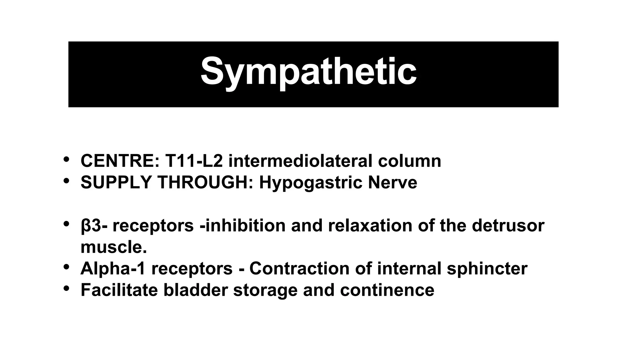
• Sympthetic (T11 - L2) [Hypogastric nerve] -Via β3-
adrenergic receptors - inhibition and relaxation of the
detrusor muscle
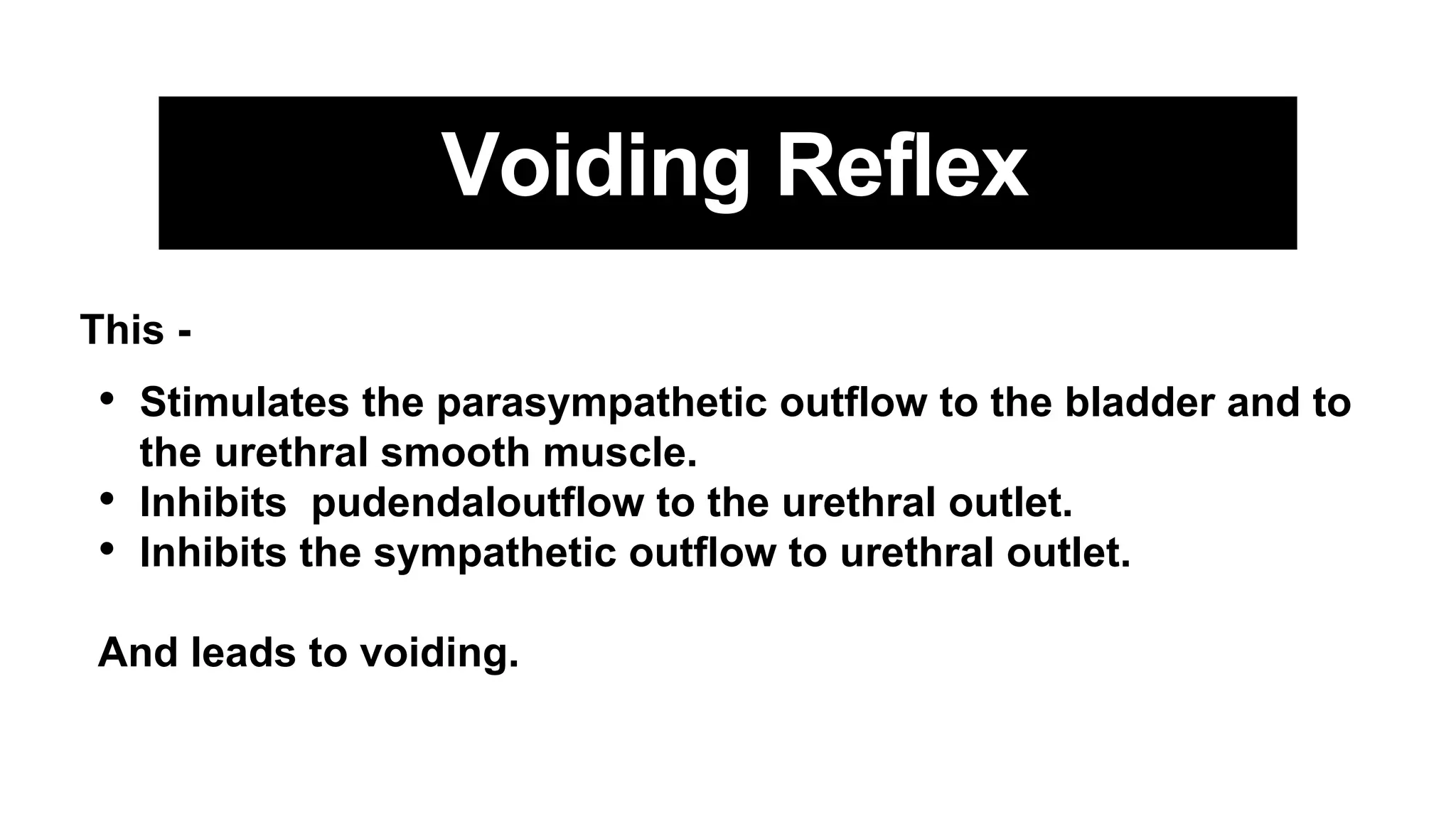
• Parasympathetic (S2,S3,S4) [Pelvic splanchnic nerve] - via
M2/M3 cholinergic receptors (NT - Ach) - contraction of the
detrusor muscle.](https://image.slidesharecdn.com/bladderpresentationreshma-240507184941-7f7492f4/75/Urinary-Bladder-neurology-presentation-pptx-3-2048.jpg)
![Internal Sphincter
• Smooth muscle
• Involuntary control
• Sympthetic (T11 - L2) [Hypogastric nerve] -Through alpha-1
receptors causes Contraction of internal sphincter](https://image.slidesharecdn.com/bladderpresentationreshma-240507184941-7f7492f4/75/Urinary-Bladder-neurology-presentation-pptx-4-2048.jpg)
![External Sphincter
• Skeletal muscle
• Voluntary control
• Innervated by Somatic nervous system (S2,S3,S4)
[Pudendal nerve] -Through Nicotinic receptors (NT - Ach)](https://image.slidesharecdn.com/bladderpresentationreshma-240507184941-7f7492f4/75/Urinary-Bladder-neurology-presentation-pptx-5-2048.jpg)