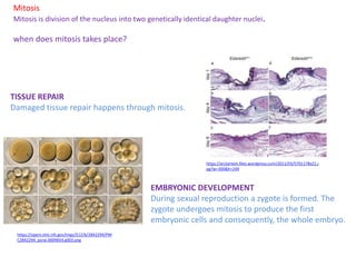
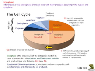
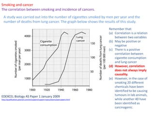
1.6. Cell division
Miltiadis-Spyridon Kitsos
Platon IB Diploma
http://www.edupic.net/Images/Mitosis/prometaphase.png
The official IB Diploma Biology guide
Essential idea: Cell division is essential but must be controlled.
https://ibpublishing.ibo.org/server2/rest/app/tsm.xql?doc=d_4_biolo_gui_1402_1_e&part=8
&chapter=1
Mitosis
Mitosis is division of the nucleus into two genetically identical daughter nuclei
Mitosis is a type of cell division happening in eukaryotic cells, which creates two
genetically identical daughter cells.
when does mitosis takes place?
TISSUE GROWTH
In multicellular organisms mitosis increases the number of
cells, and hence, the size of the organisms.
http://emp.byui.edu/wellerg/Secondary%20Growth/I
mages/Corn%20Stem.jpg
http://book.bionumbers.org/wp-
content/uploads/2014/07/115-f1-Yeast-
micrograph-11.png
ASEXUAL REPRODUCTION OF UNICELLULAR EUKARYOTES
Yeast cells, reproduce asexually, via budding. The formation
of the bud is a result of mitosis and cytokinesis.
Mitosis
Mitosis is division of the nucleus into two genetically identical daughter nuclei.
TISSUE REPAIR
Damaged tissue repair happens through mitosis.
EMBRYONIC DEVELOPMENT
During sexual reproduction a zygote is formed. The
zygote undergoes mitosis to produce the first
embryonic cells and consequently, the whole embryo.
https://arztartem.files.wordpress.com/2011/03/5701178x21.j
pg?w=300&h=249
https://openi.nlm.nih.gov/imgs/512/6/2842294/PM
C2842294_pone.0009654.g003.png
when does mitosis takes place?
Mitosis
Mitosis is division of the nucleus into two genetically identical daughter nuclei.
Slide from
Slide from
The cell cycle
Mitosis is division of the nucleus into two genetically identical daughter nuclei.
The cell cycle is a series of events between one cell division and the next. The result of the
cell cycle is the formation of two genetically identical cells.
Interphase
Interphase is a very active phase of the cell cycle with many processes occurring in the nucleus and
cytoplasm.
Slide from
Supercoiling of chromosomes
Chromosomes condense by supercoiling during mitosis.
Chromosomes (Greek: chroma (colour) + soma (body) are characteristic structures which
become visible during the different stages of mitosis. They are made of proteins and DNA
and they may gradually supercoil or uncoil.
http://www.gistsupport.org/media/cells%20%20DNA/DNA-Marina-3.jpg
https://youtu.be/OjPcT1uUZiE
Getting things straight
Chromosomes condense by supercoiling during mitosis.
This is a chromatin fiber. It is made of DNA coiled around clusters of
proteins called histones.
This is how DNA looks at the beginning of interphase. The genome is
the form of multiple chromatin fibers forming a network.
http://cdn.biologydiscussion.com/wp-content/uploads/2015/11/clip_image016_thumb4.jpg
During the S phase of interphase DNA undergoes replication. At
the end of replication each chromatin fiber has replicated. The
two identical molecules are now called sister chromatids and
they are attached to the centromere.
Getting things straight
Chromosomes condense by supercoiling during mitosis.
At the start of mitosis, sister chromatids start to supercoil with
the aid of the histones and other enzymes. Supercoiling
completes during metaphase when the chromosomes assume
their maximum degree of coiling.
http://www.wiley.com/college/trefil/04701
18547/animations/ch23/dna_coiling/09_02.
Prophase
Identification of phases of mitosis in cells viewed with a microscope.
https://upload.wikimedia.org/wikipedia/commons/5/58/Allium-Mitose06-DM100x_BL28.jpg
http://cahsbiology.weebly.com/uploads/1/3/5/0/13500838/5120946.jpg?291
Nuclear membrane
starts to
break down
Chromosomes consist
of the two sister
chromatids attached
to the centromere
Shorter and fatter.
Centrosomes (and not centromeres) start moving towards
the opposite poles of the cell forming the Microtubule
organizing centers (MTOC’s) . Microtubules start growing
from the MTOC’s
Late Prophase
Identification of phases of mitosis in cells viewed with a microscope.
https://upload.wikimedia.org/wikipedia/commons/5/58/Allium-Mitose06-DM100x_BL28.jpg
http://cahsbiology.weebly.com/uploads/1/3/5/0/13500838/5120946.jpg?291
Nuclear membrane
has disintegrated
Spindle microtubules
MTOC
Mitotic
spindle
Metaphase
Identification of phases of mitosis in cells viewed with a microscope.
https://upload.wikimedia.org/wikipedia/commons/thumb/b/b4/Mitosis_(261_03)_Pressed%3B_ro
ot_meristem_of_Vicia_faba_(cells_in_prophase,_metaphase).jpg/1280px-
Mitosis_(261_03)_Pressed%3B_root_meristem_of_Vicia_faba_(cells_in_prophase,_metaphase).jpg
https://usercontent2.hubstatic.com/5967991_f260.jpg
The centromeres of the chromosomes attach to
microtubules from the two opposing MTOC’s
Contraction of the microtubules tests if the
attachment is correct. Contraction of the
microtubules is happening through shortening of
the microtubules at the centromere. The
chromosomes align to the equator of the cell.
Anaphase
Identification of phases of mitosis in cells viewed with a microscope.
Contraction of the microtubules leads to the
breaking of centromeres.
Each of the two sister chromatids is pulled to
opposite poles.
https://upload.wikimedia.org/wikipedia/commons/thumb/1/1c/Mitosis_%28261_10%29_Pressed%3B_root_meristem_of_Vicia_faba_%
28cells_in_anaphase%2C_prophase%29.jpg/800px-
Mitosis_%28261_10%29_Pressed%3B_root_meristem_of_Vicia_faba_%28cells_in_anaphase%2C_prophase%29.jpg
http://67.media.tumblr.com/tumblr_lqftagAg5H1qg1up7o1_1280.jpg
Telophase
Identification of phases of mitosis in cells viewed with a microscope.
https://upload.wikimedia.org/wikipedia/commons/b/b1/Mitosis_(261_07
)_Pressed%3B_root_meristem_of_Vicia_faba_(cells_in_anaphase,_teloph
ase).jpg
• Chromosomes uncoil and they are no
longer distinguishable under the
microscope.
• Nuclear envelopes reform around the
tightly packed chromatids.
• Mitotic spindle disintegrates.
• Cytokinesis is about to start.
The mitotic index
Determination of a mitotic index from a micrograph.
http://www.mondadorieducation.it/media/contenuti/sfoglialibro/120900037338_cavagna_la
boratoriobiologia/files/assets/seo/page11_images/0001.jpg
Mitotic index =
Number of cells in mitosis
Total number of cells observed
In the image below you need to
(a) count the number of cells in
different stages of mitosis
(b) and then, calculate the mitotic
index.
The mitotic index
Determination of a mitotic index from a micrograph.
Cytokinesis
Cytokinesis occurs after mitosis and is different in plant and animal cells.
http://celldivisionandreproduction.weebly.com/uploads/1/9/2/4/19240233/7619115_orig.jpg
Cytokinesis is the process of cell division that follows
mitosis.
Animal cells
Plasma membrane is pulled inwards with the aid of a
ring of contractile proteins (actin and myosin).
When cleavage furrow reaches the centre, the two cells
pinch apart.
https://youtu.be/H2HmRnYLDhA
Cytokinesis
Cytokinesis occurs after mitosis and is different in plant and animal cells.
http://celldivisionandreproduction.weebly.com/uploads/1/9/2/4/19240233/7619115_orig.jpg
Cytokinesis is the process of cell division that
follows mitosis.
Plant cells
• Vesicles are transferred to the area of the
equator and two layers of membrane are
developed.
• Vesicles are transferred in the area between
the two membranes and the middle lamella is
developed.
• The two daughter cells deposit cellulose on
both sides of lamella adjacent to the cell
membrane and the two new cell walls are
formed.
Cytokinesis
Cytokinesis occurs after mitosis and is different in plant and animal cells.
http://celldivisionandreproduction.weebly.com/uploads/1/9/2/4/19240233/7619115_orig.jpg
Plant cells
Cell plate
Middle lamella
Cytokinesis
Cytokinesis occurs after mitosis and is different in plant and animal cells.
Media
https://highered.mheducation.com/sites/9834092339/
student_view0/chapter10/animation_-
_cytokinesis.html
http://www.sumanasinc.com/webcontent/animations/content/mitosis.html
https://highered.mheducation.com/sites/0072495855
/student_view0/chapter2/animation__mitosis_and_c
ytokinesis.html
http://www.johnkyrk.com/mitosis.html
Slide from
Slide from
Slide from
Slide from
Slide from
Cyclins
Cyclins are involved in the control of the cell cycle.
Cyclins are a group of proteins that ensures:
• tasks in mitosis will happen on the correct time and
• cell advances to the next stage when appropriate
Their action is concentration-dependent. In high
concentrations, they bind to specific enzymes called cyclin
dependent kinases. These enzyme will in turn add
phosphate groups to other proteins, activating them.
These activated proteins are responsibly for many tasks
during mitosis
P
CDK
Mitosis promoting factor
https://highered.mheducation.com/olcweb/cgi/pluginpop.cgi?it=sw
f::535::535::/sites/dl/free/0072437316/120082/bio34a.swf::Control
%20of%20the%20Cell%20Cycle
http://upload.wikimedia.org/wikipedia/commons/thumb/9/99/Protein_CCNE1_PDB_1w98.png/800px-Protein_CCNE1_PDB_1w98.png
Triggers cells to
move from G0 to
G1 and from G1
into S phase.
prepares the cell
for DNA
replication in S
phase.
activates DNA
replication inside
the nucleus in S
phase.
promotes the assembly
of the mitotic spindle
and other tasks in the
cytoplasm to prepare
for mitosis.
Cyclins
Cyclins are involved in the control of the cell cycle.
There are four different kinds of cyclins.
Cyclins
Serendipity and scientific discoveries: the discovery of cyclins was accidental.
Tim Hunt discovered cyclins during a series of experiments on
the synthesis of proteins in sea urchin fertilized eggs.
He traced a protein that would be synthesized for 30 minutes
and then being broken down.
Tim Hunt’s experiments initiated a series of innovative
breakthroughs that would explain the fundamental
mechanism determining cell division. For this discovery, Tim
Hunt—together with Lee Hartwell and Paul Nurse—would
receive the 2001 Nobel Prize in Physiology or Medicine
Tim Hunt has always emphasized on the effect of serendipity
on his scientific discoveries.
For example, in his Nobel prize lecture he noted that he was
lucky enough to select 10-minute intervals in his experiments.
This selection revealed the true dynamics in the protein
synthesis
http://www.cell.com/cell/pdf/S0092-8674(08)00888-X.pdfRead more
https://blogs.biomedcentral.com/on-biology/wp-
content/uploads/sites/5/2015/08/Untitled-design-
620x200.jpg
Tumour formation and cancer
Mutagens, oncogenes and metastasis are involved in the development of primary and
secondary tumours
Tumours results from the uncontrolled division of a group of cells.
Tumour cells may stay adhered to the initial tissue and may not
invade neighbouring tissues. These tumours are called benign and
most likely will not cause serious health problems.
However, in many cases, cells from the tumours travel through the
blood and lymph circulation and invade other organs or tissues
creating secondary tumours . This tumours are called malignant
and may cause severe health problems
http://ib.bioninja.com.au/_Media/metastasis_med.jpeg
https://youtu.be/ggwTjOsN0_w
Causes of tumour formation
Mutagens, oncogenes and metastasis are involved in the development of primary and
secondary tumours
These are different factors associated with tumour formation and
cancer (disease related to malignant tumours). These factos are
known as carcinogens.
• Air pollution
• Smoking (chemical produced from the combustion of
tobacco)
• Specific strains of viruses (e.g., strains of HPV)
• Different forms of radiation (X-rays, short-wave UV)
Read more here
http://www.cancer.org/cancer/cancercauses/othercarcinogens/generalinformationabo
utcarcinogens/known-and-probable-human-carcinogens
In most cases carcinogens cause mutations (this is why they are
also called mutagens).
Mutations are changes in the sequence of genes related to the
regulation of cell division (proto-oncogenes, promoting cell
division) and (Tumour suppressor genes, repressing cell division)
Smoking and cancer
The correlation between smoking and incidence of cancers.
A study was carried out into the number of cigarettes smoked by men per year and the
number of deaths from lung cancer. The graph below shows the results of this study.
EDEXCEL Biology AS Paper 1 January 2009
http://qualifications.pearson.com/en/support/support-topics/exams/past-papers.html
(a) Describe the changes in
the number of deaths from
lung cancer between 1920
and 1975.
(b) Explain the meaning of
the term correlation.
(c) Describe the evidence
shown in this graph that
suggests there is a
correlation between
cigarette smoking and the
number of deaths from lung
cancer.
(d) Give two additional
pieces of information that
would increase the validity of
any conclusions made from
this study.
Smoking and cancer
The correlation between smoking and incidence of cancers.
A study was carried out into the number of cigarettes smoked by men per year and the
number of deaths from lung cancer. The graph below shows the results of this study.
EDEXCEL Biology AS Paper 1 January 2009
http://qualifications.pearson.com/en/support/support-topics/exams/past-papers.html
Remember that:
(a) Correlation is a relation
between two variables
(b) May be positive or
negative
(c) There is a positive
correlation between
cigarette consumption
and lung cancer
(d) However, correlation
does not always imply
causality.
(e) However, in the case of
smoking 20 different
chemicals have been
identified to be causing
tumours in lab animals,
while another 40 have
been identified as
carcinogenic.
http://www.stepsinbiology.com

1.6. Cell division

  • 1.
    1.6. Cell division Miltiadis-SpyridonKitsos Platon IB Diploma http://www.edupic.net/Images/Mitosis/prometaphase.png
  • 2.
    The official IBDiploma Biology guide Essential idea: Cell division is essential but must be controlled. https://ibpublishing.ibo.org/server2/rest/app/tsm.xql?doc=d_4_biolo_gui_1402_1_e&part=8 &chapter=1
  • 3.
    Mitosis Mitosis is divisionof the nucleus into two genetically identical daughter nuclei Mitosis is a type of cell division happening in eukaryotic cells, which creates two genetically identical daughter cells. when does mitosis takes place? TISSUE GROWTH In multicellular organisms mitosis increases the number of cells, and hence, the size of the organisms. http://emp.byui.edu/wellerg/Secondary%20Growth/I mages/Corn%20Stem.jpg http://book.bionumbers.org/wp- content/uploads/2014/07/115-f1-Yeast- micrograph-11.png ASEXUAL REPRODUCTION OF UNICELLULAR EUKARYOTES Yeast cells, reproduce asexually, via budding. The formation of the bud is a result of mitosis and cytokinesis.
  • 4.
    Mitosis Mitosis is divisionof the nucleus into two genetically identical daughter nuclei. TISSUE REPAIR Damaged tissue repair happens through mitosis. EMBRYONIC DEVELOPMENT During sexual reproduction a zygote is formed. The zygote undergoes mitosis to produce the first embryonic cells and consequently, the whole embryo. https://arztartem.files.wordpress.com/2011/03/5701178x21.j pg?w=300&h=249 https://openi.nlm.nih.gov/imgs/512/6/2842294/PM C2842294_pone.0009654.g003.png when does mitosis takes place?
  • 5.
    Mitosis Mitosis is divisionof the nucleus into two genetically identical daughter nuclei. Slide from
  • 6.
    Slide from The cellcycle Mitosis is division of the nucleus into two genetically identical daughter nuclei. The cell cycle is a series of events between one cell division and the next. The result of the cell cycle is the formation of two genetically identical cells.
  • 7.
    Interphase Interphase is avery active phase of the cell cycle with many processes occurring in the nucleus and cytoplasm. Slide from
  • 8.
    Supercoiling of chromosomes Chromosomescondense by supercoiling during mitosis. Chromosomes (Greek: chroma (colour) + soma (body) are characteristic structures which become visible during the different stages of mitosis. They are made of proteins and DNA and they may gradually supercoil or uncoil. http://www.gistsupport.org/media/cells%20%20DNA/DNA-Marina-3.jpg https://youtu.be/OjPcT1uUZiE
  • 9.
    Getting things straight Chromosomescondense by supercoiling during mitosis. This is a chromatin fiber. It is made of DNA coiled around clusters of proteins called histones. This is how DNA looks at the beginning of interphase. The genome is the form of multiple chromatin fibers forming a network. http://cdn.biologydiscussion.com/wp-content/uploads/2015/11/clip_image016_thumb4.jpg During the S phase of interphase DNA undergoes replication. At the end of replication each chromatin fiber has replicated. The two identical molecules are now called sister chromatids and they are attached to the centromere.
  • 10.
    Getting things straight Chromosomescondense by supercoiling during mitosis. At the start of mitosis, sister chromatids start to supercoil with the aid of the histones and other enzymes. Supercoiling completes during metaphase when the chromosomes assume their maximum degree of coiling. http://www.wiley.com/college/trefil/04701 18547/animations/ch23/dna_coiling/09_02.
  • 11.
    Prophase Identification of phasesof mitosis in cells viewed with a microscope. https://upload.wikimedia.org/wikipedia/commons/5/58/Allium-Mitose06-DM100x_BL28.jpg http://cahsbiology.weebly.com/uploads/1/3/5/0/13500838/5120946.jpg?291 Nuclear membrane starts to break down Chromosomes consist of the two sister chromatids attached to the centromere Shorter and fatter. Centrosomes (and not centromeres) start moving towards the opposite poles of the cell forming the Microtubule organizing centers (MTOC’s) . Microtubules start growing from the MTOC’s
  • 12.
    Late Prophase Identification ofphases of mitosis in cells viewed with a microscope. https://upload.wikimedia.org/wikipedia/commons/5/58/Allium-Mitose06-DM100x_BL28.jpg http://cahsbiology.weebly.com/uploads/1/3/5/0/13500838/5120946.jpg?291 Nuclear membrane has disintegrated Spindle microtubules MTOC Mitotic spindle
  • 13.
    Metaphase Identification of phasesof mitosis in cells viewed with a microscope. https://upload.wikimedia.org/wikipedia/commons/thumb/b/b4/Mitosis_(261_03)_Pressed%3B_ro ot_meristem_of_Vicia_faba_(cells_in_prophase,_metaphase).jpg/1280px- Mitosis_(261_03)_Pressed%3B_root_meristem_of_Vicia_faba_(cells_in_prophase,_metaphase).jpg https://usercontent2.hubstatic.com/5967991_f260.jpg The centromeres of the chromosomes attach to microtubules from the two opposing MTOC’s Contraction of the microtubules tests if the attachment is correct. Contraction of the microtubules is happening through shortening of the microtubules at the centromere. The chromosomes align to the equator of the cell.
  • 14.
    Metaphase Identification of phasesof mitosis in cells viewed with a microscope. https://upload.wikimedia.org/wikipedia/commons/thumb/b/b4/Mitosis_(261_03)_Pressed%3B_ro ot_meristem_of_Vicia_faba_(cells_in_prophase,_metaphase).jpg/1280px- Mitosis_(261_03)_Pressed%3B_root_meristem_of_Vicia_faba_(cells_in_prophase,_metaphase).jpg https://usercontent2.hubstatic.com/5967991_f260.jpg The centromeres of the chromosomes attach to microtubules from the two opposing MTOC’s Contraction of the microtubules tests if the attachment is correct. Contraction of the microtubules is happening through shortening of the microtubules at the centromere. The chromosomes align to the equator of the cell.
  • 15.
    Anaphase Identification of phasesof mitosis in cells viewed with a microscope. Contraction of the microtubules leads to the breaking of centromeres. Each of the two sister chromatids is pulled to opposite poles. https://upload.wikimedia.org/wikipedia/commons/thumb/1/1c/Mitosis_%28261_10%29_Pressed%3B_root_meristem_of_Vicia_faba_% 28cells_in_anaphase%2C_prophase%29.jpg/800px- Mitosis_%28261_10%29_Pressed%3B_root_meristem_of_Vicia_faba_%28cells_in_anaphase%2C_prophase%29.jpg http://67.media.tumblr.com/tumblr_lqftagAg5H1qg1up7o1_1280.jpg
  • 16.
    Telophase Identification of phasesof mitosis in cells viewed with a microscope. https://upload.wikimedia.org/wikipedia/commons/b/b1/Mitosis_(261_07 )_Pressed%3B_root_meristem_of_Vicia_faba_(cells_in_anaphase,_teloph ase).jpg • Chromosomes uncoil and they are no longer distinguishable under the microscope. • Nuclear envelopes reform around the tightly packed chromatids. • Mitotic spindle disintegrates. • Cytokinesis is about to start.
  • 17.
    The mitotic index Determinationof a mitotic index from a micrograph. http://www.mondadorieducation.it/media/contenuti/sfoglialibro/120900037338_cavagna_la boratoriobiologia/files/assets/seo/page11_images/0001.jpg Mitotic index = Number of cells in mitosis Total number of cells observed In the image below you need to (a) count the number of cells in different stages of mitosis (b) and then, calculate the mitotic index.
  • 18.
    The mitotic index Determinationof a mitotic index from a micrograph.
  • 19.
    Cytokinesis Cytokinesis occurs aftermitosis and is different in plant and animal cells. http://celldivisionandreproduction.weebly.com/uploads/1/9/2/4/19240233/7619115_orig.jpg Cytokinesis is the process of cell division that follows mitosis. Animal cells Plasma membrane is pulled inwards with the aid of a ring of contractile proteins (actin and myosin). When cleavage furrow reaches the centre, the two cells pinch apart. https://youtu.be/H2HmRnYLDhA
  • 20.
    Cytokinesis Cytokinesis occurs aftermitosis and is different in plant and animal cells. http://celldivisionandreproduction.weebly.com/uploads/1/9/2/4/19240233/7619115_orig.jpg Cytokinesis is the process of cell division that follows mitosis. Plant cells • Vesicles are transferred to the area of the equator and two layers of membrane are developed. • Vesicles are transferred in the area between the two membranes and the middle lamella is developed. • The two daughter cells deposit cellulose on both sides of lamella adjacent to the cell membrane and the two new cell walls are formed.
  • 21.
    Cytokinesis Cytokinesis occurs aftermitosis and is different in plant and animal cells. http://celldivisionandreproduction.weebly.com/uploads/1/9/2/4/19240233/7619115_orig.jpg Plant cells Cell plate Middle lamella
  • 22.
    Cytokinesis Cytokinesis occurs aftermitosis and is different in plant and animal cells. Media https://highered.mheducation.com/sites/9834092339/ student_view0/chapter10/animation_- _cytokinesis.html http://www.sumanasinc.com/webcontent/animations/content/mitosis.html https://highered.mheducation.com/sites/0072495855 /student_view0/chapter2/animation__mitosis_and_c ytokinesis.html http://www.johnkyrk.com/mitosis.html
  • 23.
  • 24.
  • 25.
  • 26.
  • 27.
  • 28.
    Cyclins Cyclins are involvedin the control of the cell cycle. Cyclins are a group of proteins that ensures: • tasks in mitosis will happen on the correct time and • cell advances to the next stage when appropriate Their action is concentration-dependent. In high concentrations, they bind to specific enzymes called cyclin dependent kinases. These enzyme will in turn add phosphate groups to other proteins, activating them. These activated proteins are responsibly for many tasks during mitosis P CDK Mitosis promoting factor https://highered.mheducation.com/olcweb/cgi/pluginpop.cgi?it=sw f::535::535::/sites/dl/free/0072437316/120082/bio34a.swf::Control %20of%20the%20Cell%20Cycle
  • 29.
    http://upload.wikimedia.org/wikipedia/commons/thumb/9/99/Protein_CCNE1_PDB_1w98.png/800px-Protein_CCNE1_PDB_1w98.png Triggers cells to movefrom G0 to G1 and from G1 into S phase. prepares the cell for DNA replication in S phase. activates DNA replication inside the nucleus in S phase. promotes the assembly of the mitotic spindle and other tasks in the cytoplasm to prepare for mitosis. Cyclins Cyclins are involved in the control of the cell cycle. There are four different kinds of cyclins.
  • 30.
    Cyclins Serendipity and scientificdiscoveries: the discovery of cyclins was accidental. Tim Hunt discovered cyclins during a series of experiments on the synthesis of proteins in sea urchin fertilized eggs. He traced a protein that would be synthesized for 30 minutes and then being broken down. Tim Hunt’s experiments initiated a series of innovative breakthroughs that would explain the fundamental mechanism determining cell division. For this discovery, Tim Hunt—together with Lee Hartwell and Paul Nurse—would receive the 2001 Nobel Prize in Physiology or Medicine Tim Hunt has always emphasized on the effect of serendipity on his scientific discoveries. For example, in his Nobel prize lecture he noted that he was lucky enough to select 10-minute intervals in his experiments. This selection revealed the true dynamics in the protein synthesis http://www.cell.com/cell/pdf/S0092-8674(08)00888-X.pdfRead more https://blogs.biomedcentral.com/on-biology/wp- content/uploads/sites/5/2015/08/Untitled-design- 620x200.jpg
  • 31.
    Tumour formation andcancer Mutagens, oncogenes and metastasis are involved in the development of primary and secondary tumours Tumours results from the uncontrolled division of a group of cells. Tumour cells may stay adhered to the initial tissue and may not invade neighbouring tissues. These tumours are called benign and most likely will not cause serious health problems. However, in many cases, cells from the tumours travel through the blood and lymph circulation and invade other organs or tissues creating secondary tumours . This tumours are called malignant and may cause severe health problems http://ib.bioninja.com.au/_Media/metastasis_med.jpeg https://youtu.be/ggwTjOsN0_w
  • 32.
    Causes of tumourformation Mutagens, oncogenes and metastasis are involved in the development of primary and secondary tumours These are different factors associated with tumour formation and cancer (disease related to malignant tumours). These factos are known as carcinogens. • Air pollution • Smoking (chemical produced from the combustion of tobacco) • Specific strains of viruses (e.g., strains of HPV) • Different forms of radiation (X-rays, short-wave UV) Read more here http://www.cancer.org/cancer/cancercauses/othercarcinogens/generalinformationabo utcarcinogens/known-and-probable-human-carcinogens In most cases carcinogens cause mutations (this is why they are also called mutagens). Mutations are changes in the sequence of genes related to the regulation of cell division (proto-oncogenes, promoting cell division) and (Tumour suppressor genes, repressing cell division)
  • 33.
    Smoking and cancer Thecorrelation between smoking and incidence of cancers. A study was carried out into the number of cigarettes smoked by men per year and the number of deaths from lung cancer. The graph below shows the results of this study. EDEXCEL Biology AS Paper 1 January 2009 http://qualifications.pearson.com/en/support/support-topics/exams/past-papers.html (a) Describe the changes in the number of deaths from lung cancer between 1920 and 1975. (b) Explain the meaning of the term correlation. (c) Describe the evidence shown in this graph that suggests there is a correlation between cigarette smoking and the number of deaths from lung cancer. (d) Give two additional pieces of information that would increase the validity of any conclusions made from this study.
  • 34.
    Smoking and cancer Thecorrelation between smoking and incidence of cancers. A study was carried out into the number of cigarettes smoked by men per year and the number of deaths from lung cancer. The graph below shows the results of this study. EDEXCEL Biology AS Paper 1 January 2009 http://qualifications.pearson.com/en/support/support-topics/exams/past-papers.html Remember that: (a) Correlation is a relation between two variables (b) May be positive or negative (c) There is a positive correlation between cigarette consumption and lung cancer (d) However, correlation does not always imply causality. (e) However, in the case of smoking 20 different chemicals have been identified to be causing tumours in lab animals, while another 40 have been identified as carcinogenic.
  • 35.