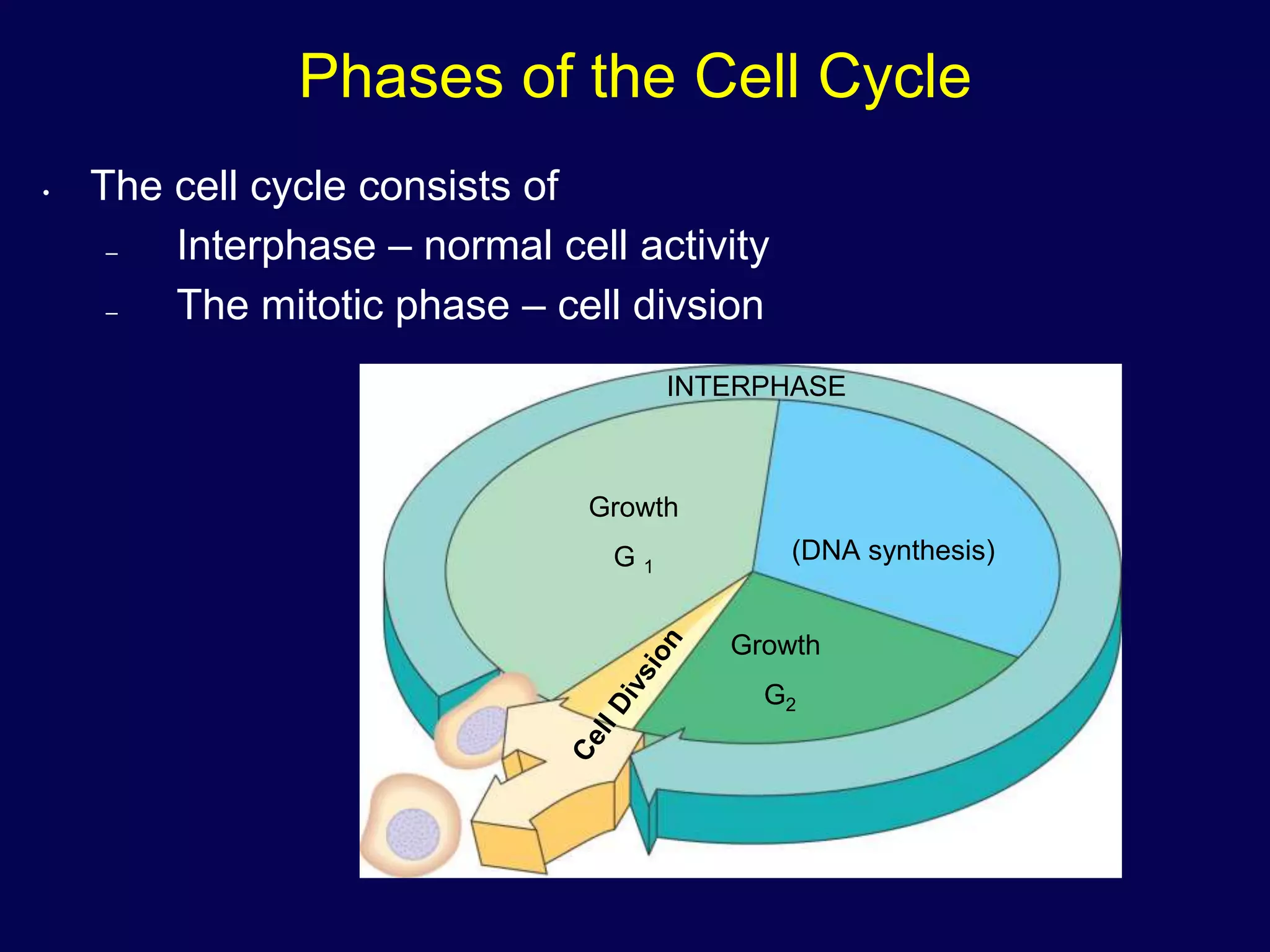
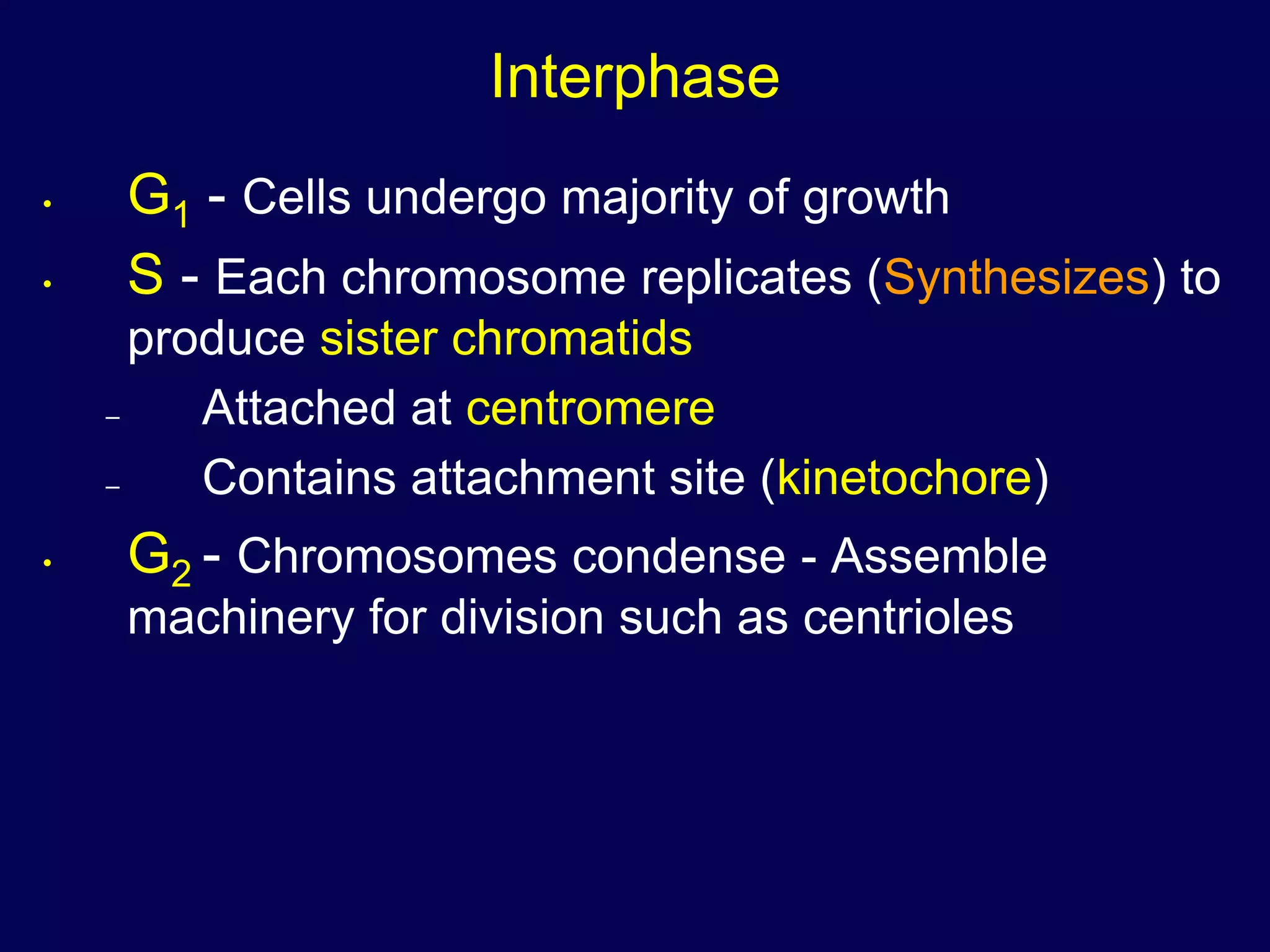
The Cell Cycle and How Cells Divide
Phases of the Cell Cycle
• The cell cycle consists of
– Interphase – normal cell activity
– The mitotic phase – cell divsion
INTERPHASE
Growth
G 1
(DNA synthesis)
Growth
G2
Functions of Cell Division
20 µm100 µm 200 µm
(a) Reproduction. An amoeba,
a single-celled eukaryote, is
dividing into two cells. Each
new cell will be an individual
organism (LM).
(b) Growth and development.
This micrograph shows a
sand dollar embryo shortly after
the fertilized egg divided, forming
two cells (LM).
(c) Tissue renewal. These dividing
bone marrow cells (arrow) will
give rise to new blood cells (LM).
Cell Division
• An integral part of the cell cycle
• Results in genetically identical daughter cells
• Cells duplicate their genetic material
– Before they divide, ensuring that each
daughter cell receives an exact copy of the
genetic material, DNA
DNA
• Genetic information - genome
• Packaged into chromosomes
50 µm
Figure 12.3
DNA And Chromosomes
• An average eukaryotic cell has about 1,000
times more DNA then an average
prokaryotic cell.
• The DNA in a eukaryotic cell is organized
into several linear chromosomes, whose
organization is much more complex than the
single, circular DNA molecule in a
prokaryotic cell
Chromosomes
• All eukaryotic cells store genetic information
in chromosomes.
– Most eukaryotes have between 10 and 50 chromosomes in their body cells.
– Human cells have 46 chromosomes.
– 23 nearly-identical pairs
Structure of Chromosomes
• Chromosomes are composed of a
complex of DNA and protein called
chromatin that condenses during cell
division
• DNA exists as a single, long, double-
stranded fiber extending chromosome’s
entire length.
• Each unduplicated chromosome
contains one DNA molecule, which may
be several inches long
 Every 200 nucleotide pairs, the DNA wraps twice around a
group of 8 histone proteins to form a nucleosome.
 Higher order coiling and supercoiling also help condense
and package the chromatin inside the nucleus:
Structure of Chromosomes
5 µm
Pair of homologous
chromosomes
Centromere
Sister
chromatids
Karyotype
• An ordered, visual representation of the chromosomes in a cell
• Chromosomes are photographed when they are highly condensed, then photos of
the individual chromosomes are arranged in order of decreasing size:
• In humans each somatic cell has 46 chromosomes, made up of two sets, one set
of chromosomes comes from each parent
Chromosomes
• Non-homologous chromosomes
– Look different
– Control different traits
• Sex chromosomes
– Are distinct from each other in their
characteristics
– Are represented as X and Y
– Determine the sex of the individual, XX being
female, XY being male
• In a diploid cell, the chromosomes occur in pairs.
The 2 members of each pair are called homologous
chromosomes or homologues.
Chromosomes
• A diploid cell has two sets of each of its chromosomes
• A human has 46 chromosomes (2n = 46)
• In a cell in which DNA synthesis has occurred all the chromosomes are
duplicated and thus each consists of two identical sister chromatids
Maternal set of
chromosomes (n = 3)
Paternal set of
chromosomes (n = 3)
2n = 6
Two sister chromatids
of one replicated
chromosome
Two nonsister
chromatids in
a homologous pair
Pair of homologous
chromosomes
(one from each set)
Centromere
Homologues
• Homologous chromosomes:
• Look the same
• Control the same traits
• May code for different forms of each trait
• Independent origin - each one was inherited
from a different parent
Chromosome Duplication
0.5 µm
Chromosome
duplication
(including DNA
synthesis)
Centromere
Separation
of sister
chromatids
Sister
chromatids
Centrometers Sister chromatids
A eukaryotic cell has multiple
chromosomes, one of which is
represented here. Before
duplication, each chromosome
has a single DNA molecule.
Once duplicated, a chromosome
consists of two sister chromatids
connected at the centromere. Each
chromatid contains a copy of the
DNA molecule.
Mechanical processes separate
the sister chromatids into two
chromosomes and distribute
them to two daughter cells.
• In preparation for cell division, DNA is replicated and the chromosomes condense
• Each duplicated chromosome has two sister chromatids, which separate during cell division
• Because of duplication, each condensed chromosome
consists of 2 identical chromatids joined by a centromere.
• Each duplicated chromosome contains 2 identical DNA
molecules (unless a mutation occurred), one in each
chromatid:
Chromosome Duplication
Copyright © The McGraw-Hill Companies, Inc. Permission required for reproduction or display.
Two unduplicated
chromosomes
Centromere
Sister
chromatids
Sister
chromatids
Duplication
Non-sister
chromatids
Two duplicated chromosomes
Structure of Chromosomes
• The centromere is a constricted region of the chromosome containing a
specific DNA sequence, to which is bound 2 discs of protein called
kinetochores.
• Kinetochores serve as points of attachment for microtubules that move the
chromosomes during cell division:
Copyright © The McGraw-Hill Companies, Inc. Permission required for reproduction or display.
Metaphase chromosome
Kinetochore
Kinetochore
microtubules
Centromere
region of
chromosome
Sister Chromatids
Structure of Chromosomes
– Diploid - A cell possessing two copies of each
chromosome (human body cells).
 Homologous chromosomes are made up of sister
chromatids joined at the centromere.
– Haploid - A cell possessing a single copy of each
chromosome (human sex cells).
Phases of the Cell Cycle
• Interphase
– G1 - primary growth
– S - genome replicated
– G2 - secondary growth
• M - mitosis
• C - cytokinesis
Interphase
• G1 - Cells undergo majority of growth
• S - Each chromosome replicates (Synthesizes) to
produce sister chromatids
– Attached at centromere
– Contains attachment site (kinetochore)
• G2 - Chromosomes condense - Assemble
machinery for division such as centrioles
Mitosis
 Some haploid & diploid cells divide by mitosis.
 Each new cell receives one copy of every
chromosome that was present in the original cell.
 Produces 2 new cells that are both genetically
identical to the original cell.
DNA duplication
during interphase
Mitosis
Diploid Cell
Mitotic Division of an Animal Cell
G2 OF INTERPHASE PROPHASE PROMETAPHASE
Centrosomes
(with centriole pairs) Chromatin
(duplicated)
Early mitotic
spindle
Aster
Centromere
Fragments
of nuclear
envelope
Kinetochore
Nucleolus Nuclear
envelope
Plasma
membrane
Chromosome, consisting
of two sister chromatids
Kinetochore
microtubule
Nonkinetochore
microtubules
METAPHASE ANAPHASE TELOPHASE AND CYTOKINESIS
Spindle
Metaphase
plate Nucleolus
forming
Cleavage
furrow
Nuclear
envelope
formingCentrosome at
one spindle pole
Daughter
chromosomes
Mitotic Division of an Animal Cell
G2 of Interphase
• A nuclear envelope bounds
the nucleus.
• The nucleus contains one or
more nucleoli (singular,
nucleolus).
• Two centrosomes have
formed by replication of a
single centrosome.
• In animal cells, each
centrosome features two
centrioles.
• Chromosomes, duplicated
during S phase, cannot be
seen individually because
they have not yet condensed.
The light micrographs show dividing lung cells
from a newt, which has 22 chromosomes in its
somatic cells (chromosomes appear blue,
microtubules green, intermediate filaments
red). For simplicity, the drawings show only
four chromosomes.
G2 OF INTERPHASE
Centrosomes
(with centriole pairs) Chromatin
(duplicated)
Nucleolus Nuclear
envelope
Plasma
membrane
Prophase
• The chromatin fibers become
more tightly coiled, condensing
into discrete chromosomes
observable with a light
microscope.
• The nucleoli disappear.
• Each duplicated chromosome
appears as two identical sister
chromatids joined together.
• The mitotic spindle begins to form.
It is composed of the centrosomes
and the microtubules that extend
from them. The radial arrays of
shorter microtubules that extend
from the centrosomes are called
asters (“stars”).
• The centrosomes move away from
each other, apparently propelled
by the lengthening microtubules
between them.
PROPHASE
Early mitotic
spindle
Aster
Centromere
Chromosome, consisting
of two sister chromatids
Metaphase
• Metaphase is the longest stage of
mitosis, lasting about 20 minutes.
• The centrosomes are now at
opposite ends of the cell.
•The chromosomes convene on the
metaphase plate, an imaginary
plane that is equidistant between
the spindle’s two poles. The
chromosomes’ centromeres lie on
the metaphase plate.
• For each chromosome, the
kinetochores of the sister
chromatids are attached to
kinetochore microtubules coming
from opposite poles.
• The entire apparatus of
microtubules is called the spindle
because of its shape.
METAPHASE
Spindle
Metaphase
plate
Centrosome at
one spindle pole
The Mitotic Spindle
• The spindle includes the centrosomes, the spindle
microtubules, and the asters
• The apparatus of microtubules controls
chromosome movement during mitosis
• The centrosome replicates, forming two
centrosomes that migrate to opposite ends of the
cell
• Assembly of spindle microtubules begins in the
centrosome, the microtubule organizing center
• An aster (a radial array of short microtubules)
extends from each centrosome
• Some spindle microtubules attach to the kinetochores of
chromosomes and move the chromosomes to the
metaphase plate
• In anaphase, sister chromatids separate and move along the
kinetochore microtubules toward opposite ends of the cell
Microtubules Chromosomes
Sister
chromatids
Aster
Centrosome
Metaphase
plate
Kineto-
chores
Kinetochore
microtubules
0.5 µm
Overlapping
nonkinetochore
microtubules
1 µmCentrosome
The Mitotic Spindle
Anaphase
• Anaphase is the shortest stage of
mitosis, lasting only a few minutes.
• Anaphase begins when the two sister
chromatids of each pair suddenly part.
Each chromatid thus becomes a full-
fledged chromosome.
• The two liberated chromosomes begin
moving toward opposite ends of the cell,
as their kinetochore microtubules
shorten. Because these microtubules are
attached at the centromere region, the
chromosomes move centromere first (at
about 1 µm/min).
• The cell elongates as the
nonkinetochore microtubules lengthen.
• By the end of anaphase, the two ends of
the cell have equivalent—and
complete—collections of chromosomes.
ANAPHASE
Daughter
chromosomes
Telophase
• Two daughter nuclei begin to
form in the cell.
• Nuclear envelopes arise from
the fragments of the parent
cell’s nuclear envelope and
other portions of the
endomembrane system.
• The chromosomes become
less condensed.
• Mitosis, the division of one
nucleus into two genetically
identical nuclei, is now
complete.
TELOPHASE AND CYTOKINESIS
Nucleolus
forming
Cleavage
furrow
Nuclear
envelope
forming
Mitosis in a plant cell
1 Prophase.
The chromatin
is condensing.
The nucleolus is
beginning to
disappear.
Although not
yet visible
in the micrograph,
the mitotic spindle is
staring to from.
Prometaphase.
We now see discrete
chromosomes; each
consists of two
identical sister
chromatids. Later
in prometaphase, the
nuclear envelop will
fragment.
Metaphase. The
spindle is complete,
and the chromosomes,
attached to microtubules
at their kinetochores,
are all at the metaphase
plate.
Anaphase. The
chromatids of each
chromosome have
separated, and the
daughter chromosomes
are moving to the ends
of cell as their
kinetochore
microtubles shorten.
Telophase. Daughter
nuclei are forming.
Meanwhile, cytokinesis
has started: The cell
plate, which will
divided the cytoplasm
in two, is growing
toward the perimeter
of the parent cell.
2 3 4 5
Nucleus
Nucleolus
ChromosomeChromatine
condensing
Cytokinesis
• Cleavage of cell into
two halves
– Animal cells
 Constriction belt
of actin filaments
– Plant cells
 Cell plate
– Fungi and protists
 Mitosis occurs
within the nucleus
Cytokinesis In Animal And Plant Cells
Daughter cells
Cleavage furrow
Contractile ring of
microfilaments
Daughter cells
100 µm
1 µmVesicles
forming
cell plate
Wall of
patent cell Cell plate
New cell wall
(a) Cleavage of an animal cell (SEM) (b) Cell plate formation in a plant cell (SEM)
Mitosis ppt

Mitosis ppt

  • 1.
    The Cell Cycleand How Cells Divide
  • 2.
    Phases of theCell Cycle • The cell cycle consists of – Interphase – normal cell activity – The mitotic phase – cell divsion INTERPHASE Growth G 1 (DNA synthesis) Growth G2
  • 3.
    Functions of CellDivision 20 µm100 µm 200 µm (a) Reproduction. An amoeba, a single-celled eukaryote, is dividing into two cells. Each new cell will be an individual organism (LM). (b) Growth and development. This micrograph shows a sand dollar embryo shortly after the fertilized egg divided, forming two cells (LM). (c) Tissue renewal. These dividing bone marrow cells (arrow) will give rise to new blood cells (LM).
  • 4.
    Cell Division • Anintegral part of the cell cycle • Results in genetically identical daughter cells • Cells duplicate their genetic material – Before they divide, ensuring that each daughter cell receives an exact copy of the genetic material, DNA
  • 5.
    DNA • Genetic information- genome • Packaged into chromosomes 50 µm Figure 12.3
  • 6.
    DNA And Chromosomes •An average eukaryotic cell has about 1,000 times more DNA then an average prokaryotic cell. • The DNA in a eukaryotic cell is organized into several linear chromosomes, whose organization is much more complex than the single, circular DNA molecule in a prokaryotic cell
  • 7.
    Chromosomes • All eukaryoticcells store genetic information in chromosomes. – Most eukaryotes have between 10 and 50 chromosomes in their body cells. – Human cells have 46 chromosomes. – 23 nearly-identical pairs
  • 8.
    Structure of Chromosomes •Chromosomes are composed of a complex of DNA and protein called chromatin that condenses during cell division • DNA exists as a single, long, double- stranded fiber extending chromosome’s entire length. • Each unduplicated chromosome contains one DNA molecule, which may be several inches long
  • 9.
     Every 200nucleotide pairs, the DNA wraps twice around a group of 8 histone proteins to form a nucleosome.  Higher order coiling and supercoiling also help condense and package the chromatin inside the nucleus: Structure of Chromosomes
  • 10.
    5 µm Pair ofhomologous chromosomes Centromere Sister chromatids Karyotype • An ordered, visual representation of the chromosomes in a cell • Chromosomes are photographed when they are highly condensed, then photos of the individual chromosomes are arranged in order of decreasing size: • In humans each somatic cell has 46 chromosomes, made up of two sets, one set of chromosomes comes from each parent
  • 11.
    Chromosomes • Non-homologous chromosomes –Look different – Control different traits • Sex chromosomes – Are distinct from each other in their characteristics – Are represented as X and Y – Determine the sex of the individual, XX being female, XY being male • In a diploid cell, the chromosomes occur in pairs. The 2 members of each pair are called homologous chromosomes or homologues.
  • 12.
    Chromosomes • A diploidcell has two sets of each of its chromosomes • A human has 46 chromosomes (2n = 46) • In a cell in which DNA synthesis has occurred all the chromosomes are duplicated and thus each consists of two identical sister chromatids Maternal set of chromosomes (n = 3) Paternal set of chromosomes (n = 3) 2n = 6 Two sister chromatids of one replicated chromosome Two nonsister chromatids in a homologous pair Pair of homologous chromosomes (one from each set) Centromere
  • 13.
    Homologues • Homologous chromosomes: •Look the same • Control the same traits • May code for different forms of each trait • Independent origin - each one was inherited from a different parent
  • 14.
    Chromosome Duplication 0.5 µm Chromosome duplication (includingDNA synthesis) Centromere Separation of sister chromatids Sister chromatids Centrometers Sister chromatids A eukaryotic cell has multiple chromosomes, one of which is represented here. Before duplication, each chromosome has a single DNA molecule. Once duplicated, a chromosome consists of two sister chromatids connected at the centromere. Each chromatid contains a copy of the DNA molecule. Mechanical processes separate the sister chromatids into two chromosomes and distribute them to two daughter cells. • In preparation for cell division, DNA is replicated and the chromosomes condense • Each duplicated chromosome has two sister chromatids, which separate during cell division
  • 15.
    • Because ofduplication, each condensed chromosome consists of 2 identical chromatids joined by a centromere. • Each duplicated chromosome contains 2 identical DNA molecules (unless a mutation occurred), one in each chromatid: Chromosome Duplication Copyright © The McGraw-Hill Companies, Inc. Permission required for reproduction or display. Two unduplicated chromosomes Centromere Sister chromatids Sister chromatids Duplication Non-sister chromatids Two duplicated chromosomes
  • 16.
    Structure of Chromosomes •The centromere is a constricted region of the chromosome containing a specific DNA sequence, to which is bound 2 discs of protein called kinetochores. • Kinetochores serve as points of attachment for microtubules that move the chromosomes during cell division: Copyright © The McGraw-Hill Companies, Inc. Permission required for reproduction or display. Metaphase chromosome Kinetochore Kinetochore microtubules Centromere region of chromosome Sister Chromatids
  • 17.
    Structure of Chromosomes –Diploid - A cell possessing two copies of each chromosome (human body cells).  Homologous chromosomes are made up of sister chromatids joined at the centromere. – Haploid - A cell possessing a single copy of each chromosome (human sex cells).
  • 18.
    Phases of theCell Cycle • Interphase – G1 - primary growth – S - genome replicated – G2 - secondary growth • M - mitosis • C - cytokinesis
  • 19.
    Interphase • G1 -Cells undergo majority of growth • S - Each chromosome replicates (Synthesizes) to produce sister chromatids – Attached at centromere – Contains attachment site (kinetochore) • G2 - Chromosomes condense - Assemble machinery for division such as centrioles
  • 20.
    Mitosis  Some haploid& diploid cells divide by mitosis.  Each new cell receives one copy of every chromosome that was present in the original cell.  Produces 2 new cells that are both genetically identical to the original cell. DNA duplication during interphase Mitosis Diploid Cell
  • 21.
    Mitotic Division ofan Animal Cell G2 OF INTERPHASE PROPHASE PROMETAPHASE Centrosomes (with centriole pairs) Chromatin (duplicated) Early mitotic spindle Aster Centromere Fragments of nuclear envelope Kinetochore Nucleolus Nuclear envelope Plasma membrane Chromosome, consisting of two sister chromatids Kinetochore microtubule Nonkinetochore microtubules
  • 22.
    METAPHASE ANAPHASE TELOPHASEAND CYTOKINESIS Spindle Metaphase plate Nucleolus forming Cleavage furrow Nuclear envelope formingCentrosome at one spindle pole Daughter chromosomes Mitotic Division of an Animal Cell
  • 23.
    G2 of Interphase •A nuclear envelope bounds the nucleus. • The nucleus contains one or more nucleoli (singular, nucleolus). • Two centrosomes have formed by replication of a single centrosome. • In animal cells, each centrosome features two centrioles. • Chromosomes, duplicated during S phase, cannot be seen individually because they have not yet condensed. The light micrographs show dividing lung cells from a newt, which has 22 chromosomes in its somatic cells (chromosomes appear blue, microtubules green, intermediate filaments red). For simplicity, the drawings show only four chromosomes. G2 OF INTERPHASE Centrosomes (with centriole pairs) Chromatin (duplicated) Nucleolus Nuclear envelope Plasma membrane
  • 24.
    Prophase • The chromatinfibers become more tightly coiled, condensing into discrete chromosomes observable with a light microscope. • The nucleoli disappear. • Each duplicated chromosome appears as two identical sister chromatids joined together. • The mitotic spindle begins to form. It is composed of the centrosomes and the microtubules that extend from them. The radial arrays of shorter microtubules that extend from the centrosomes are called asters (“stars”). • The centrosomes move away from each other, apparently propelled by the lengthening microtubules between them. PROPHASE Early mitotic spindle Aster Centromere Chromosome, consisting of two sister chromatids
  • 25.
    Metaphase • Metaphase isthe longest stage of mitosis, lasting about 20 minutes. • The centrosomes are now at opposite ends of the cell. •The chromosomes convene on the metaphase plate, an imaginary plane that is equidistant between the spindle’s two poles. The chromosomes’ centromeres lie on the metaphase plate. • For each chromosome, the kinetochores of the sister chromatids are attached to kinetochore microtubules coming from opposite poles. • The entire apparatus of microtubules is called the spindle because of its shape. METAPHASE Spindle Metaphase plate Centrosome at one spindle pole
  • 26.
    The Mitotic Spindle •The spindle includes the centrosomes, the spindle microtubules, and the asters • The apparatus of microtubules controls chromosome movement during mitosis • The centrosome replicates, forming two centrosomes that migrate to opposite ends of the cell • Assembly of spindle microtubules begins in the centrosome, the microtubule organizing center • An aster (a radial array of short microtubules) extends from each centrosome
  • 27.
    • Some spindlemicrotubules attach to the kinetochores of chromosomes and move the chromosomes to the metaphase plate • In anaphase, sister chromatids separate and move along the kinetochore microtubules toward opposite ends of the cell Microtubules Chromosomes Sister chromatids Aster Centrosome Metaphase plate Kineto- chores Kinetochore microtubules 0.5 µm Overlapping nonkinetochore microtubules 1 µmCentrosome The Mitotic Spindle
  • 28.
    Anaphase • Anaphase isthe shortest stage of mitosis, lasting only a few minutes. • Anaphase begins when the two sister chromatids of each pair suddenly part. Each chromatid thus becomes a full- fledged chromosome. • The two liberated chromosomes begin moving toward opposite ends of the cell, as their kinetochore microtubules shorten. Because these microtubules are attached at the centromere region, the chromosomes move centromere first (at about 1 µm/min). • The cell elongates as the nonkinetochore microtubules lengthen. • By the end of anaphase, the two ends of the cell have equivalent—and complete—collections of chromosomes. ANAPHASE Daughter chromosomes
  • 29.
    Telophase • Two daughternuclei begin to form in the cell. • Nuclear envelopes arise from the fragments of the parent cell’s nuclear envelope and other portions of the endomembrane system. • The chromosomes become less condensed. • Mitosis, the division of one nucleus into two genetically identical nuclei, is now complete. TELOPHASE AND CYTOKINESIS Nucleolus forming Cleavage furrow Nuclear envelope forming
  • 30.
    Mitosis in aplant cell 1 Prophase. The chromatin is condensing. The nucleolus is beginning to disappear. Although not yet visible in the micrograph, the mitotic spindle is staring to from. Prometaphase. We now see discrete chromosomes; each consists of two identical sister chromatids. Later in prometaphase, the nuclear envelop will fragment. Metaphase. The spindle is complete, and the chromosomes, attached to microtubules at their kinetochores, are all at the metaphase plate. Anaphase. The chromatids of each chromosome have separated, and the daughter chromosomes are moving to the ends of cell as their kinetochore microtubles shorten. Telophase. Daughter nuclei are forming. Meanwhile, cytokinesis has started: The cell plate, which will divided the cytoplasm in two, is growing toward the perimeter of the parent cell. 2 3 4 5 Nucleus Nucleolus ChromosomeChromatine condensing
  • 31.
    Cytokinesis • Cleavage ofcell into two halves – Animal cells  Constriction belt of actin filaments – Plant cells  Cell plate – Fungi and protists  Mitosis occurs within the nucleus
  • 32.
    Cytokinesis In AnimalAnd Plant Cells Daughter cells Cleavage furrow Contractile ring of microfilaments Daughter cells 100 µm 1 µmVesicles forming cell plate Wall of patent cell Cell plate New cell wall (a) Cleavage of an animal cell (SEM) (b) Cell plate formation in a plant cell (SEM)