Download to read offline
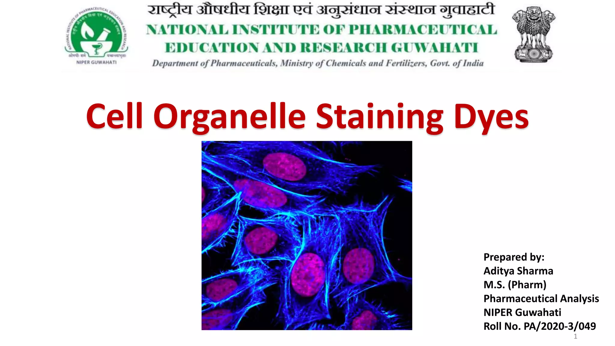

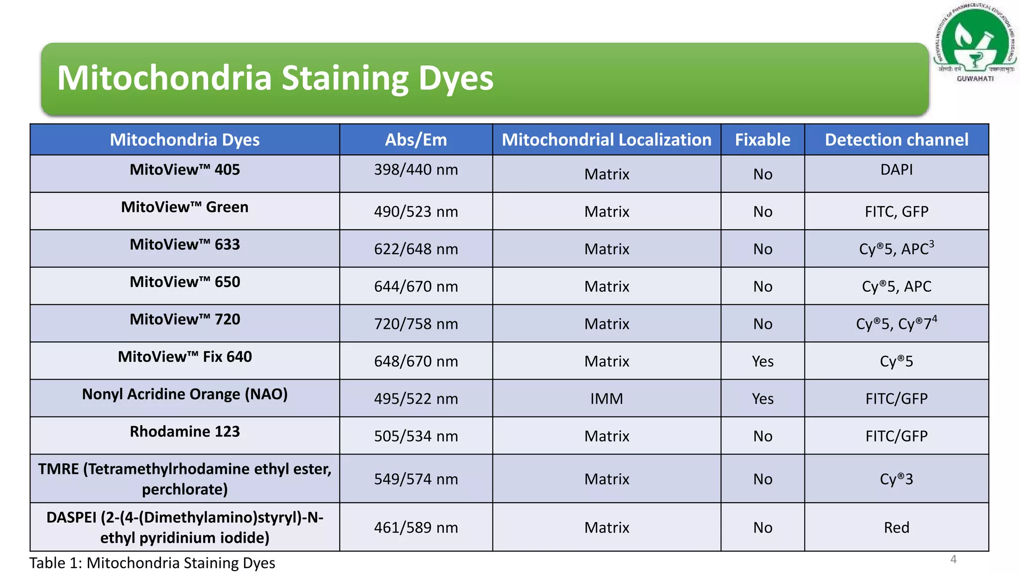
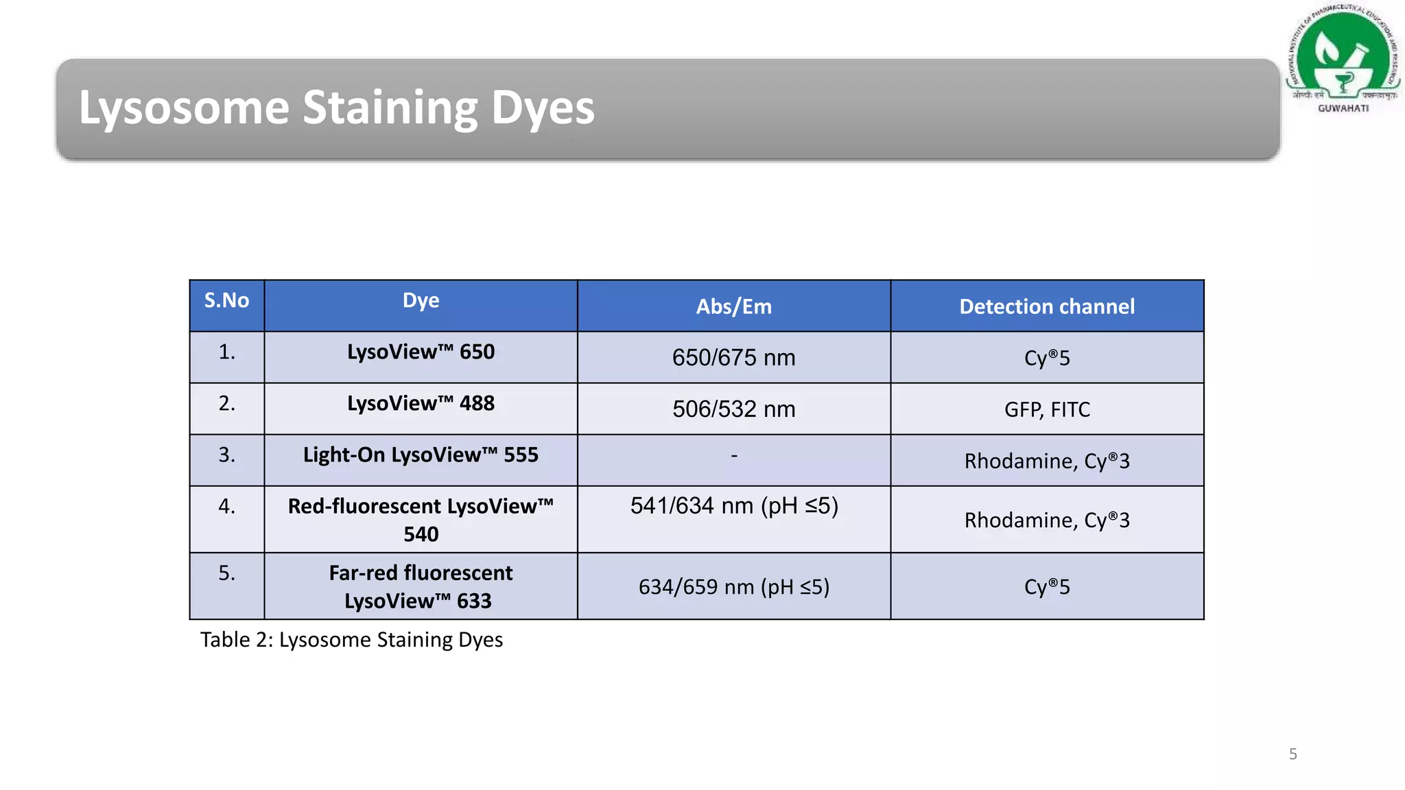
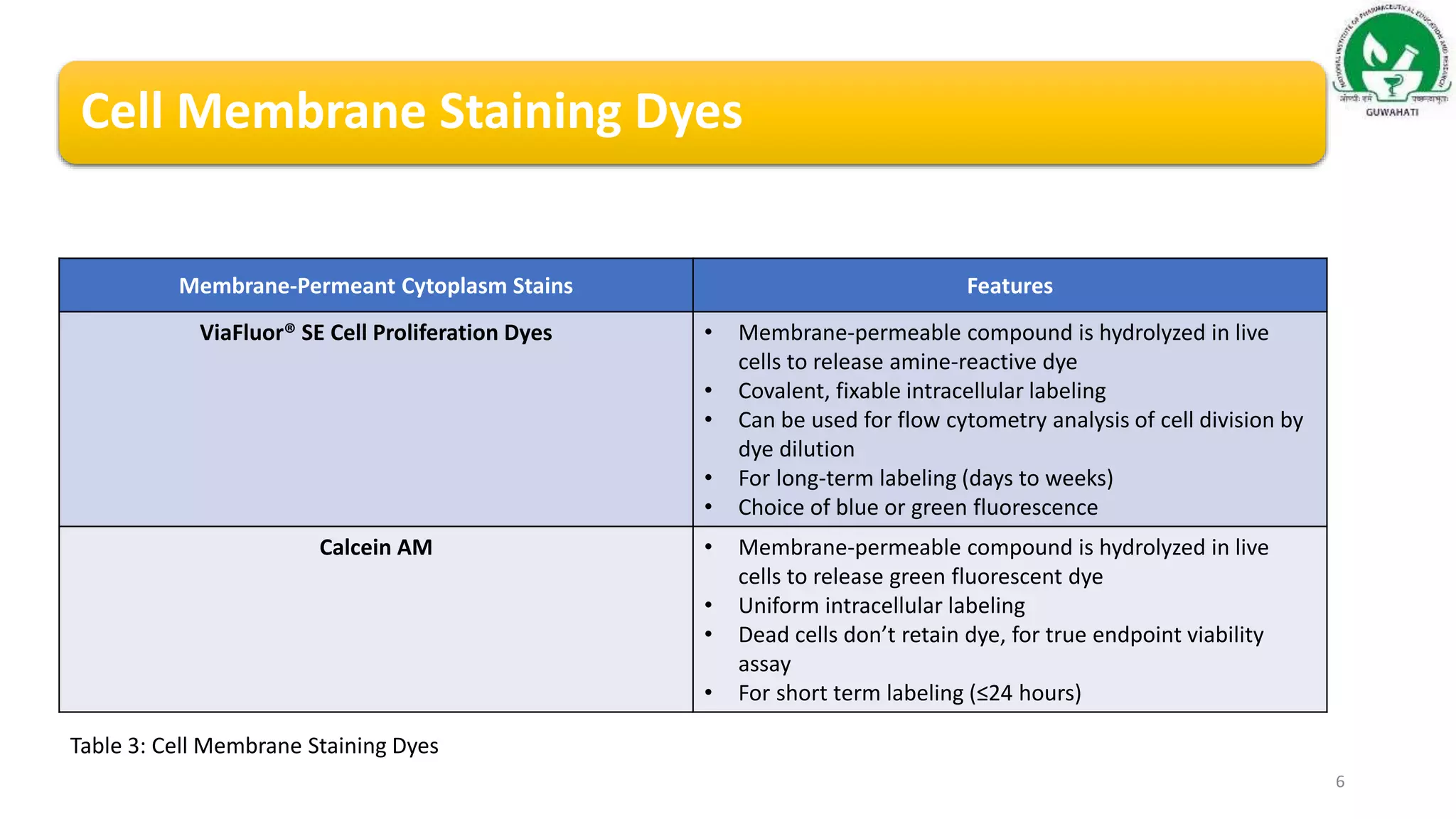
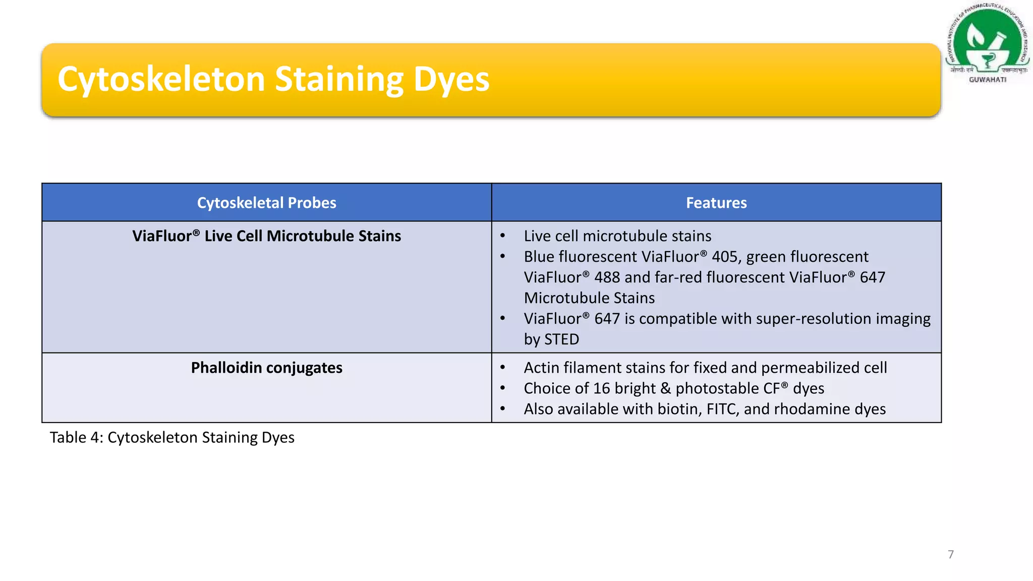


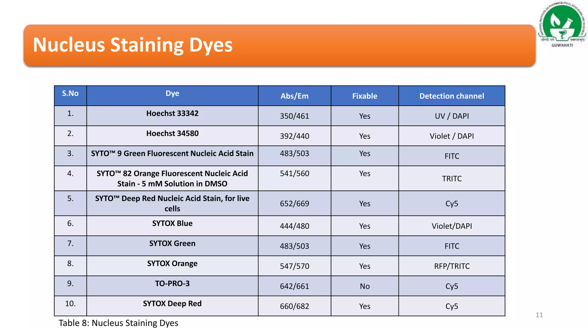

This document discusses various dyes used for staining cell organelles in eukaryotic cells, which aids in cell biology studies by allowing visualization of structures like mitochondria, lysosomes, and the nucleus. It provides detailed tables of fluorescent dyes specific to each organelle, their absorption/emission properties, and applications. The conclusion emphasizes the importance of live cell fluorescent organelle dyes for studying cellular processes without increasing cytotoxicity.


