Downloaded 104 times
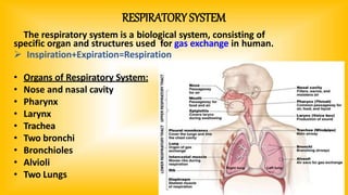
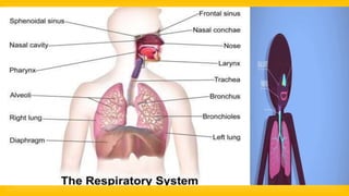
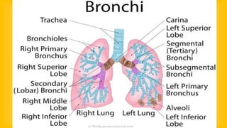
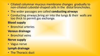
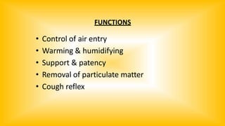
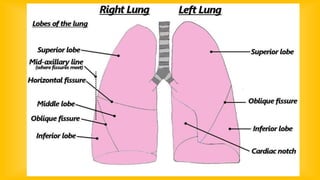


The document provides a comprehensive overview of the respiratory system, detailing its anatomy, including the organ structure from the nose to the lungs, and the functions of each component. It explains the processes of breathing (inspiration and expiration), gas exchange, and the roles of various muscles and tissues in respiratory function. Additionally, it covers the lung structure, gas transport in the bloodstream, and the nervous system's control over respiration.