SlideShare a Scribd company logo
LAB 3
                                     Bacterial Staining Techniques II
I. Differential Stains: Gram Stain and Acid-fast Stain
II. Morphological Unknown

I. DIFFERENTIAL STAINS
    A. Gram Stain
    B. Acid-fast Stain

A. Gram Stain
   The previous lab introduced simple staining techniques that enable microbiologists to observe the
   morphological characteristics of bacteria. Although simple stains are useful, they do not reveal details
   about the bacteria other than morphology and arrangement. The Gram stain is a differential stain
   commonly used in the microbiology laboratory that differentiates bacteria on the basis of their cell wall
   structure. Most bacteria can be divided into two groups based on the composition of their cell wall:

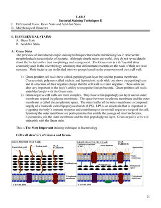
       1) Gram-positive cell walls have a thick peptidoglycan layer beyond the plasma membrane.
          Characteristic polymers called teichoic and lipoteichoic acids stick out above the peptidoglycan
          and it is because of their negative charge that the cell wall is overall negative. These acids are
          also very important in the body’s ability to recognize foreign bacteria. Gram-positive cell walls
          stain blue/purple with the Gram stain.
       2) Gram-negative cell walls are more complex. They have a thin peptidoglycan layer and an outer
          membrane beyond the plasma membrane. The space between the plasma membrane and the outer
          membrane is called the periplasmic space. The outer leaflet of the outer membrane is composed
          largely of a molecule called lipopolysaccharide (LPS). LPS is an endotoxin that is important in
          triggering the body’s immune response and contributing to the overall negative charge of the cell.
          Spanning the outer membrane are porin proteins that enable the passage of small molecules.
          Lipoproteins join the outer membrane and the thin peptidoglycan layer. Gram-negative cells will
          stain pink with the Gram stain.

   This is The Most Important staining technique in Bacteriology.

   Cell wall structure of Gram+ and Gram-


                                                                                                               21
GRAM STAIN
                                                                 Cell Color
Procedure                 Reagent                    Gram Positive            Gram Negative
Fixed cells on slide                                 COLORLESS                COLORLESS
Primary stain             Crystal Violet             PURPLE                   PURPLE
Mordant                   Iodine                     PURPLE                   PURPLE
Decolorizer               Alcohol                    PURPLE                   COLORLESS
Counterstain              Safranin                   PURPLE                   RED

An easy way to remember the steps of the Gram stain is...


                                                                                              22
PROCEDURE: (EACH STUDENT)
1. Using a sterile inoculating loop, add 1 drop of sterile water to the slide. Prepare a mixed smear of
    Escherichia coli (G- rod) and Staphylococcus epidermidis (G+ coccus).
2. Air dry and Heat fix.
3. Cover the smear with Crystal Violet (primary stain) for 1 min.
4. Gently wash off the slide with water.
5. Add Gram’s Iodine (mordant) for 1 min.
6. Wash with water.
7. Decolorize with 95% ethanol. This is the "tricky" step. Stop decolorizing with alcohol as soon as the
    purple color has stopped leaching off the slide (time will vary depending on thickness of smear).
    Immediately wash with water. Be sure to dispose of all ethanol waste in the appropriately labeled waste
    container.
8. Cover the smear with Safranin for 30 seconds.
9. Wash both the top & the bottom of the slide with water.
10. Blot the slide with bibulous paper.
11. Using the 10X objective lens, focus first on the line and then on the smear. Follow the focusing
    procedure in Lab #1. Use the focusing procedure in Lab #2 to view the smear using the 100X (oil
    immersion lens).
                                        Focus line
                                                               E. coli


                                                                            S. epidermidis


Note: Escherichia coli is a tiny pink (Gram-) rod. Staphylococcus epidermidis is a purple (Gram+) sphere
or coccus.

Draw a picture of a typical microscopic field and identify both Escherichia coli and Staphylococcus
epidermidis. Record this in the results section for this lab. Colored pencils are available throughout the room
on the chalkboard trays.


                                                                                                             23
B. Acid-fast Stain
   Mycobacterium and many Nocardia species are called acid-fast because during an acid-fast staining
   procedure they retain the primary dye carbol fuchsin despite decolorization with the powerful solvent
   acid-alcohol (95% ethanol with 3% HCl). Nearly all other genera of bacteria are nonacid-fast. The acid-
   fast genera have the waxy hydroxy-lipid called mycolic acid in their cell walls. It is assumed that
   mycolic acid prevents acid-alcohol from decolorizing protoplasm. The acid-fast stain is a differential
   stain.

   Ziehl Neelsen Acid-fast stain

                                   ACID-FAST STAIN
                                                         Cell Color
Procedure          Reagent                  Acid-fast Bacteria      Nonacid-fast Bacteria
Primary dye        Carbolfuchsin            RED                     RED
Decolorizer        Acid-alcohol             RED                     COLORLESS
Counterstain       Methylene blue           RED                     BLUE

PROCEDURE - (EACH STUDENT)
1. Add one loopful of sterile water to a microscope slide.
2. Make a heavy smear of Mycobacterium smegmatis. Mix thoroughly with your loop. Then transfer a
    small amount of Staphylococcus epidermidis to the same drop of water.
    You will now have a mixture of M. smegmatis and S. epidermidis.
3. Air dry and heat fix well.
4. Cover the smear with carbolfuchsin dye. Place a piece of paper towel on top of the dye. Be sure the
    paper towel is saturated with the dye. Carbolfuchsin is a potential carcinogen. Please wear gloves when
    working with this dye.
5. Place the slide on the rack over dry heat for 2 minutes.
6. Cool and rinse with water.
7. Decolorize by placing a drop of acid alcohol on the slide and allowing it to sit for 15 seconds.
8. Wash the top and bottom of slide with water and clean the slide bottom well.
9. Counterstain with Methylene Blue for 30 seconds to 1 minute.
10. Wash and blot the slide with bibulous paper.
11. Focus 10X - then use oil immersion.
                                                     M. smegmatis (heavy)


                                                      Focus line
                          S . epidermidis
Draw a typical microscopic field and record in Results Lab 3.
Note: The acid-fast Mycobacterium retains carbolfuchsin and stains hot pink. The Staphylococcus
epidermidis is decolorized and the counterstain colors them blue. See table for acid-fast stain.


                                                                                                              24
II.   Morphological Unknown: continue your investigation of your morphological unknown.
1.    Collect your unknown from the side bench. Verify that the # matches that used for your direct stain.
2.    Perform a Gram stain and an Acid-fast stain as described in the above sections.
3.    Record your observations in the Results section of Lab #4.


                                                                                                             25
NOTES


        26
LAB 3 RESULTS
I. DIFFERENTIAL STAINS
A. Gram Stain Draw and label examples of Escherichia coli and Staphylococcus epidermidis.


B. Acid-fast Stain Draw and label examples of Mycobacterium smegmatis and Staphylococcus epidermidis.
                      Mycobacterium      &    Staphylococcus
                      smegmatis               epidermidis


QUESTIONS:
1. What is the difference between a simple and a differential stain?__________________________
 _______________________________________________________________________________

 _______________________________________________________________________________
2. Describe the function of each of the following in the Gram stain
   Mordant: __________________________________________________________________
   Primary stain:_______________________________________________________________
   Decolorizer: ________________________________________________________________
   Counterstain: _______________________________________________________________
3. Which step in the Gram stain is most likely to cause poor results if done incorrectly? _________
 _______________________________________________________________________________

4. Why must fresh bacterial cultures be used in a Gram stain? ______________________________

 _______________________________________________________________________________


                                                                                                      27
5. Briefly describe the mechanism of Gram staining. _____________________________________

 _______________________________________________________________________________

 _______________________________________________________________________________

6. What is the primary stain used in the acid-fast staining procedure? ________________________

 _______________________________________________________________________________
7. What is the purpose of the heat/steam during the acid-fast staining procedure? _______________

 _______________________________________________________________________________

 _______________________________________________________________________________
8. In a clinical microbiology laboratory, the acid-fast stain would be used for diagnosis of what diseases?

 _______________________________________________________________________________
9. What makes a microorganism nonacid-fast? __________________________________________

 _______________________________________________________________________________


Organisms introduced in this lab:
Mycobacterium smegmatis
Mycobacterium tuberculosis
Mycobacterium leprae
Staphylococcus epidermidis


                                                                                                              28
GENERAL MICROBIOLOGY 2210: GRAM STAIN REPORT (15 PTS)
ASSIGNMENT
Each student will have the opportunity to submit one example of a Gram stain with a brief written report
describing the staining procedure and the stain itself. A good report will likely necessitate a minimum of 2
pages double-spaced and is limited to a 3-page maximum.
OVERALL FORMAT
Please include the following in your report:
A. Title Page with the following information: (1 point) __________
        --Title: A title should be brief, creative, but descriptive of the work that was done.
        --Your Name
        --Course and Lab Section
        --Date
B. Introduction: (4 points) __________
   An introduction should include pertinent background information. In this case, the history of the Gram
   stain and its significance in microbiology are relevant (1 point). A discussion of the mechanism of Gram
   staining and how it differentiates bacteria on the basis of their cell wall structure should also be included
   (1 point). The purpose and objectives of the experiment should be stated and a hypothesis should be
   made (2 points). Remember that a hypothesis need not be correct, but it must be tested by the procedure.
   Good hypotheses are also grounded in background knowledge and clearly state the predicted results.
   Hypotheses can be of several types. 1 We will discuss two types here:
        1. Manipulated hypotheses
        This type of hypothesis is used when one variable (the independent variable) is manipulated by the
        experimenter and the effects on a second variable (the dependent variable) are observed. This type of
        hypothesis is generally written as an if, then statement (e.g. If the smear of the unknown bacterium is
        decolorized as according to the above procedure (neither over- nor under-decolorized), then all the
        cells will stain a single color.) In this example, decolorization is the independent variable (the
        variable controlled by the experimenter) and the color of the cells is the dependent variable (the
        variable affected by the changes in the independent variable).
        2. Observational hypotheses
        States something about the nature of an organism (e.g. The unknown bacterium will stain Gram-
        positive.)
C. Materials and Methods: (4 points) __________
   The Materials and Methods section should include a description of the procedure/steps in the Gram stain,
   including the preparation of a bacterial smear and heat fixation (3 points). This section must be written
   in complete sentences and in paragraph form (1 point). Illustrations may assist in clarifying points.
D. Results: (1 point) __________
   In the results section, describe your Gram stain/s. Include at least one illustration of your own
   creation.
E. Discussion: (3 points) __________
   What features of your Gram stain exemplify the Gram stain theory (1 point)? If there are features of the
   Gram stain that are not as expected, what possible sources or error may be responsible (e.g. poor quality
   of the smear preparation)? (1 point) Discuss whether the hypothesis made in the introduction was tested
   and if so was it supported? (1 point)


1
 For a discussion of hypotheses formation please visit
http://www.utas.edu.au/sciencelinks/exdesign/HF2C.HTM
                                                                                                               29
OVERALL QUALITY
Students have the opportunity to earn points for reports that are well written, organized and edited.
(2 points) __________

DUE DATE: TAs will inform you of the due date.
*Please tear this grading key out of your lab book and attach it to your lab report at the time of
submission. Please also write your name and lab section on the frosted portion of your Gram stain
slide and hand it in with your written report.

TA comments: __________________________________________________________________________
_______________________________________________________________________________________
_______________________________________________________________________________________

TA’s suggestions for improving Gram stain: ___________________________________________________
_______________________________________________________________________________________
_______________________________________________________________________________________


Points earned out of 15 possible: ____________


                                                                                                        30

More Related Content

What's hot

3. Antimicrobial Effectiveness Final.pptx
3. Antimicrobial Effectiveness Final.pptx3. Antimicrobial Effectiveness Final.pptx
3. Antimicrobial Effectiveness Final.pptx
Rushikesh Tamhane
 
Sterility tests
Sterility testsSterility tests
Sterility tests
Karumazzi Lohitha
 
Haemocytometer
HaemocytometerHaemocytometer
Haemocytometer
Aswathy Vijayan
 
Mc farland standards
Mc farland standardsMc farland standards
Mc farland standards
Erhovwon Aggreh
 
Lab 2 disk diffusion testing
Lab 2 disk diffusion testingLab 2 disk diffusion testing
Lab 2 disk diffusion testing
Dr. Samira Fattah
 
Microbial limit tests I.P By Dr.P.Srinivas Jangaon institute of pharmaceutica...
Microbial limit tests I.P By Dr.P.Srinivas Jangaon institute of pharmaceutica...Microbial limit tests I.P By Dr.P.Srinivas Jangaon institute of pharmaceutica...
Microbial limit tests I.P By Dr.P.Srinivas Jangaon institute of pharmaceutica...
cnupogu
 
Laboratory Decontamination
Laboratory DecontaminationLaboratory Decontamination
Laboratory Decontamination
Triumvirate Environmental
 
Aspergillus niger (FUNGUS)
Aspergillus niger  (FUNGUS)Aspergillus niger  (FUNGUS)
Aspergillus niger (FUNGUS)
HannaLawrencePereira
 
Methods of measuring microbial growth
Methods of measuring microbial growthMethods of measuring microbial growth
Methods of measuring microbial growth
Abdul Rehman
 
Incubators types and applications
Incubators  types and applicationsIncubators  types and applications
Incubators types and applications
Dhamodharan Paramasivam
 
Culture of larval stage of nematodes
Culture of larval stage of nematodesCulture of larval stage of nematodes
Culture of larval stage of nematodes
ssuser2cecdc
 
The preservation of bacterial and fungal strains
The preservation of bacterial and fungal strainsThe preservation of bacterial and fungal strains
The preservation of bacterial and fungal strains
Dr. Ishan Y. Pandya
 
Microtome
MicrotomeMicrotome
Microtome
George saweris
 
Bacterial isolation
Bacterial isolation Bacterial isolation
Bacterial isolation
Avijit Pramanik
 
Laminar air flow
Laminar air flowLaminar air flow
Laminar air flow
Bhavya Sree
 
Glassware apparatus and their uses(pdf)
Glassware apparatus and their uses(pdf)Glassware apparatus and their uses(pdf)
Glassware apparatus and their uses(pdf)
Science Equip
 
Glassware
GlasswareGlassware
Glassware
anjali thakur
 
Microbial Culture Preservation and its Methods
Microbial Culture Preservation and its MethodsMicrobial Culture Preservation and its Methods
Microbial Culture Preservation and its Methods
DENNISMMONDAH1
 
Butter analysis
Butter analysis Butter analysis
Butter analysis
MehulJain143
 
Stainings
StainingsStainings
Stainings
Mohmmad Savarkar
 

What's hot (20)

3. Antimicrobial Effectiveness Final.pptx
3. Antimicrobial Effectiveness Final.pptx3. Antimicrobial Effectiveness Final.pptx
3. Antimicrobial Effectiveness Final.pptx
 
Sterility tests
Sterility testsSterility tests
Sterility tests
 
Haemocytometer
HaemocytometerHaemocytometer
Haemocytometer
 
Mc farland standards
Mc farland standardsMc farland standards
Mc farland standards
 
Lab 2 disk diffusion testing
Lab 2 disk diffusion testingLab 2 disk diffusion testing
Lab 2 disk diffusion testing
 
Microbial limit tests I.P By Dr.P.Srinivas Jangaon institute of pharmaceutica...
Microbial limit tests I.P By Dr.P.Srinivas Jangaon institute of pharmaceutica...Microbial limit tests I.P By Dr.P.Srinivas Jangaon institute of pharmaceutica...
Microbial limit tests I.P By Dr.P.Srinivas Jangaon institute of pharmaceutica...
 
Laboratory Decontamination
Laboratory DecontaminationLaboratory Decontamination
Laboratory Decontamination
 
Aspergillus niger (FUNGUS)
Aspergillus niger  (FUNGUS)Aspergillus niger  (FUNGUS)
Aspergillus niger (FUNGUS)
 
Methods of measuring microbial growth
Methods of measuring microbial growthMethods of measuring microbial growth
Methods of measuring microbial growth
 
Incubators types and applications
Incubators  types and applicationsIncubators  types and applications
Incubators types and applications
 
Culture of larval stage of nematodes
Culture of larval stage of nematodesCulture of larval stage of nematodes
Culture of larval stage of nematodes
 
The preservation of bacterial and fungal strains
The preservation of bacterial and fungal strainsThe preservation of bacterial and fungal strains
The preservation of bacterial and fungal strains
 
Microtome
MicrotomeMicrotome
Microtome
 
Bacterial isolation
Bacterial isolation Bacterial isolation
Bacterial isolation
 
Laminar air flow
Laminar air flowLaminar air flow
Laminar air flow
 
Glassware apparatus and their uses(pdf)
Glassware apparatus and their uses(pdf)Glassware apparatus and their uses(pdf)
Glassware apparatus and their uses(pdf)
 
Glassware
GlasswareGlassware
Glassware
 
Microbial Culture Preservation and its Methods
Microbial Culture Preservation and its MethodsMicrobial Culture Preservation and its Methods
Microbial Culture Preservation and its Methods
 
Butter analysis
Butter analysis Butter analysis
Butter analysis
 
Stainings
StainingsStainings
Stainings
 

Viewers also liked

Micro lab book 07
Micro lab book 07Micro lab book 07
Micro lab book 07
leroy walker
 
Exp1 microbial growth
Exp1 microbial growthExp1 microbial growth
Exp1 microbial growth
Rione Drevale
 
Lab Report: Isolation of Pure Culture, Gram-staining, and Microscopic Observa...
Lab Report: Isolation of Pure Culture, Gram-staining, and Microscopic Observa...Lab Report: Isolation of Pure Culture, Gram-staining, and Microscopic Observa...
Lab Report: Isolation of Pure Culture, Gram-staining, and Microscopic Observa...
Annisa Hayatunnufus
 
Microbiology (lab report 1 format)
Microbiology (lab report 1 format)Microbiology (lab report 1 format)
Microbiology (lab report 1 format)
MBBS IMS MSU
 
Lab 4
Lab 4Lab 4
Microbiology lab 2
Microbiology lab 2Microbiology lab 2
Microbiology lab 2
MBBS IMS MSU
 
Gram stain lab report - by asem shadid
Gram stain lab report - by asem shadidGram stain lab report - by asem shadid
Gram stain lab report - by asem shadid
imam univarsity , college of medicine .
 
Types of water demand
Types of water demandTypes of water demand
Types of water demand
Koi Bdk Pahang
 
Lab #4
Lab #4Lab #4
Laboratory reports by carolina montañez
Laboratory reports by carolina montañezLaboratory reports by carolina montañez
Laboratory reports by carolina montañez
tmiranda1
 
Practical lab 2-dr saleh
Practical lab 2-dr salehPractical lab 2-dr saleh
Practical lab 2-dr saleh
MBBS IMS MSU
 
Microbiology lab practical
Microbiology   lab practicalMicrobiology   lab practical
Microbiology lab practical
MBBS IMS MSU
 
Pure culture technic
Pure culture technicPure culture technic
Pure culture technic
Ritesh ranjan
 
Gram Stains
Gram StainsGram Stains
Gram Stains
Margie Morgan
 
Pure culture techniques
Pure culture techniquesPure culture techniques
Pure culture techniques
Ayush Singh
 
Practical handbook of microbiology
Practical handbook of microbiologyPractical handbook of microbiology
Practical handbook of microbiology
Patricia Costa
 
Guila real
 Guila real Guila real
Guila real
Max Hesse
 
School magazine contents page
School magazine contents pageSchool magazine contents page
School magazine contents page
hanzypoo
 

Viewers also liked (18)

Micro lab book 07
Micro lab book 07Micro lab book 07
Micro lab book 07
 
Exp1 microbial growth
Exp1 microbial growthExp1 microbial growth
Exp1 microbial growth
 
Lab Report: Isolation of Pure Culture, Gram-staining, and Microscopic Observa...
Lab Report: Isolation of Pure Culture, Gram-staining, and Microscopic Observa...Lab Report: Isolation of Pure Culture, Gram-staining, and Microscopic Observa...
Lab Report: Isolation of Pure Culture, Gram-staining, and Microscopic Observa...
 
Microbiology (lab report 1 format)
Microbiology (lab report 1 format)Microbiology (lab report 1 format)
Microbiology (lab report 1 format)
 
Lab 4
Lab 4Lab 4
Lab 4
 
Microbiology lab 2
Microbiology lab 2Microbiology lab 2
Microbiology lab 2
 
Gram stain lab report - by asem shadid
Gram stain lab report - by asem shadidGram stain lab report - by asem shadid
Gram stain lab report - by asem shadid
 
Types of water demand
Types of water demandTypes of water demand
Types of water demand
 
Lab #4
Lab #4Lab #4
Lab #4
 
Laboratory reports by carolina montañez
Laboratory reports by carolina montañezLaboratory reports by carolina montañez
Laboratory reports by carolina montañez
 
Practical lab 2-dr saleh
Practical lab 2-dr salehPractical lab 2-dr saleh
Practical lab 2-dr saleh
 
Microbiology lab practical
Microbiology   lab practicalMicrobiology   lab practical
Microbiology lab practical
 
Pure culture technic
Pure culture technicPure culture technic
Pure culture technic
 
Gram Stains
Gram StainsGram Stains
Gram Stains
 
Pure culture techniques
Pure culture techniquesPure culture techniques
Pure culture techniques
 
Practical handbook of microbiology
Practical handbook of microbiologyPractical handbook of microbiology
Practical handbook of microbiology
 
Guila real
 Guila real Guila real
Guila real
 
School magazine contents page
School magazine contents pageSchool magazine contents page
School magazine contents page
 

Similar to bacterial different staining technique

Staining Techniques in Microbiology
Staining Techniques in MicrobiologyStaining Techniques in Microbiology
Staining Techniques in Microbiology
Guddeti Prashanth Kumar
 
403149684 stainingtechniquesgp-150203151421-conversion-gate02-pdf
403149684 stainingtechniquesgp-150203151421-conversion-gate02-pdf403149684 stainingtechniquesgp-150203151421-conversion-gate02-pdf
403149684 stainingtechniquesgp-150203151421-conversion-gate02-pdf
ssuserd72fc5
 
5. Staining techniques (Microbiology)
5. Staining techniques (Microbiology)5. Staining techniques (Microbiology)
5. Staining techniques (Microbiology)
Jay Khaniya
 
Microbiology 2 unit (1).pptx
Microbiology 2 unit (1).pptxMicrobiology 2 unit (1).pptx
Microbiology 2 unit (1).pptx
Sanjeevan College of Pharmacy Dausa
 
Staining.pptx
Staining.pptxStaining.pptx
Staining.pptx
sneha_pharmacist
 
GRAM STAINING OF BACTERIA
GRAM STAINING OF BACTERIAGRAM STAINING OF BACTERIA
GRAM STAINING OF BACTERIA
Dr. Anindya Das
 
Gram stain by manoj
Gram stain by manojGram stain by manoj
Gram stain by manoj
Manoj Mahato
 
Staining of Bacteria
Staining of BacteriaStaining of Bacteria
Staining of Bacteria
Arshad Khan Pathan
 
Staining
Staining Staining
Staining techniques
Staining techniquesStaining techniques
Staining techniques
RESHMASOMAN3
 
Bacterial staining
Bacterial stainingBacterial staining
Bacterial staining
drsameh16
 
INTRODUCTION TO MICRO LAB, STAINING TECHNIQUES & MORPHOLOGY OF BACTERIA
INTRODUCTION TO MICRO LAB, STAINING TECHNIQUES & MORPHOLOGY OF BACTERIAINTRODUCTION TO MICRO LAB, STAINING TECHNIQUES & MORPHOLOGY OF BACTERIA
INTRODUCTION TO MICRO LAB, STAINING TECHNIQUES & MORPHOLOGY OF BACTERIA
DrBhavikapatel
 
Chapter 2 Staining.pptx
Chapter 2 Staining.pptxChapter 2 Staining.pptx
Chapter 2 Staining.pptx
drcmhabibullah
 
Staining techniques
Staining techniques Staining techniques
Identification of bacteria
Identification of bacteriaIdentification of bacteria
Identification of bacteria
Dhanashri Mali
 
bacterial-staining.pptx
bacterial-staining.pptxbacterial-staining.pptx
gram staining
gram staininggram staining
gram staining
Harsha2711
 
identificationof bacteria.pptx
identificationof bacteria.pptxidentificationof bacteria.pptx
identificationof bacteria.pptx
obedcudjoe1
 
Staining techniques
Staining techniquesStaining techniques
Staining techniques
Maninderjitkaur4
 
unti 2 staining...pptx
unti 2 staining...pptxunti 2 staining...pptx
unti 2 staining...pptx
PragyatiwariItmunive
 

Similar to bacterial different staining technique (20)

Staining Techniques in Microbiology
Staining Techniques in MicrobiologyStaining Techniques in Microbiology
Staining Techniques in Microbiology
 
403149684 stainingtechniquesgp-150203151421-conversion-gate02-pdf
403149684 stainingtechniquesgp-150203151421-conversion-gate02-pdf403149684 stainingtechniquesgp-150203151421-conversion-gate02-pdf
403149684 stainingtechniquesgp-150203151421-conversion-gate02-pdf
 
5. Staining techniques (Microbiology)
5. Staining techniques (Microbiology)5. Staining techniques (Microbiology)
5. Staining techniques (Microbiology)
 
Microbiology 2 unit (1).pptx
Microbiology 2 unit (1).pptxMicrobiology 2 unit (1).pptx
Microbiology 2 unit (1).pptx
 
Staining.pptx
Staining.pptxStaining.pptx
Staining.pptx
 
GRAM STAINING OF BACTERIA
GRAM STAINING OF BACTERIAGRAM STAINING OF BACTERIA
GRAM STAINING OF BACTERIA
 
Gram stain by manoj
Gram stain by manojGram stain by manoj
Gram stain by manoj
 
Staining of Bacteria
Staining of BacteriaStaining of Bacteria
Staining of Bacteria
 
Staining
Staining Staining
Staining
 
Staining techniques
Staining techniquesStaining techniques
Staining techniques
 
Bacterial staining
Bacterial stainingBacterial staining
Bacterial staining
 
INTRODUCTION TO MICRO LAB, STAINING TECHNIQUES & MORPHOLOGY OF BACTERIA
INTRODUCTION TO MICRO LAB, STAINING TECHNIQUES & MORPHOLOGY OF BACTERIAINTRODUCTION TO MICRO LAB, STAINING TECHNIQUES & MORPHOLOGY OF BACTERIA
INTRODUCTION TO MICRO LAB, STAINING TECHNIQUES & MORPHOLOGY OF BACTERIA
 
Chapter 2 Staining.pptx
Chapter 2 Staining.pptxChapter 2 Staining.pptx
Chapter 2 Staining.pptx
 
Staining techniques
Staining techniques Staining techniques
Staining techniques
 
Identification of bacteria
Identification of bacteriaIdentification of bacteria
Identification of bacteria
 
bacterial-staining.pptx
bacterial-staining.pptxbacterial-staining.pptx
bacterial-staining.pptx
 
gram staining
gram staininggram staining
gram staining
 
identificationof bacteria.pptx
identificationof bacteria.pptxidentificationof bacteria.pptx
identificationof bacteria.pptx
 
Staining techniques
Staining techniquesStaining techniques
Staining techniques
 
unti 2 staining...pptx
unti 2 staining...pptxunti 2 staining...pptx
unti 2 staining...pptx
 

Recently uploaded

Role of Mukta Pishti in the Management of Hyperthyroidism
Role of Mukta Pishti in the Management of HyperthyroidismRole of Mukta Pishti in the Management of Hyperthyroidism
Role of Mukta Pishti in the Management of Hyperthyroidism
Dr. Jyothirmai Paindla
 
Part II - Body Grief: Losing parts of ourselves and our identity before, duri...
Part II - Body Grief: Losing parts of ourselves and our identity before, duri...Part II - Body Grief: Losing parts of ourselves and our identity before, duri...
Part II - Body Grief: Losing parts of ourselves and our identity before, duri...
bkling
 
Histololgy of Female Reproductive System.pptx
Histololgy of Female Reproductive System.pptxHistololgy of Female Reproductive System.pptx
Histololgy of Female Reproductive System.pptx
AyeshaZaid1
 
Efficacy of Avartana Sneha in Ayurveda
Efficacy of Avartana Sneha in AyurvedaEfficacy of Avartana Sneha in Ayurveda
Efficacy of Avartana Sneha in Ayurveda
Dr. Jyothirmai Paindla
 
Ear and its clinical correlations By Dr. Rabia Inam Gandapore.pptx
Ear and its clinical correlations By Dr. Rabia Inam Gandapore.pptxEar and its clinical correlations By Dr. Rabia Inam Gandapore.pptx
Ear and its clinical correlations By Dr. Rabia Inam Gandapore.pptx
Dr. Rabia Inam Gandapore
 
The Best Ayurvedic Antacid Tablets in India
The Best Ayurvedic Antacid Tablets in IndiaThe Best Ayurvedic Antacid Tablets in India
The Best Ayurvedic Antacid Tablets in India
Swastik Ayurveda
 
Top 10 Best Ayurvedic Kidney Stone Syrups in India
Top 10 Best Ayurvedic Kidney Stone Syrups in IndiaTop 10 Best Ayurvedic Kidney Stone Syrups in India
Top 10 Best Ayurvedic Kidney Stone Syrups in India
Swastik Ayurveda
 
Dehradun #ℂall #gIRLS Oyo Hotel 8107221448 #ℂall #gIRL in Dehradun
Dehradun #ℂall #gIRLS Oyo Hotel 8107221448 #ℂall #gIRL in DehradunDehradun #ℂall #gIRLS Oyo Hotel 8107221448 #ℂall #gIRL in Dehradun
Dehradun #ℂall #gIRLS Oyo Hotel 8107221448 #ℂall #gIRL in Dehradun
chandankumarsmartiso
 
Basavarajeeyam - Ayurvedic heritage book of Andhra pradesh
Basavarajeeyam - Ayurvedic heritage book of Andhra pradeshBasavarajeeyam - Ayurvedic heritage book of Andhra pradesh
Basavarajeeyam - Ayurvedic heritage book of Andhra pradesh
Dr. Madduru Muni Haritha
 
Chapter 11 Nutrition and Chronic Diseases.pptx
Chapter 11 Nutrition and Chronic Diseases.pptxChapter 11 Nutrition and Chronic Diseases.pptx
Chapter 11 Nutrition and Chronic Diseases.pptx
Earlene McNair
 
Osteoporosis - Definition , Evaluation and Management .pdf
Osteoporosis - Definition , Evaluation and Management .pdfOsteoporosis - Definition , Evaluation and Management .pdf
Osteoporosis - Definition , Evaluation and Management .pdf
Jim Jacob Roy
 
#cALL# #gIRLS# In Dehradun ꧁❤8107221448❤꧂#cALL# #gIRLS# Service In Dehradun W...
#cALL# #gIRLS# In Dehradun ꧁❤8107221448❤꧂#cALL# #gIRLS# Service In Dehradun W...#cALL# #gIRLS# In Dehradun ꧁❤8107221448❤꧂#cALL# #gIRLS# Service In Dehradun W...
#cALL# #gIRLS# In Dehradun ꧁❤8107221448❤꧂#cALL# #gIRLS# Service In Dehradun W...
chandankumarsmartiso
 
Journal Article Review on Rasamanikya
Journal Article Review on RasamanikyaJournal Article Review on Rasamanikya
Journal Article Review on Rasamanikya
Dr. Jyothirmai Paindla
 
Top-Vitamin-Supplement-Brands-in-India List
Top-Vitamin-Supplement-Brands-in-India ListTop-Vitamin-Supplement-Brands-in-India List
Top-Vitamin-Supplement-Brands-in-India List
SwisschemDerma
 
Top Effective Soaps for Fungal Skin Infections in India
Top Effective Soaps for Fungal Skin Infections in IndiaTop Effective Soaps for Fungal Skin Infections in India
Top Effective Soaps for Fungal Skin Infections in India
SwisschemDerma
 
REGULATION FOR COMBINATION PRODUCTS AND MEDICAL DEVICES.pptx
REGULATION FOR COMBINATION PRODUCTS AND MEDICAL DEVICES.pptxREGULATION FOR COMBINATION PRODUCTS AND MEDICAL DEVICES.pptx
REGULATION FOR COMBINATION PRODUCTS AND MEDICAL DEVICES.pptx
LaniyaNasrink
 
Cell Therapy Expansion and Challenges in Autoimmune Disease
Cell Therapy Expansion and Challenges in Autoimmune DiseaseCell Therapy Expansion and Challenges in Autoimmune Disease
Cell Therapy Expansion and Challenges in Autoimmune Disease
Health Advances
 
Thyroid Gland- Gross Anatomy by Dr. Rabia Inam Gandapore.pptx
Thyroid Gland- Gross Anatomy by Dr. Rabia Inam Gandapore.pptxThyroid Gland- Gross Anatomy by Dr. Rabia Inam Gandapore.pptx
Thyroid Gland- Gross Anatomy by Dr. Rabia Inam Gandapore.pptx
Dr. Rabia Inam Gandapore
 
Vestibulocochlear Nerve by Dr. Rabia Inam Gandapore.pptx
Vestibulocochlear Nerve by Dr. Rabia Inam Gandapore.pptxVestibulocochlear Nerve by Dr. Rabia Inam Gandapore.pptx
Vestibulocochlear Nerve by Dr. Rabia Inam Gandapore.pptx
Dr. Rabia Inam Gandapore
 
CHEMOTHERAPY_RDP_CHAPTER 1_ANTI TB DRUGS.pdf
CHEMOTHERAPY_RDP_CHAPTER 1_ANTI TB DRUGS.pdfCHEMOTHERAPY_RDP_CHAPTER 1_ANTI TB DRUGS.pdf
CHEMOTHERAPY_RDP_CHAPTER 1_ANTI TB DRUGS.pdf
rishi2789
 

Recently uploaded (20)

Role of Mukta Pishti in the Management of Hyperthyroidism
Role of Mukta Pishti in the Management of HyperthyroidismRole of Mukta Pishti in the Management of Hyperthyroidism
Role of Mukta Pishti in the Management of Hyperthyroidism
 
Part II - Body Grief: Losing parts of ourselves and our identity before, duri...
Part II - Body Grief: Losing parts of ourselves and our identity before, duri...Part II - Body Grief: Losing parts of ourselves and our identity before, duri...
Part II - Body Grief: Losing parts of ourselves and our identity before, duri...
 
Histololgy of Female Reproductive System.pptx
Histololgy of Female Reproductive System.pptxHistololgy of Female Reproductive System.pptx
Histololgy of Female Reproductive System.pptx
 
Efficacy of Avartana Sneha in Ayurveda
Efficacy of Avartana Sneha in AyurvedaEfficacy of Avartana Sneha in Ayurveda
Efficacy of Avartana Sneha in Ayurveda
 
Ear and its clinical correlations By Dr. Rabia Inam Gandapore.pptx
Ear and its clinical correlations By Dr. Rabia Inam Gandapore.pptxEar and its clinical correlations By Dr. Rabia Inam Gandapore.pptx
Ear and its clinical correlations By Dr. Rabia Inam Gandapore.pptx
 
The Best Ayurvedic Antacid Tablets in India
The Best Ayurvedic Antacid Tablets in IndiaThe Best Ayurvedic Antacid Tablets in India
The Best Ayurvedic Antacid Tablets in India
 
Top 10 Best Ayurvedic Kidney Stone Syrups in India
Top 10 Best Ayurvedic Kidney Stone Syrups in IndiaTop 10 Best Ayurvedic Kidney Stone Syrups in India
Top 10 Best Ayurvedic Kidney Stone Syrups in India
 
Dehradun #ℂall #gIRLS Oyo Hotel 8107221448 #ℂall #gIRL in Dehradun
Dehradun #ℂall #gIRLS Oyo Hotel 8107221448 #ℂall #gIRL in DehradunDehradun #ℂall #gIRLS Oyo Hotel 8107221448 #ℂall #gIRL in Dehradun
Dehradun #ℂall #gIRLS Oyo Hotel 8107221448 #ℂall #gIRL in Dehradun
 
Basavarajeeyam - Ayurvedic heritage book of Andhra pradesh
Basavarajeeyam - Ayurvedic heritage book of Andhra pradeshBasavarajeeyam - Ayurvedic heritage book of Andhra pradesh
Basavarajeeyam - Ayurvedic heritage book of Andhra pradesh
 
Chapter 11 Nutrition and Chronic Diseases.pptx
Chapter 11 Nutrition and Chronic Diseases.pptxChapter 11 Nutrition and Chronic Diseases.pptx
Chapter 11 Nutrition and Chronic Diseases.pptx
 
Osteoporosis - Definition , Evaluation and Management .pdf
Osteoporosis - Definition , Evaluation and Management .pdfOsteoporosis - Definition , Evaluation and Management .pdf
Osteoporosis - Definition , Evaluation and Management .pdf
 
#cALL# #gIRLS# In Dehradun ꧁❤8107221448❤꧂#cALL# #gIRLS# Service In Dehradun W...
#cALL# #gIRLS# In Dehradun ꧁❤8107221448❤꧂#cALL# #gIRLS# Service In Dehradun W...#cALL# #gIRLS# In Dehradun ꧁❤8107221448❤꧂#cALL# #gIRLS# Service In Dehradun W...
#cALL# #gIRLS# In Dehradun ꧁❤8107221448❤꧂#cALL# #gIRLS# Service In Dehradun W...
 
Journal Article Review on Rasamanikya
Journal Article Review on RasamanikyaJournal Article Review on Rasamanikya
Journal Article Review on Rasamanikya
 
Top-Vitamin-Supplement-Brands-in-India List
Top-Vitamin-Supplement-Brands-in-India ListTop-Vitamin-Supplement-Brands-in-India List
Top-Vitamin-Supplement-Brands-in-India List
 
Top Effective Soaps for Fungal Skin Infections in India
Top Effective Soaps for Fungal Skin Infections in IndiaTop Effective Soaps for Fungal Skin Infections in India
Top Effective Soaps for Fungal Skin Infections in India
 
REGULATION FOR COMBINATION PRODUCTS AND MEDICAL DEVICES.pptx
REGULATION FOR COMBINATION PRODUCTS AND MEDICAL DEVICES.pptxREGULATION FOR COMBINATION PRODUCTS AND MEDICAL DEVICES.pptx
REGULATION FOR COMBINATION PRODUCTS AND MEDICAL DEVICES.pptx
 
Cell Therapy Expansion and Challenges in Autoimmune Disease
Cell Therapy Expansion and Challenges in Autoimmune DiseaseCell Therapy Expansion and Challenges in Autoimmune Disease
Cell Therapy Expansion and Challenges in Autoimmune Disease
 
Thyroid Gland- Gross Anatomy by Dr. Rabia Inam Gandapore.pptx
Thyroid Gland- Gross Anatomy by Dr. Rabia Inam Gandapore.pptxThyroid Gland- Gross Anatomy by Dr. Rabia Inam Gandapore.pptx
Thyroid Gland- Gross Anatomy by Dr. Rabia Inam Gandapore.pptx
 
Vestibulocochlear Nerve by Dr. Rabia Inam Gandapore.pptx
Vestibulocochlear Nerve by Dr. Rabia Inam Gandapore.pptxVestibulocochlear Nerve by Dr. Rabia Inam Gandapore.pptx
Vestibulocochlear Nerve by Dr. Rabia Inam Gandapore.pptx
 
CHEMOTHERAPY_RDP_CHAPTER 1_ANTI TB DRUGS.pdf
CHEMOTHERAPY_RDP_CHAPTER 1_ANTI TB DRUGS.pdfCHEMOTHERAPY_RDP_CHAPTER 1_ANTI TB DRUGS.pdf
CHEMOTHERAPY_RDP_CHAPTER 1_ANTI TB DRUGS.pdf
 

bacterial different staining technique

  • 1. LAB 3 Bacterial Staining Techniques II I. Differential Stains: Gram Stain and Acid-fast Stain II. Morphological Unknown I. DIFFERENTIAL STAINS A. Gram Stain B. Acid-fast Stain A. Gram Stain The previous lab introduced simple staining techniques that enable microbiologists to observe the morphological characteristics of bacteria. Although simple stains are useful, they do not reveal details about the bacteria other than morphology and arrangement. The Gram stain is a differential stain commonly used in the microbiology laboratory that differentiates bacteria on the basis of their cell wall structure. Most bacteria can be divided into two groups based on the composition of their cell wall: 1) Gram-positive cell walls have a thick peptidoglycan layer beyond the plasma membrane. Characteristic polymers called teichoic and lipoteichoic acids stick out above the peptidoglycan and it is because of their negative charge that the cell wall is overall negative. These acids are also very important in the body’s ability to recognize foreign bacteria. Gram-positive cell walls stain blue/purple with the Gram stain. 2) Gram-negative cell walls are more complex. They have a thin peptidoglycan layer and an outer membrane beyond the plasma membrane. The space between the plasma membrane and the outer membrane is called the periplasmic space. The outer leaflet of the outer membrane is composed largely of a molecule called lipopolysaccharide (LPS). LPS is an endotoxin that is important in triggering the body’s immune response and contributing to the overall negative charge of the cell. Spanning the outer membrane are porin proteins that enable the passage of small molecules. Lipoproteins join the outer membrane and the thin peptidoglycan layer. Gram-negative cells will stain pink with the Gram stain. This is The Most Important staining technique in Bacteriology. Cell wall structure of Gram+ and Gram- 21
  • 2. GRAM STAIN Cell Color Procedure Reagent Gram Positive Gram Negative Fixed cells on slide COLORLESS COLORLESS Primary stain Crystal Violet PURPLE PURPLE Mordant Iodine PURPLE PURPLE Decolorizer Alcohol PURPLE COLORLESS Counterstain Safranin PURPLE RED An easy way to remember the steps of the Gram stain is... 22
  • 3. PROCEDURE: (EACH STUDENT) 1. Using a sterile inoculating loop, add 1 drop of sterile water to the slide. Prepare a mixed smear of Escherichia coli (G- rod) and Staphylococcus epidermidis (G+ coccus). 2. Air dry and Heat fix. 3. Cover the smear with Crystal Violet (primary stain) for 1 min. 4. Gently wash off the slide with water. 5. Add Gram’s Iodine (mordant) for 1 min. 6. Wash with water. 7. Decolorize with 95% ethanol. This is the "tricky" step. Stop decolorizing with alcohol as soon as the purple color has stopped leaching off the slide (time will vary depending on thickness of smear). Immediately wash with water. Be sure to dispose of all ethanol waste in the appropriately labeled waste container. 8. Cover the smear with Safranin for 30 seconds. 9. Wash both the top & the bottom of the slide with water. 10. Blot the slide with bibulous paper. 11. Using the 10X objective lens, focus first on the line and then on the smear. Follow the focusing procedure in Lab #1. Use the focusing procedure in Lab #2 to view the smear using the 100X (oil immersion lens). Focus line E. coli S. epidermidis Note: Escherichia coli is a tiny pink (Gram-) rod. Staphylococcus epidermidis is a purple (Gram+) sphere or coccus. Draw a picture of a typical microscopic field and identify both Escherichia coli and Staphylococcus epidermidis. Record this in the results section for this lab. Colored pencils are available throughout the room on the chalkboard trays. 23
  • 4. B. Acid-fast Stain Mycobacterium and many Nocardia species are called acid-fast because during an acid-fast staining procedure they retain the primary dye carbol fuchsin despite decolorization with the powerful solvent acid-alcohol (95% ethanol with 3% HCl). Nearly all other genera of bacteria are nonacid-fast. The acid- fast genera have the waxy hydroxy-lipid called mycolic acid in their cell walls. It is assumed that mycolic acid prevents acid-alcohol from decolorizing protoplasm. The acid-fast stain is a differential stain. Ziehl Neelsen Acid-fast stain ACID-FAST STAIN Cell Color Procedure Reagent Acid-fast Bacteria Nonacid-fast Bacteria Primary dye Carbolfuchsin RED RED Decolorizer Acid-alcohol RED COLORLESS Counterstain Methylene blue RED BLUE PROCEDURE - (EACH STUDENT) 1. Add one loopful of sterile water to a microscope slide. 2. Make a heavy smear of Mycobacterium smegmatis. Mix thoroughly with your loop. Then transfer a small amount of Staphylococcus epidermidis to the same drop of water. You will now have a mixture of M. smegmatis and S. epidermidis. 3. Air dry and heat fix well. 4. Cover the smear with carbolfuchsin dye. Place a piece of paper towel on top of the dye. Be sure the paper towel is saturated with the dye. Carbolfuchsin is a potential carcinogen. Please wear gloves when working with this dye. 5. Place the slide on the rack over dry heat for 2 minutes. 6. Cool and rinse with water. 7. Decolorize by placing a drop of acid alcohol on the slide and allowing it to sit for 15 seconds. 8. Wash the top and bottom of slide with water and clean the slide bottom well. 9. Counterstain with Methylene Blue for 30 seconds to 1 minute. 10. Wash and blot the slide with bibulous paper. 11. Focus 10X - then use oil immersion. M. smegmatis (heavy) Focus line S . epidermidis Draw a typical microscopic field and record in Results Lab 3. Note: The acid-fast Mycobacterium retains carbolfuchsin and stains hot pink. The Staphylococcus epidermidis is decolorized and the counterstain colors them blue. See table for acid-fast stain. 24
  • 5. II. Morphological Unknown: continue your investigation of your morphological unknown. 1. Collect your unknown from the side bench. Verify that the # matches that used for your direct stain. 2. Perform a Gram stain and an Acid-fast stain as described in the above sections. 3. Record your observations in the Results section of Lab #4. 25
  • 6. NOTES 26
  • 7. LAB 3 RESULTS I. DIFFERENTIAL STAINS A. Gram Stain Draw and label examples of Escherichia coli and Staphylococcus epidermidis. B. Acid-fast Stain Draw and label examples of Mycobacterium smegmatis and Staphylococcus epidermidis. Mycobacterium & Staphylococcus smegmatis epidermidis QUESTIONS: 1. What is the difference between a simple and a differential stain?__________________________ _______________________________________________________________________________ _______________________________________________________________________________ 2. Describe the function of each of the following in the Gram stain Mordant: __________________________________________________________________ Primary stain:_______________________________________________________________ Decolorizer: ________________________________________________________________ Counterstain: _______________________________________________________________ 3. Which step in the Gram stain is most likely to cause poor results if done incorrectly? _________ _______________________________________________________________________________ 4. Why must fresh bacterial cultures be used in a Gram stain? ______________________________ _______________________________________________________________________________ 27
  • 8. 5. Briefly describe the mechanism of Gram staining. _____________________________________ _______________________________________________________________________________ _______________________________________________________________________________ 6. What is the primary stain used in the acid-fast staining procedure? ________________________ _______________________________________________________________________________ 7. What is the purpose of the heat/steam during the acid-fast staining procedure? _______________ _______________________________________________________________________________ _______________________________________________________________________________ 8. In a clinical microbiology laboratory, the acid-fast stain would be used for diagnosis of what diseases? _______________________________________________________________________________ 9. What makes a microorganism nonacid-fast? __________________________________________ _______________________________________________________________________________ Organisms introduced in this lab: Mycobacterium smegmatis Mycobacterium tuberculosis Mycobacterium leprae Staphylococcus epidermidis 28
  • 9. GENERAL MICROBIOLOGY 2210: GRAM STAIN REPORT (15 PTS) ASSIGNMENT Each student will have the opportunity to submit one example of a Gram stain with a brief written report describing the staining procedure and the stain itself. A good report will likely necessitate a minimum of 2 pages double-spaced and is limited to a 3-page maximum. OVERALL FORMAT Please include the following in your report: A. Title Page with the following information: (1 point) __________ --Title: A title should be brief, creative, but descriptive of the work that was done. --Your Name --Course and Lab Section --Date B. Introduction: (4 points) __________ An introduction should include pertinent background information. In this case, the history of the Gram stain and its significance in microbiology are relevant (1 point). A discussion of the mechanism of Gram staining and how it differentiates bacteria on the basis of their cell wall structure should also be included (1 point). The purpose and objectives of the experiment should be stated and a hypothesis should be made (2 points). Remember that a hypothesis need not be correct, but it must be tested by the procedure. Good hypotheses are also grounded in background knowledge and clearly state the predicted results. Hypotheses can be of several types. 1 We will discuss two types here: 1. Manipulated hypotheses This type of hypothesis is used when one variable (the independent variable) is manipulated by the experimenter and the effects on a second variable (the dependent variable) are observed. This type of hypothesis is generally written as an if, then statement (e.g. If the smear of the unknown bacterium is decolorized as according to the above procedure (neither over- nor under-decolorized), then all the cells will stain a single color.) In this example, decolorization is the independent variable (the variable controlled by the experimenter) and the color of the cells is the dependent variable (the variable affected by the changes in the independent variable). 2. Observational hypotheses States something about the nature of an organism (e.g. The unknown bacterium will stain Gram- positive.) C. Materials and Methods: (4 points) __________ The Materials and Methods section should include a description of the procedure/steps in the Gram stain, including the preparation of a bacterial smear and heat fixation (3 points). This section must be written in complete sentences and in paragraph form (1 point). Illustrations may assist in clarifying points. D. Results: (1 point) __________ In the results section, describe your Gram stain/s. Include at least one illustration of your own creation. E. Discussion: (3 points) __________ What features of your Gram stain exemplify the Gram stain theory (1 point)? If there are features of the Gram stain that are not as expected, what possible sources or error may be responsible (e.g. poor quality of the smear preparation)? (1 point) Discuss whether the hypothesis made in the introduction was tested and if so was it supported? (1 point) 1 For a discussion of hypotheses formation please visit http://www.utas.edu.au/sciencelinks/exdesign/HF2C.HTM 29
  • 10. OVERALL QUALITY Students have the opportunity to earn points for reports that are well written, organized and edited. (2 points) __________ DUE DATE: TAs will inform you of the due date. *Please tear this grading key out of your lab book and attach it to your lab report at the time of submission. Please also write your name and lab section on the frosted portion of your Gram stain slide and hand it in with your written report. TA comments: __________________________________________________________________________ _______________________________________________________________________________________ _______________________________________________________________________________________ TA’s suggestions for improving Gram stain: ___________________________________________________ _______________________________________________________________________________________ _______________________________________________________________________________________ Points earned out of 15 possible: ____________ 30