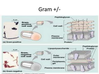
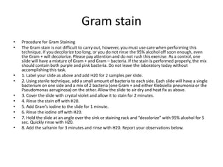
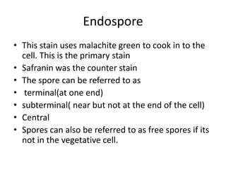
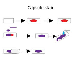
Lab 4

Microlab 2421
quiz
• what structure is the gram stain targeted or
  stain?
• Why do you not heat fix on the capsule stain.
quiz
• what structure is the gram stain targeted or
  stain?
peptidoglycan Cell wall
• Why do you not heat fix on the capsule stain.
Because it might shrink the cell where it looks
  like a non capsulated cell, look capsulated, Or
  it destroys the capsule.
Bacillus anthracis
• This is sporulating microorganism that it occurs most
  commonly in soil.
• This organism has been use countless in bioterrorist attacks
  including Amerithrax where Bacillus anthracis was sent in
  the mail to a senator, and several reporters .

• This organism usually attacks a individual thought the
  breathing in spores where it will cross the lungs epithelium
  in to the organism blood where it then will be intake by a
  macrophage, where it will then become a vegetative cell
  where it starts to replicate inside the macrophage.
Answers
• Questions
• 1. How many colonies did you count on your “spread plate”?
   Calculate how many bacteria were in the mix prior to your dilution?
   (Hint: remember the volume that you added to the plate and the
   dilution you carried out. Give your answer in the format of colony
   forming units/ml (CFU/ml). Count the colonies from all of your
   dilutions if possible. Your calculations should give you the same
   answer or very close. The equation that is used
• Colonies =X
X/10^-8=_Y____ cfu/(1/10)ml
Y/.10=cfu/ml
• 2. Why did you test 3 different dilutions of culture? Do not write
   that the instructions told you to do so.
• This was to see how the different dilutions will have different
   isolated colonies
•   3. Did you have isolated colonies of all three types on your streak for isolation? Did
    you have isolated colonies of all three types on your spread plate? Which method
    of isolation worked better streaking for isolation or spread plate? Do you know
    why?
• 4. Why is a colony considered “pure”?
• Because all the organism in the colony are clones of each other, because they are
    all derive from each cell
• 5. Why did you heat the inoculating loop in between streaking for isolation?
• To kill the bacteria so each time you streaked it would then dilute the bacteria
• 6. What is the difference between a “contaminated” culture and a “mixed”
    culture? What was the mixture that you were given?
• A contaminated culture the organism is not known in a bacteria of 2 or more.
    Mixed is where 2 or more bacteria are known.
• 7. Why did you use sterile saline to dilute your bacteria culture? (Hint: what would
    have happened if you used pure water?)
This was because if you had pure water then it would lyses the cell thought osmotic
    pressure increasing and breaking open the cell.
Today
•   Three different stains that will be used today.
•   Gram stain
•   Capsule stain
•   Spore stain
Gram stain
• This is a differential stain
• What is a differential stain ?
• This is a stain that differentials some structure
  of a bacteria.
• Was invented by Hans Christian Gram.
• Cristal violet is the primary stain
• Grams iodine is the mordant
• safranin secondary stain.
Gram stains
• Why is the Gram stain important?
• Because the gram stain tells doctors how to
  treat the infection. It also tells microbiologist
  how to work with the bacteria.
Gram +/-
Gram stain
•   Procedure for Gram Staining
•   The Gram stain is not difficult to carry out, however, you must use care when performing this
    technique. If you decolorize too long, or you do not rinse the 95% alcohol off soon enough, even
    the Gram + will decolorize. Please pay attention and do not rush this exercise. As a control, one
    slide will have a mixture of Gram + and Gram – bacteria. If the stain is performed properly, the mix
    should contain both purple and pink bacteria. Do not leave the laboratory today without
    accomplishing this task.
•   1. Label your slide as above and add H20 for 2 samples per slide.
•   2. Using sterile technique, add a small amount of bacteria to each side. Each slide will have a single
    bacterium on one side and a mix of 2 bacteria (one Gram + and either Klebsiella pneumonia or the
    Pseudomonas aeruginosa) on the other. Allow the slide to air dry and heat fix as above.
•   3. Cover the slide with crystal violet and allow it to stain for 2 minutes.
•   4. Rinse the stain off with H20.
•   5. Add Gram’s iodine to the slide for 1 minute.
•   6. Rinse the iodine off with H20.
•   7. Hold the slide at an angle over the sink or staining rack and “decolorize” with 95% alcohol for 5
    sec. Quickly rinse with H20.
•   8. Add the safranin for 3 minutes and rinse with H20. Report your observations below.
Gram stain
Endospore
• This stain uses malachite green to cook in to the
  cell. This is the primary stain
• Safranin was the counter stain
• The spore can be referred to as
• terminal(at one end)
• subterminal( near but not at the end of the cell)
• Central
• Spores can also be referred to as free spores if its
  not in the vegetative cell.
Endospore stain
•   Procedure for endospore staining:
•   1. Slides have already been prepared by the aseptic transfer of a sample of Bacillus
    brevis and a sample of Pseudomonas aeruginosa to a slide. The samples were air
    dried but you NEED to HEAT fix the specimen.
•   2. Tear a paper towel just smaller than the size of the slide and cover the heat-
    fixed slide with the toweling. Flood the slide with malachite green, a basic stain
    which will stain the vegetative cells as well as the endospores.
•   3. CAREFULLY, using the clothes pins, hold your slide over the Bunsen burner just
    out of reach of the flame. Alternatively, have a beaker of boiling water and use this
    as a heat source. Do not make the dye boil. If you notice that the malachite green
    is drying up, add more. Heat the slide for 5 minutes.
•   4. Cool the slide before DESTAINING gently with H20 for 30 sec.This will decolorize
    the vegetative cells.
•   5. Counterstain the cells with safranin for 2 minutes. The counterstain is also called
    the secondary stain.
•   6. Rinse the slide with H20, blot dry and observe with the oil immersion objective.
    Draw your observations. Where are the endospores; subterminal, terminal, etc. Do
    you see free endsopores?
Endospore stain
Capsule Stain
• There are two different stains the Maneval and the
  Anthony stain.
• What one are we using today
       Manevel
This stain uses Congo red as a ph indicator s when you
  add Maneval solution to the Congo red it changes
  Congo red to a purple or blue.
• What are two reasons why the capsule is beneficial to a
  bacteria?
• It allows for adhesion, and doesn’t allow for a
  macrophage to phagocytosis.
Capsule stain
• Procedure for CAPSULE stain
• 1. Place a few drops of Congo red on a clean slide. Do NOT use a
  water drop in this sample preparation. 2. Mix in a small amount of
  Klebsiella pneumonia. 3. AIR DRY. DO NOT HEAT FIX. (Heating the
  slide will destroy the polysaccharide capsule and cause non-
  encapsulated cells to shrink and appear encapsulated) 4. Place the
  slide on a rack of the staining tray. Gently cover the smear with the
  Maneval solution and wait 5 minutes. 5. Lift the slide and gently
  pour off excess stain and GENTLY rinse with water. If you hit the
  slide with a strong force of water, you may remove the cells and
  stain. 6. Place slide on Kimwipes to soak up the excess water. Do
  not blot, the slide needs to air dry.
• 7. Examine the slide with an oil immersion lens at 1,000X
  magnification. Repeat with E. coli.
Capsule stain
Next week
• Study for next weeks quiz

Lab 4

  • 1.
  • 2.
    quiz • what structureis the gram stain targeted or stain? • Why do you not heat fix on the capsule stain.
  • 3.
    quiz • what structureis the gram stain targeted or stain? peptidoglycan Cell wall • Why do you not heat fix on the capsule stain. Because it might shrink the cell where it looks like a non capsulated cell, look capsulated, Or it destroys the capsule.
  • 4.
    Bacillus anthracis • Thisis sporulating microorganism that it occurs most commonly in soil. • This organism has been use countless in bioterrorist attacks including Amerithrax where Bacillus anthracis was sent in the mail to a senator, and several reporters . • This organism usually attacks a individual thought the breathing in spores where it will cross the lungs epithelium in to the organism blood where it then will be intake by a macrophage, where it will then become a vegetative cell where it starts to replicate inside the macrophage.
  • 5.
    Answers • Questions • 1.How many colonies did you count on your “spread plate”? Calculate how many bacteria were in the mix prior to your dilution? (Hint: remember the volume that you added to the plate and the dilution you carried out. Give your answer in the format of colony forming units/ml (CFU/ml). Count the colonies from all of your dilutions if possible. Your calculations should give you the same answer or very close. The equation that is used • Colonies =X X/10^-8=_Y____ cfu/(1/10)ml Y/.10=cfu/ml • 2. Why did you test 3 different dilutions of culture? Do not write that the instructions told you to do so. • This was to see how the different dilutions will have different isolated colonies
  • 6.
    • 3. Did you have isolated colonies of all three types on your streak for isolation? Did you have isolated colonies of all three types on your spread plate? Which method of isolation worked better streaking for isolation or spread plate? Do you know why? • 4. Why is a colony considered “pure”? • Because all the organism in the colony are clones of each other, because they are all derive from each cell • 5. Why did you heat the inoculating loop in between streaking for isolation? • To kill the bacteria so each time you streaked it would then dilute the bacteria • 6. What is the difference between a “contaminated” culture and a “mixed” culture? What was the mixture that you were given? • A contaminated culture the organism is not known in a bacteria of 2 or more. Mixed is where 2 or more bacteria are known. • 7. Why did you use sterile saline to dilute your bacteria culture? (Hint: what would have happened if you used pure water?) This was because if you had pure water then it would lyses the cell thought osmotic pressure increasing and breaking open the cell.
  • 7.
    Today • Three different stains that will be used today. • Gram stain • Capsule stain • Spore stain
  • 8.
    Gram stain • Thisis a differential stain • What is a differential stain ? • This is a stain that differentials some structure of a bacteria. • Was invented by Hans Christian Gram. • Cristal violet is the primary stain • Grams iodine is the mordant • safranin secondary stain.
  • 9.
    Gram stains • Whyis the Gram stain important? • Because the gram stain tells doctors how to treat the infection. It also tells microbiologist how to work with the bacteria.
  • 10.
  • 11.
    Gram stain • Procedure for Gram Staining • The Gram stain is not difficult to carry out, however, you must use care when performing this technique. If you decolorize too long, or you do not rinse the 95% alcohol off soon enough, even the Gram + will decolorize. Please pay attention and do not rush this exercise. As a control, one slide will have a mixture of Gram + and Gram – bacteria. If the stain is performed properly, the mix should contain both purple and pink bacteria. Do not leave the laboratory today without accomplishing this task. • 1. Label your slide as above and add H20 for 2 samples per slide. • 2. Using sterile technique, add a small amount of bacteria to each side. Each slide will have a single bacterium on one side and a mix of 2 bacteria (one Gram + and either Klebsiella pneumonia or the Pseudomonas aeruginosa) on the other. Allow the slide to air dry and heat fix as above. • 3. Cover the slide with crystal violet and allow it to stain for 2 minutes. • 4. Rinse the stain off with H20. • 5. Add Gram’s iodine to the slide for 1 minute. • 6. Rinse the iodine off with H20. • 7. Hold the slide at an angle over the sink or staining rack and “decolorize” with 95% alcohol for 5 sec. Quickly rinse with H20. • 8. Add the safranin for 3 minutes and rinse with H20. Report your observations below.
  • 12.
  • 13.
    Endospore • This stainuses malachite green to cook in to the cell. This is the primary stain • Safranin was the counter stain • The spore can be referred to as • terminal(at one end) • subterminal( near but not at the end of the cell) • Central • Spores can also be referred to as free spores if its not in the vegetative cell.
  • 14.
    Endospore stain • Procedure for endospore staining: • 1. Slides have already been prepared by the aseptic transfer of a sample of Bacillus brevis and a sample of Pseudomonas aeruginosa to a slide. The samples were air dried but you NEED to HEAT fix the specimen. • 2. Tear a paper towel just smaller than the size of the slide and cover the heat- fixed slide with the toweling. Flood the slide with malachite green, a basic stain which will stain the vegetative cells as well as the endospores. • 3. CAREFULLY, using the clothes pins, hold your slide over the Bunsen burner just out of reach of the flame. Alternatively, have a beaker of boiling water and use this as a heat source. Do not make the dye boil. If you notice that the malachite green is drying up, add more. Heat the slide for 5 minutes. • 4. Cool the slide before DESTAINING gently with H20 for 30 sec.This will decolorize the vegetative cells. • 5. Counterstain the cells with safranin for 2 minutes. The counterstain is also called the secondary stain. • 6. Rinse the slide with H20, blot dry and observe with the oil immersion objective. Draw your observations. Where are the endospores; subterminal, terminal, etc. Do you see free endsopores?
  • 15.
  • 16.
    Capsule Stain • Thereare two different stains the Maneval and the Anthony stain. • What one are we using today Manevel This stain uses Congo red as a ph indicator s when you add Maneval solution to the Congo red it changes Congo red to a purple or blue. • What are two reasons why the capsule is beneficial to a bacteria? • It allows for adhesion, and doesn’t allow for a macrophage to phagocytosis.
  • 17.
    Capsule stain • Procedurefor CAPSULE stain • 1. Place a few drops of Congo red on a clean slide. Do NOT use a water drop in this sample preparation. 2. Mix in a small amount of Klebsiella pneumonia. 3. AIR DRY. DO NOT HEAT FIX. (Heating the slide will destroy the polysaccharide capsule and cause non- encapsulated cells to shrink and appear encapsulated) 4. Place the slide on a rack of the staining tray. Gently cover the smear with the Maneval solution and wait 5 minutes. 5. Lift the slide and gently pour off excess stain and GENTLY rinse with water. If you hit the slide with a strong force of water, you may remove the cells and stain. 6. Place slide on Kimwipes to soak up the excess water. Do not blot, the slide needs to air dry. • 7. Examine the slide with an oil immersion lens at 1,000X magnification. Repeat with E. coli.
  • 18.
  • 19.
    Next week • Studyfor next weeks quiz