1. Culture of Larval Stage of Nematodes
-Nematodes infections giving rise to larval stages that hatch in soil or tissue may
be diagnosed by using certain fecal culture methods to concentrate them more
readily.
-These techniques are especially useful for hook worm, Strongyloides, and
Trichostrongyle infections.
-The rearing of infective-stage nematode larvae also serves to aid the specific
diagnosis of hookworm, and Trichostrongyle infections.
-The eggs of many of these species are identical and specific identifications are
based on larval morphology
-Culture of feces for larvae is useful to:
(a) Reveal the presence of larvae when they are too scanty to be detected by
concentration methods.
(b) Distinguish whether the infection is due to Strongyloides or hookworm on the
basis of rhabditiform larval morphology by allowing hook worm eggs to hatch and
release first-stage larvae,
(c)Allow development of larvae into the filariform stage for further differentiation.
(d)Also culture techniques are useful for obtaining a large number of infective-
stage larvae for research
Purposes.
1-Harada-Mori filter paper culture.
2-Baermann technique
3-Charcoal culture.
4-Filter paper/slant culture
2. 1-Harada-Mori filter paper culture
-To detect light infections of hookworm, Strongyloides and Trichostrongylus
spp.
-The technique employs a filter paper to which fecal material is added and a
test tube into which the filter paper is inserted.
-Moisture is provided by adding water to the tube.
-The water continuously soaks the filter paper by capillary action.
-Incubation under suitable conditions favors hatching of ova and/or
development of larvae.
-Fecal specimens to be cultured should not be refrigerated, since some
parasites (especially Ancylostoma spp.) are susceptible to cold and may fail to
develop after refrigeration.
-The specimen must be fresh stool that has not been refrigerated.
Procedure:
-Cut a narrow (3/8 by 5 in.) strip of filter paper, and taper it slightly at one end.
-Smear 0.5 to 1 g of feces in the center of the strip.
-Add 3 - 4 ml of distilled water to a 15-ml conical centrifuge tube.
-Insert the filter paper strip into the tube so that the tapered end is near the bottom
of the tube.
-The water level should be slightly (0.5 in.) below the fecal spot.
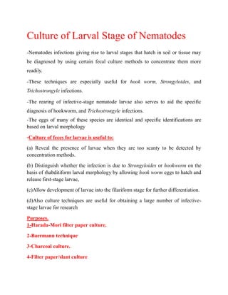
-It is not necessary to cap the tube. However, a cork stopper or a cotton plug may
be used (see Fig.1).
-Allow the tube to stand upright in a rack at 25 - 28 Co. Add distilled water to
maintain the original level.
3. -Keep the tube for 10 days, and check daily by with drawing a small amount of
fluid from the bottom of the tube.
-Prepare a smear on a glass slide, cover with a coverslip, and examine with the
10X objective.
-Examine the larvae for motility and typical morphological features to reveal
whether hookworm, Strongyloides, or Trichostrongylus larvae are present.
Harada-Mori filter paper culture Fig. 1
RESULTS
A. Larval nematodes (hookworm, Strongyloides spp., or Trichostrongylus spp.)
may be recovered.
B. If Strongyloides spp.are present, freeliving stages and larvae may be found after
several days in culture.
4. PROCEDURE NOTES :
1- It is often difficult to observe details in rapidly moving larvae. If desired, use
slight heating or a drop of iodine or formalin to kill the larvae.
2- Infective larvae may be found anytime after day 4 or even on day 1 in a heavy
infection.
3- Since infective larvae may migrate upward as well as downward on the filter
paper strip, be careful when handling the fluid and the paper strip itself to prevent
infection.
Handle the filter paper with forceps.
4- It is important to maintain the original water level to keep optimum humidity.
5- Preserved fecal specimens or specimens obtained after a barium meal are not
suitable for processing by this method.
2-Baermann technique
-Strongyloides spp. larvae are usually the only larvae found in stool specimens.
-Depending on bowel transit time and the condition of the patient, rhabditiform and
rarely, filariform larvae may be present.
-If there is delay in examination of the stool, then embryonated ova and larvae of
hookworm may be present.
-If possible, use a fresh fecal specimen that has been obtained after administration
of a mild saline cathartic, not a stool softener. Soft stool is recommended, but any
fresh fecal specimen is acceptable.
-Set up a clamp supporting a 6-in.-mouth glass funnel.
-Attach rubber tubing and a pinch clamp to the bottom of the funnel
-Place a collection beaker underneath (see the Fig.2).
5. Baermann technique Fig.2
Procedure :
- Place wire gauze or a nylon filter over the top of the funnel (or resting within the
funnel), and then place a pad consisting of two layers of gauze over that.
-Close the pinch clamp at the bottom of the tubing, and fill the funnel with tap
water until it just soaks the gauze padding.
-Spread a large amount of fecal material on the gauze padding so the specimen is
in contact with water. If the fecal material is very firm, emulsify in water.
-Allow the apparatus to stand for 2 or more hours, draw off 10 ml of fluid into the
beaker by releasing the pinch clamp, centrifuge for 2 min at 500 g, and examine
the sediment under the microscope (100X and 400X) for the presence of motile
larvae.
-Make sure that the end of the tubing is well inside the beaker before slowly
releasing the pinch clamp.
6. Infective larvae may be present; wear gloves when performing this procedure.
-If there are no larvae seen, allow the apparatus to stand at room temperature for 8
- 12 h and examine additional fluid (after centrifugation).
Discard after 12 h.