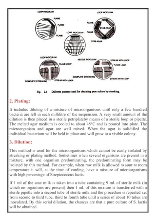
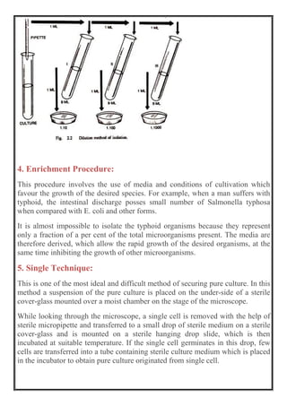
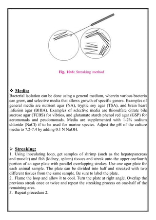
BACTERIA ISOLATION
 Introduction:
There are ap proximately 10,000 named species of microbes. It is estimated that
there are between 10,000 and 100,000 more unidentified species for every
identified one. Not only are there many types of bacteria, there are a lot of
individual bacteria. A single spoonful of soil can have 100 million individual
bacteria. A scraping of your gums can yield 1 million bacteria per cm2 (a cm2 is
about the size of your little fingernail). The bacteria in and on our bodies makes
up about 10% of our dry body weight.
Most of the currently known species of bacteria have been identified using
traditional microbiological techniques such as the gram stain reaction,
morphology, and metabolic reactions. Bacteria rarely live alone but in
communities with other bacteria. This is true both in the environment and in and
on our bodies. This class focuses on the role of bacteria in disease. Isolating a
single bacterium species is the first step in identifying the bacteria possibly
responsible for a disease process.
The first requirement for physically isolating a bacterium is that it can be
cultured in the laboratory. This requires knowledge of optimal temperature for
growth, optimal oxygen requirements, and optimal nutritional needs. We work
with a very limited number of bacteria in this course. The bacteria we work with
are also very easy to culture in the lab.
 Isolation of Microorganisms:
Microorganisms occur in natural environment like soil. They are mixed with
several other forms of life. Many microbes are pathogenic. They cause a number
of diseases with a variety of symptoms, depending on how they interact with the
patient. The isolation and growth of suspected microbe in pure culture is essential
for the identification and control the infectious agent.
The primary culture from natural source will normally be a mixed culture
containing microbes of different kinds. But in laboratory, the various species may
be isolated from one another. A culture which contains just one species of
microorganism is called a pure culture. The process of obtaining a pure culture
by separating one species of microbe from a mixture of other species, is known
as isolation of the organisms.
 Methods of Isolation:
There are special techniques employed to obtain pure cultures of microorganisms.
In few cases it is possible to secure pure culture by direct isolation or direct
transfer. This can be done only in those situations in which pure culture occurs
naturally. Kinds of specimens taken for culturing will depend on the nature and
habitat of microbes.
Different pathogens can be isolated from body tissues and fluids such as blood,
urine, sputum, pus, faces, spinal fluid, bile, pleural fluids, stomach fluids etc. In
the blood stream of a patient suffering with typhoid fever, the bacteria
Salmonella typhosa may be present.
A pure culture of this bacterium may be obtained by drawing blood sample using
a sterilized hypodermic syringe and treating the blood with anticoagulant such as
heparin and potassium oxalate. The presence of the anticoagulant prevents the
pathogenic microbe from entrapping in fibrin clot. The sample of the blood may
be inoculated into a suitable medium.
Following isolation methods are employed to isolate microbes from mixed
cultures:
1. Streaking
2. Plating
3. Dilution
4. Enriched procedure, and
5. Single cell technique.
1. Streaking:
This is most widely used method of isolation. The technique consists of pouring
a suitable sterile medium into sterile petriplate and allowing the medium to
solidify. By means of a sterile loope or straight needle or a sterile bent glass-rod
a small amount of growth preferably from a broth culture or bacterial suspension
is streaked back and forth across the surface of agar until about one third of the
diameter of the plate has been covered.
The needle is then flamed and streaking in done at right angles to and across the
first streak. This serves to drag bacteria out in a long line from the initial streak.
When this streaking is completed the needle is again flamed and streaking is
done at right angles to the second streak and parallel to the first.
2. Plating:
It includes diluting of a mixture of microorganisms until only a few hundred
bacteria are left in each millilitre of the suspension. A very small amount of the
dilution is then placed in a sterile petriplateby means of a sterile loop or pipette.
The melted agar medium is cooled to about 45°C and is poured into plate. The
microorganism and agar are well mixed. When the agar is solidified the
individual bacterium will be held in place and will grow to a visible colony.
3. Dilution:
This method is used for the microorganisms which cannot be easily isolated by
streaking or plating method. Sometimes when several organisms are present in a
mixture, with one organism predominating, the predominating form may be
isolated by this method. For example, when raw milk is allowed to sour at room
temperature it will, at the time of curding, have a mixture of microorganisms
with high percentage of Streptococcus lactis.
If 1 ml of the sour milk is taken into a tube containing 9 ml. of sterile milk (in
which no organisms are present) then 1 ml. of this mixture is transferred with a
sterile pipette into a second tube of sterile milk and the procedure is repeated i.e.
from second to third tube, third to fourth tube until a series of about 10 tubes are
inoculated. By this serial dilution, the chances are that a pure culture of S. lactis
will be obtained.
4. Enrichment Procedure:
This procedure involves the use of media and conditions of cultivation which
favour the growth of the desired species. For example, when a man suffers with
typhoid, the intestinal discharge posses small number of Salmonella typhosa
when compared with E. coli and other forms.
It is almost impossible to isolate the typhoid organisms because they represent
only a fraction of a per cent of the total microorganisms present. The media are
therefore derived, which allow the rapid growth of the desired organisms, at the
same time inhibiting the growth of other microorganisms.
5. Single Technique:
This is one of the most ideal and difficult method of securing pure culture. In this
method a suspension of the pure culture is placed on the under-side of a sterile
cover-glass mounted over a moist chamber on the stage of the microscope.
While looking through the microscope, a single cell is removed with the help of
sterile micropipette and transferred to a small drop of sterile medium on a sterile
cover-glass and is mounted on a sterile hanging drop slide, which is then
incubated at suitable temperature. If the single cell germinates in this drop, few
cells are transferred into a tube containing sterile culture medium which is placed
in the incubator to obtain pure culture originated from single cell.
Other methods:
The isolation of anaerobic microorganisms is very difficult. There are certain
special techniques by which these organisms are isolated.
 Cultivation of Microorganisms:
For cultivating microbes in laboratory, we require culture media. The various
mixtures of nutritive substances used for the laboratory cultivation of
microorganisms are collectively known as culture media. The culture media
serve as soil in which bacteria are planted for the purpose of study.
 Culture Media:
Culture media must contain all the essential nutrients required by the organism
for its growth and reproduction. A suitable source of energy, building materials
and growth factors must be supplied in adequate amounts.
So a culture medium must contain:
Since microorganisms show a considerable variation in their nutritional
requirements, no single medium is suitable for growth of all of them.
The different types of culture media employed fall into three groups:
1. Defined or synthetic media:
These are the media prepared from chemical compounds. They are highly
purified and specific, an investigator working in another laboratory can duplicate
them.
2. Complex or non-synthetic media:
Media that are prepared from ingredients that have not been precisely defined. It
contains hydrolysed proteins and vitamin extracts. This type of medium cannot
be duplicated by another worker in another laboratory. Peptone is usually
produced by boiling beef, by the hydrolysis of its protein. Casein peptone and
milk peptone are also used in complex media as the source of amino acids and
nitrogen.
All liquid media, whether complex or synthetic may be converted to solid media
by adding either gelat in (a protein) or agar-agar, (a complex polysaccharide)
extracted from red marine algae. The use of agar has an advantage. The most
bacteria are unable to hydrolyze this molecule into more simple components.
Since gelatin is a liquid at room temperature, the use agar allows the medium to
remain in a solid form while microbes are growing on its surface.
3. Living cells:
These are used for the cultivation of viruses. For example, fertilized eggs
incubated for 8 to 12 days are able to support the growth of many viruses.
In another classification culture media are grouped into following four
types:
1. Natural media:
Includes substances occurring in nature, as 1) Milk 2) Eggs 3) Blood 4) Extract
of plant and animal tissues.
2. Derived media:
Includes known substances but the chemical composition of each is unknown.
Examples are 1. Nutrient broth (prepared by boiling beef to extract nutrients and
adding an amino acid-nitrogen source.) 2. Nutrient agar 3. Nutrient gelatin.
3. Chemically defined media:
Exact chemical composition known.
4. Special media:
Include combinations of the other three types of media.
There are four categories of media used in laboratory:
They are:
1. Enrichment
2. Selective
3. Differential and
4. Propagation.
1. Enrichment media:
They are prepared with ingredients that will enhance the growth of certain
microbes. Enrichment media encourage the growth of the suspected pathogen so
that it will become the most pre-dominant type of microbe in the culture. Two
types of enrichment media are blood agar and chocolate agar.
2. Selective media:
They are prepared with ingredients that inhibit the growth of unwanted microbes
which might be in the specimen. The inhibitor may be an antibiotic, salt or other
chemical. Mixed culture of microbes originally grown in enrichment media may
be inoculated into selective media to isolate the desired microbe.
3. Differential media:
They are designed to differentiate among microbes. Different bacterial species
may produce dissimilar colony colours when grown on differential agar. While in
differential broth cultures, the media change colour. Differential media are used
to confirm the identity of a microbe that has already been isolated by culturing in
enrichment and selective media.
4. Propagation media:
They are used to propagate, or keep microbes growing for a long lime. Samples
grown on these media may be taken for analysis. The most common propagation
media are nutrient broth and agar.
 Preparation of Media:
There are three main steps in the preparation of media:
(a) Preparation as solutions of chemicals and adjusting the pH.
(b) Dispensing the media, and
(c) Sterilization.
A broth is prepared by dissolving the appropriate amount of the components in
distilled water and pH is adjusted by the addition of either dilute NaOH or Hcl.
The broth is dispensed into clean rimless ‘Pyrex’ test tubes which are plugged
with non-absorbant cotton wool plugs. The test tubes are placed in wire baskets
which are covered with grease proof paper.
The media are sterilized by autoclaving at a temperature of 121 °C and a pressure
of 151 b/in2 for 15 minutes. But medium containing heat- sensitive substances
are sterilized either by filtering the solution at room temperature, using
bacteria-proof filter or by a process called Tyndallization.
In this method, the liquids are steamed for one hour a day on three consecutive
days and the liquids are incubated at 25-30°C. During the first steaming, all the
heat sensitive vegetative cells are killed, leaving only the spores. During the first
incubation period, most of the spores germinate in to vegetative cells. These
vegetative cells are killed by the second steam period.
In the second incubation period, the rest of the spores germinate into vegetative
cells which are killed by the third steaming period. In this way, the liquids are
sterilized without temperature rising above 100°C.
 Maintenance of Pure Culture:
After obtaining the pure culture of a particular microbe, it may be grown
and maintained as a pure culture in different ways:
1. The most common practice is to grow the culture on suitable medium until it
reaches the stationary phase of growth, and then store in a refrigerator. If they are
to be kept alive for a long period all culture must be transferred to a fresh sterile
medium. Thus by successive transfer, a culture may be kept for an indefinite
period.
2. A second method involves freezing of young culture and desiccating it under
vaccum. The cells of a pure culture will remain viable for a long period of time if
they are mixed with sterile blood serum, sterile skimmed milk, before freezing
and drying. They dried cultures are kept in the sealed, evacuated tubes and are
stored in cool places.
3. This method of maintaining pure culture is most suitable for spore forming
species. The microorganisms are grown in pure culture in suitable media. A
suspension of microorganisms is then transferred to cotton stoppered tubes of
sterilized dry soil. The spores remain viable, though dormant, for long periods of
time, in dry soil. The organism can be grown after a desired period, by
transferring the soil into a suitable medium and incubating it under suitable
temperature.
 PRINCIPLE:
Bacterial isolation, purification and identification are the first steps to
bacteriological studies. Isolation is done to obtain pure bacterial cultures.
Bacteria are usually isolated from fish kidney and spleen; and from the
hepatopancreas, lymphoid organ and muscles of shrimp. These tissues are
monitor organs that usually harbor the disease-causing bacteria during infection.
To obtain a pure bacterial culture is the first step to bacterial identification. Pure
culture is essential in the study of the morphology, physiology, biochemical
characteristics, and susceptibility to antimicrobial agents of a particular bacterial
strain. Pure cultures are best obtained by using solid media, by streak plate or
pour plate method. Streak plate, if properly done, is the most practical method. In
the streak plate method, a loopful of the inoculum is placed near the periphery of
the plate with agar medium and spread or streaked on the upper portion of the
plate with parallel overlapping strokes. The inoculum is streaked over other
portions of the plate so that isolated colonies could be observed in the last
streaked area. The identification of a bacterial pathogen is important in fish
diagnosis. Treatment could be implemented only after the causative agent or the
bacterium has been identified. Bacterial species differ in morphological,
physiological and biochemical characteristics and those can be used when coding
or labelling them (Appendix 1.1). Therefore, identification is accomplished by
performing several morphological, physiological and biochemical tests. Results
of these tests are compared to established taxa or identification schemes
(Appendix 1.2). Bacterial cultures should be preserved for future study. Storing
in appropriate medium preserves bacterial cultures. The simplest method is by
sub-culturing or by transferring the organism to fresh solid medium that has a
minimal nutrient content to prevent bacterial overgrowth. The bacteria are
allowed to grow before storing in the refrigerator or are covered with paraffin oil
and stored at room temperature in the dark. Another simple method is by
deep-freezing of the bacterial culture, stocked in a broth medium with glycerol.
Glycerol is added to prevent the dessication of bacterial cells. Bacterial cultures
may also be preserved by freeze-drying or lyophilization. In this method, water is
removed from the frozen bacterial suspension by sublimation under vacuum.
Bacterial cultures should be properly labeled or coded before storage. It is
important to label the tube or vial for storing bacterial cultures with an indelible
ink. The label or code should include the reference number and other pertinent
information such as source of sample (host animal, location), date of isolation,
special properties, identification, name of the person who isolated the organism
and the date of preparation of the stock culture.
 OBJECTIVES:
 After reading this chapter, you will be able to :
 Expalin the steps involved in the isolation of bacteria.
 describe the significance of Specimen collection.
 describe the significance of Preservation and transportation of specimen.
 explain the role of microscopy in isolation of bacteria.
 explain various methods for isolation of bacteria.

 ISOLATION OF BACTERIA:
Isolation of bacteria forms a very significant step in the diagnosis and
management of the illness. Isolation of bacteria involves various steps
 Specimen collection
 Preservation and transportation of specimen
 Microscopic examination of sample
 Various methods used for isolation of bacteria
Specimen collection Many different specimens are sent for microbiological
examination from patients with suspected bacterial infection. Common
specimens include urine, faeces, wound swabs, throat swabs, vaginal swabs,
sputum, and blood. Less common, but important specimens include cerebrospinal
fluid, pleural fluid, joint aspirates, tissue, bone and prosthetic material (e.g. line
tips).
Some types of specimen are normally sterile e.g. blood, CSF. These samples are
usually obtained via a percutaneous route with needle and syringe, using
appropriate skin disinfection and an aseptic technique. The culture of bacteria
from such specimens is usually indicative of definite infection except if they are
skin contaminants (bacteria inhabitants of normal skin).
In contrast, many microbiological specimens are obtained from non-sterile sites
e.g. vaginal or throat swabs, urine sample, stool sample. Such samples often
contain bacteria of no clinical relevance in addition to possible pathogens,
making the interpretation of culture results more difficult. In general it is
preferable to send samples from sterile sites if available.
It is preferred to obtain the samples for bacteriological culture before antibiotic
therapy is started. This maximizes the sensitivity of the investigations and
reduces false-negative results. Similarly, samples of tissue or pus are preferred
over swabs, to maximize the recovery of bacteria in the laboratory.
Specimens must be accurately labelled and accompanied by a properly
completed requisition form, indicating the nature of the specimen, the date of
sample collection, relevant clinical information, the investigations required, and
details of antibiotic therapy, if any.
This allows the laboratory to perform the correct range of tests, and helps in the
interpretation of results and reporting. Along with clinical specimens, medical
microbiology laboratories also process samples of food, water and other
environmental samples (e.g. air sampling from operating theatres) as part of
infection control procedures.
 High-risk samples :
Certain bacterial infections are a particular hazard to laboratory staff, and
specimens that might contain these pathogens should be labelled as ‘high risk’ to
allow for additional safety measures if necessary. For example - blood cultures
from suspected typhoid (Salmonella typhi) or brucellosis (Brucella species), and
samples from suspected Mycobacterium tuberculosis.
 Preservation and Transport of specimen:
Most specimens are sent to the laboratory in sterile universal containers. Swabs
are placed in a suitable transport medium (eg. charcoal medium) otherwise it
leads to false negative reporting.
Specimens should be transported as soon as possible to the laboratory. In case a
delay is anticipated the specimen should be stored at 4° C.
Immediate transport is necessary in order to:
(i) Preserve the viability of the ‘delicate’ bacteria, such as Streptococcus
pneumoniae or Haemophilus influenzae (delays in processing can cause
false-negative culture results);
(ii) (ii) Minimize the multiplication of bacteria (e.g. coliforms) within
specimens before they reach the laboratory. In particular urine and other
specimens that utilize a semiquantitative culture technique for thier detection, as
delays in transport can give rise to falsely high bacterial counts when the
specimen is processed.
 Microscopy:
A Gram stain helps with the visualization of bacteria, and gives an indication of
the type of bacteria present, based on the shape of the bacteria and the staining
properties (Gram positive: purple; Gram negative: pink/red).
A Gram stain also helps to identify mixtures of bacteria, helps to determine the
appropriate range of agar plates to be used for subsequent culture, and helps with
the interpretation of culture results.
For liquid specimens e.g. CSF, the sample is first centrifuged to concentrate any
bacterial cells in the deposit, and Gram stain and culture is performed from the
deposit after the supernatant is decanted. This helps increase the sensitivity of
both microscopy and culture.
Ziehl-Neelsen (ZN) stain is used to demonstrate the presence of Mycobacteria.
Mycobacteria can also be visualized using the fluorescent dye auramine and a
fluorescence microscope. Direct immunofluorescence is employed to detect
certain pathogens (e.g. Legionella, Pneumocystis) using specific antibodies
conjugated to a fluorescent dye.
Another microscopic technique is dark ground microscopy. This is mainly used
to detect the thin spirochaetal cells of Treponema pallidum (syphilis bacteria).
 METHODS OF ISOLATION OF BACTERIA:
Methods of isolation of bacteria can be broadly classified into two
 Culture methods
 On Solid media
 On Liquid media
 Automated systems
 Non-culture methods
 Culture methods :
The specimens received in the laboratory are plated on the culture media. The
appropriate culture media is selected depending upon the bacteria suspected. The
following precautions need to be taken into consideration when the culture
methods are processed
 Optimal atmospheric conditions
 Optimal temperature
 Growth requirement of the bacteria
Atmospheric conditions: Colonies of bacteria are usually large enough to
identify after 18–24 hours of incubation (usually at 37°C), but for some bacteria
longer incubation times are required (from 2 days to several weeks). Culture
plates are incubated (1) in air, (2) in air with added carbon dioxide (5%), (3)
anaerobically (without oxygen) or (4) micro-aerophilically (a trace of oxygen)
according to the requirements of the different types of bacteria that may be
present in specimens.
In case of Mycobacteria especially the scotochromogen the culture bottles are
placed in dark or the bottles are covered with black paper and kept for incubation
at 37°C. Temperature: Most of the bacteria requires a temperature of 37°C for
optimal growth. This temperature is provided placing the inoculated culture
plates in the incubator set at 37°C temperature.
 Growth requirement of the bacteria:
Different bacteria have different growth requirements. For eg Streptococcus
pneumoniae requires factor V and factor X for its growth, which are found in
chocolate agar. Thus for sample suspected of S. pneumoniae the samples are
plated on chocolate agar. Similarly depending upon the growth requirements the
appropriate culture media are used.
 CULTURE ON SOLID MEDIA:
The principal method for the detection of bacteria from clinical specimens is by
culture on solid culture media. Bacteria grow on the surface of culture media to
produce distinct colonies.
Different bacteria produce different but characteristic colonies, allowing for early
presumptive identification and easy identification of mixed cultures. There are
many different types of culture media. Agar is used as the gelling agent to which
is added a variety of nutrients (e.g. blood, peptone and sugars) and other factors
(e.g. buffers, salts and indicators).
Some culture media are nonselective (e.g. blood agar, nutrient agar) and these
will grow a wide variety of bacteria. While some e.g. MacConkey agar are more
selective (in this case through the addition of bile salts selecting for the
‘biletolerant’ bacteria found in the large intestine such as Escherichia coli and
Enterococcus faecalis). MacConkey agar also contains lactose and an indicator
system that identifies lactose-fermenting coliforms (e.g. Escherichia coli,
Klebsiella) from lactose-non fermenting coliforms (e.g. Morganella Salmonella).
Media can be made even more selective by the addition of antibiotics or other
inhibitory substances, and sophisticated indicator systems can allow for the easy
detection of defined bacteria from mixed populations.
 Method of inoculating the solid culture media:
Method used for inoculating the solid media depends upon the purpose of
inoculation- whether to have isolated colonies or to know the bacterial load of the
sample (quantitative analysis).
For obtaining the isolated colonies streaking method is used, the most common
method of inoculating an agar plate is streaking.
 Media:
Bacterial isolation can be done using a general medium, wherein various bacteria
can grow, and selective media that allows growth of specific genera. Examples of
general media are nutrient agar (NA), tryptic soy agar (TSA), and brain heart
infusion agar (BHIA). Examples of selective media are thiosulfate citrate bile
sucrose agar (TCBS) for vibrios, and glutamate starch phenol red agar (GSP) for
aeromonads and peudomonads. Media are supplemented with 1-2% sodium
chloride (NaCl) if to be used for marine species. Adjust the pH of the culture
media to 7.2-7.4 by adding 0.1 N NaOH.
 Streaking:
1. Using inoculating loop, get samples of shrimp (such as the hepatopancreas
and muscle) and fish (kidney, spleen) tissues and streak onto the upper onefourth
portion of an agar plate with parallel overlapping strokes. Use one agar plate for
each animal sample. The plate can be divided into half and streaked with two
different tissues from the same sample. Be sure to label the plate.
2. Flame the loop and allow it to cool. Turn the plate at right angle. Overlap the
previous streak once or twice and repeat the streaking process on one-half of the
remaining area.
3. Repeat procedure 2.
4. Incubate plates overnight at 30°C. Photo at right shows a streaked plate after
incubation.
5. After incubation for 16-20 hours, check for bacterial growth. Check for
luminescence under dark conditions, marking the luminous colonies on the plate
with a pentel pen. Isolated colonies should be observed in the last streaked area.
6. Select representative bacterial colonies based on the difference in shape, size
and color. Mark selected colonies from each plate. Subculture onto trypticase soy
agar (TSA) plate and incubate overnight.
 Storage:
1. Observe the colonies on the agar plate to determine the purity of the culture.
Pure cultures should show the same colony characteristics and not overlapping.
2. Select a pure wellisolated colony. Stab each strain into 2 tubes of 1.2% TSA,
label and incubate. These will serve as stock cultures.
3. Keep the stock cultures in the lowest compartment of the refrigerator (8-12°C)
or at room temperature until use. Do not stock cultures in these conditions for
over 6 months.
4. Purified bacterial cultures may also be stocked in nutrient broth with 20%
glycerol and stored at –80°C until use. Bacterial cultures may be stocked in this
condition for 2 years.
 IDENTIFICATION:
Bacterial isolates may be identified using conventional methods based on their
morphological, biochemical and physiological characteristics. The following are
important biochemical tests for the identification of bacterial genera that are
important in aquaculture:
1. Gram reaction
2. Oxidase test
3. Motility
4. Oxidation and fermentation test
5. O/129 sensitivity test 6. Sensitivity to novobiocin
Further biochemical characterization must be carried out if there is a need to
identify up to the species level.
 Culture in liquid media:
Bacteria can also be grown in liquid media (broth). Like agar plates, broth
cultures may be non selective or selective. Bacterial growth is easy to detect as
the clear liquid turns turbid, usually within 24–48 hr, but incubation may need to
be extended to 14 days or more.
The advantage of broth culture is that it is significantly more sensitive than direct
culture on agar. The disadvantage is that, by itself, it is not easy to determine the
type of bacteria present or whether a mixed growth has occurred, and in most
cases the broth must be subcultured onto solid agar plates. This causes an
additional delay in culture results. Broth cultures are also prone to contamination
Broth enrichment media are used when high sensitivity is required e.g. for
detection of bacteria from CSF, or to detect small numbers of Salmonella in a
stool sample containing many millions of other bacteria.
 Automated system:
Automated blood culture systems eg. BACTEC, BacteAlert utilize liquid culture.
Bacterial growth may be detected by a variety of methods (e.g. detection of
bacterial CO2 production).
Automated liquid culture systems are also available for the culture of
Mycobacteria, and similar technology can be used to automate sensitivity.
The advantage of automated system are Rapidity : they aid in faster growth of
bacteria.Thus less time consuming.
The incidence of contamination during the processing of sample are minimised.
Real time monitoring of the growth.
One of the main limitations is the commercial viability.
 Non culture methods
Isolation of bacteria can also be carried out by non-culture methods. In particular
the more advanced Amplification techniques like Polymerase chain reaction
(PCR), ligase chain reaction (LCR), strand displacement amplification (SDA),
and nucleic acid sequence based amplification (NASBA) are being used in
clinical laboratories for isolation and identification of bacteria.
The following are some of the factors that are considered in interpreting
bacteriological culture results:
 type of specimen z any delays in processing
 types of bacteria recovered
 knowledge of the normal human flora at different sites
 clinical information provided on the request form
 details of recent antibiotic therapy
There must be good liaison between healthcare workers and the microbiology
laboratory, in order to ensure that the most appropriate investigations are
performed, results are interpreted correctly, and clinically relevant
bacteriological reports are produced.
 REFERENCES:
1. Bailey, W.R. and E.G. Scott. 1966. Diagnostic Microbiology, Second Edition.
Toppan Company Ltd., Japan, 342 pp.
2. Tonguthai, K., S. Chinabut, T. Somsiri, P. Chanratchakol and S.
Kanchanakhan. 1999. Diagnostic Procedures for Finfish Diseases. Aquatic
Animal Health Research Institute, Bangkok, Thailand.
3. Isolation and Cultivation of Microorganisms,Article Shared by Akshay
Sisodiya

Bacterial isolation

  • 1.
    BACTERIA ISOLATION  Introduction: Thereare ap proximately 10,000 named species of microbes. It is estimated that there are between 10,000 and 100,000 more unidentified species for every identified one. Not only are there many types of bacteria, there are a lot of individual bacteria. A single spoonful of soil can have 100 million individual bacteria. A scraping of your gums can yield 1 million bacteria per cm2 (a cm2 is about the size of your little fingernail). The bacteria in and on our bodies makes up about 10% of our dry body weight. Most of the currently known species of bacteria have been identified using traditional microbiological techniques such as the gram stain reaction, morphology, and metabolic reactions. Bacteria rarely live alone but in communities with other bacteria. This is true both in the environment and in and on our bodies. This class focuses on the role of bacteria in disease. Isolating a single bacterium species is the first step in identifying the bacteria possibly responsible for a disease process. The first requirement for physically isolating a bacterium is that it can be cultured in the laboratory. This requires knowledge of optimal temperature for growth, optimal oxygen requirements, and optimal nutritional needs. We work with a very limited number of bacteria in this course. The bacteria we work with are also very easy to culture in the lab.  Isolation of Microorganisms: Microorganisms occur in natural environment like soil. They are mixed with several other forms of life. Many microbes are pathogenic. They cause a number of diseases with a variety of symptoms, depending on how they interact with the patient. The isolation and growth of suspected microbe in pure culture is essential for the identification and control the infectious agent. The primary culture from natural source will normally be a mixed culture containing microbes of different kinds. But in laboratory, the various species may be isolated from one another. A culture which contains just one species of microorganism is called a pure culture. The process of obtaining a pure culture by separating one species of microbe from a mixture of other species, is known as isolation of the organisms.  Methods of Isolation:
  • 2.
    There are specialtechniques employed to obtain pure cultures of microorganisms. In few cases it is possible to secure pure culture by direct isolation or direct transfer. This can be done only in those situations in which pure culture occurs naturally. Kinds of specimens taken for culturing will depend on the nature and habitat of microbes. Different pathogens can be isolated from body tissues and fluids such as blood, urine, sputum, pus, faces, spinal fluid, bile, pleural fluids, stomach fluids etc. In the blood stream of a patient suffering with typhoid fever, the bacteria Salmonella typhosa may be present. A pure culture of this bacterium may be obtained by drawing blood sample using a sterilized hypodermic syringe and treating the blood with anticoagulant such as heparin and potassium oxalate. The presence of the anticoagulant prevents the pathogenic microbe from entrapping in fibrin clot. The sample of the blood may be inoculated into a suitable medium. Following isolation methods are employed to isolate microbes from mixed cultures: 1. Streaking 2. Plating 3. Dilution 4. Enriched procedure, and 5. Single cell technique. 1. Streaking: This is most widely used method of isolation. The technique consists of pouring a suitable sterile medium into sterile petriplate and allowing the medium to solidify. By means of a sterile loope or straight needle or a sterile bent glass-rod a small amount of growth preferably from a broth culture or bacterial suspension is streaked back and forth across the surface of agar until about one third of the diameter of the plate has been covered. The needle is then flamed and streaking in done at right angles to and across the first streak. This serves to drag bacteria out in a long line from the initial streak. When this streaking is completed the needle is again flamed and streaking is done at right angles to the second streak and parallel to the first.
  • 3.
    2. Plating: It includesdiluting of a mixture of microorganisms until only a few hundred bacteria are left in each millilitre of the suspension. A very small amount of the dilution is then placed in a sterile petriplateby means of a sterile loop or pipette. The melted agar medium is cooled to about 45°C and is poured into plate. The microorganism and agar are well mixed. When the agar is solidified the individual bacterium will be held in place and will grow to a visible colony. 3. Dilution: This method is used for the microorganisms which cannot be easily isolated by streaking or plating method. Sometimes when several organisms are present in a mixture, with one organism predominating, the predominating form may be isolated by this method. For example, when raw milk is allowed to sour at room temperature it will, at the time of curding, have a mixture of microorganisms with high percentage of Streptococcus lactis. If 1 ml of the sour milk is taken into a tube containing 9 ml. of sterile milk (in which no organisms are present) then 1 ml. of this mixture is transferred with a sterile pipette into a second tube of sterile milk and the procedure is repeated i.e. from second to third tube, third to fourth tube until a series of about 10 tubes are inoculated. By this serial dilution, the chances are that a pure culture of S. lactis will be obtained.
  • 4.
    4. Enrichment Procedure: Thisprocedure involves the use of media and conditions of cultivation which favour the growth of the desired species. For example, when a man suffers with typhoid, the intestinal discharge posses small number of Salmonella typhosa when compared with E. coli and other forms. It is almost impossible to isolate the typhoid organisms because they represent only a fraction of a per cent of the total microorganisms present. The media are therefore derived, which allow the rapid growth of the desired organisms, at the same time inhibiting the growth of other microorganisms. 5. Single Technique: This is one of the most ideal and difficult method of securing pure culture. In this method a suspension of the pure culture is placed on the under-side of a sterile cover-glass mounted over a moist chamber on the stage of the microscope. While looking through the microscope, a single cell is removed with the help of sterile micropipette and transferred to a small drop of sterile medium on a sterile cover-glass and is mounted on a sterile hanging drop slide, which is then incubated at suitable temperature. If the single cell germinates in this drop, few cells are transferred into a tube containing sterile culture medium which is placed in the incubator to obtain pure culture originated from single cell.
  • 5.
    Other methods: The isolationof anaerobic microorganisms is very difficult. There are certain special techniques by which these organisms are isolated.  Cultivation of Microorganisms: For cultivating microbes in laboratory, we require culture media. The various mixtures of nutritive substances used for the laboratory cultivation of microorganisms are collectively known as culture media. The culture media serve as soil in which bacteria are planted for the purpose of study.  Culture Media: Culture media must contain all the essential nutrients required by the organism for its growth and reproduction. A suitable source of energy, building materials and growth factors must be supplied in adequate amounts. So a culture medium must contain:
  • 6.
    Since microorganisms showa considerable variation in their nutritional requirements, no single medium is suitable for growth of all of them. The different types of culture media employed fall into three groups: 1. Defined or synthetic media: These are the media prepared from chemical compounds. They are highly purified and specific, an investigator working in another laboratory can duplicate them. 2. Complex or non-synthetic media: Media that are prepared from ingredients that have not been precisely defined. It contains hydrolysed proteins and vitamin extracts. This type of medium cannot be duplicated by another worker in another laboratory. Peptone is usually produced by boiling beef, by the hydrolysis of its protein. Casein peptone and milk peptone are also used in complex media as the source of amino acids and nitrogen. All liquid media, whether complex or synthetic may be converted to solid media by adding either gelat in (a protein) or agar-agar, (a complex polysaccharide) extracted from red marine algae. The use of agar has an advantage. The most bacteria are unable to hydrolyze this molecule into more simple components. Since gelatin is a liquid at room temperature, the use agar allows the medium to remain in a solid form while microbes are growing on its surface. 3. Living cells: These are used for the cultivation of viruses. For example, fertilized eggs incubated for 8 to 12 days are able to support the growth of many viruses. In another classification culture media are grouped into following four types: 1. Natural media: Includes substances occurring in nature, as 1) Milk 2) Eggs 3) Blood 4) Extract of plant and animal tissues. 2. Derived media: Includes known substances but the chemical composition of each is unknown. Examples are 1. Nutrient broth (prepared by boiling beef to extract nutrients and adding an amino acid-nitrogen source.) 2. Nutrient agar 3. Nutrient gelatin.
  • 7.
    3. Chemically definedmedia: Exact chemical composition known. 4. Special media: Include combinations of the other three types of media. There are four categories of media used in laboratory: They are: 1. Enrichment 2. Selective 3. Differential and 4. Propagation. 1. Enrichment media: They are prepared with ingredients that will enhance the growth of certain microbes. Enrichment media encourage the growth of the suspected pathogen so that it will become the most pre-dominant type of microbe in the culture. Two types of enrichment media are blood agar and chocolate agar. 2. Selective media: They are prepared with ingredients that inhibit the growth of unwanted microbes which might be in the specimen. The inhibitor may be an antibiotic, salt or other chemical. Mixed culture of microbes originally grown in enrichment media may be inoculated into selective media to isolate the desired microbe. 3. Differential media: They are designed to differentiate among microbes. Different bacterial species may produce dissimilar colony colours when grown on differential agar. While in differential broth cultures, the media change colour. Differential media are used to confirm the identity of a microbe that has already been isolated by culturing in enrichment and selective media. 4. Propagation media: They are used to propagate, or keep microbes growing for a long lime. Samples grown on these media may be taken for analysis. The most common propagation media are nutrient broth and agar.
  • 8.
     Preparation ofMedia: There are three main steps in the preparation of media: (a) Preparation as solutions of chemicals and adjusting the pH. (b) Dispensing the media, and (c) Sterilization. A broth is prepared by dissolving the appropriate amount of the components in distilled water and pH is adjusted by the addition of either dilute NaOH or Hcl. The broth is dispensed into clean rimless ‘Pyrex’ test tubes which are plugged with non-absorbant cotton wool plugs. The test tubes are placed in wire baskets which are covered with grease proof paper. The media are sterilized by autoclaving at a temperature of 121 °C and a pressure of 151 b/in2 for 15 minutes. But medium containing heat- sensitive substances are sterilized either by filtering the solution at room temperature, using bacteria-proof filter or by a process called Tyndallization. In this method, the liquids are steamed for one hour a day on three consecutive days and the liquids are incubated at 25-30°C. During the first steaming, all the heat sensitive vegetative cells are killed, leaving only the spores. During the first incubation period, most of the spores germinate in to vegetative cells. These vegetative cells are killed by the second steam period. In the second incubation period, the rest of the spores germinate into vegetative cells which are killed by the third steaming period. In this way, the liquids are sterilized without temperature rising above 100°C.  Maintenance of Pure Culture: After obtaining the pure culture of a particular microbe, it may be grown and maintained as a pure culture in different ways: 1. The most common practice is to grow the culture on suitable medium until it reaches the stationary phase of growth, and then store in a refrigerator. If they are to be kept alive for a long period all culture must be transferred to a fresh sterile medium. Thus by successive transfer, a culture may be kept for an indefinite period. 2. A second method involves freezing of young culture and desiccating it under vaccum. The cells of a pure culture will remain viable for a long period of time if they are mixed with sterile blood serum, sterile skimmed milk, before freezing
  • 9.
    and drying. Theydried cultures are kept in the sealed, evacuated tubes and are stored in cool places. 3. This method of maintaining pure culture is most suitable for spore forming species. The microorganisms are grown in pure culture in suitable media. A suspension of microorganisms is then transferred to cotton stoppered tubes of sterilized dry soil. The spores remain viable, though dormant, for long periods of time, in dry soil. The organism can be grown after a desired period, by transferring the soil into a suitable medium and incubating it under suitable temperature.  PRINCIPLE: Bacterial isolation, purification and identification are the first steps to bacteriological studies. Isolation is done to obtain pure bacterial cultures. Bacteria are usually isolated from fish kidney and spleen; and from the hepatopancreas, lymphoid organ and muscles of shrimp. These tissues are monitor organs that usually harbor the disease-causing bacteria during infection. To obtain a pure bacterial culture is the first step to bacterial identification. Pure culture is essential in the study of the morphology, physiology, biochemical characteristics, and susceptibility to antimicrobial agents of a particular bacterial strain. Pure cultures are best obtained by using solid media, by streak plate or pour plate method. Streak plate, if properly done, is the most practical method. In the streak plate method, a loopful of the inoculum is placed near the periphery of the plate with agar medium and spread or streaked on the upper portion of the plate with parallel overlapping strokes. The inoculum is streaked over other portions of the plate so that isolated colonies could be observed in the last streaked area. The identification of a bacterial pathogen is important in fish diagnosis. Treatment could be implemented only after the causative agent or the bacterium has been identified. Bacterial species differ in morphological, physiological and biochemical characteristics and those can be used when coding or labelling them (Appendix 1.1). Therefore, identification is accomplished by performing several morphological, physiological and biochemical tests. Results of these tests are compared to established taxa or identification schemes (Appendix 1.2). Bacterial cultures should be preserved for future study. Storing in appropriate medium preserves bacterial cultures. The simplest method is by sub-culturing or by transferring the organism to fresh solid medium that has a minimal nutrient content to prevent bacterial overgrowth. The bacteria are allowed to grow before storing in the refrigerator or are covered with paraffin oil and stored at room temperature in the dark. Another simple method is by deep-freezing of the bacterial culture, stocked in a broth medium with glycerol. Glycerol is added to prevent the dessication of bacterial cells. Bacterial cultures
  • 10.
    may also bepreserved by freeze-drying or lyophilization. In this method, water is removed from the frozen bacterial suspension by sublimation under vacuum. Bacterial cultures should be properly labeled or coded before storage. It is important to label the tube or vial for storing bacterial cultures with an indelible ink. The label or code should include the reference number and other pertinent information such as source of sample (host animal, location), date of isolation, special properties, identification, name of the person who isolated the organism and the date of preparation of the stock culture.  OBJECTIVES:  After reading this chapter, you will be able to :  Expalin the steps involved in the isolation of bacteria.  describe the significance of Specimen collection.  describe the significance of Preservation and transportation of specimen.  explain the role of microscopy in isolation of bacteria.  explain various methods for isolation of bacteria.   ISOLATION OF BACTERIA: Isolation of bacteria forms a very significant step in the diagnosis and management of the illness. Isolation of bacteria involves various steps  Specimen collection  Preservation and transportation of specimen  Microscopic examination of sample  Various methods used for isolation of bacteria Specimen collection Many different specimens are sent for microbiological examination from patients with suspected bacterial infection. Common specimens include urine, faeces, wound swabs, throat swabs, vaginal swabs, sputum, and blood. Less common, but important specimens include cerebrospinal fluid, pleural fluid, joint aspirates, tissue, bone and prosthetic material (e.g. line tips). Some types of specimen are normally sterile e.g. blood, CSF. These samples are usually obtained via a percutaneous route with needle and syringe, using appropriate skin disinfection and an aseptic technique. The culture of bacteria from such specimens is usually indicative of definite infection except if they are skin contaminants (bacteria inhabitants of normal skin).
  • 11.
    In contrast, manymicrobiological specimens are obtained from non-sterile sites e.g. vaginal or throat swabs, urine sample, stool sample. Such samples often contain bacteria of no clinical relevance in addition to possible pathogens, making the interpretation of culture results more difficult. In general it is preferable to send samples from sterile sites if available. It is preferred to obtain the samples for bacteriological culture before antibiotic therapy is started. This maximizes the sensitivity of the investigations and reduces false-negative results. Similarly, samples of tissue or pus are preferred over swabs, to maximize the recovery of bacteria in the laboratory. Specimens must be accurately labelled and accompanied by a properly completed requisition form, indicating the nature of the specimen, the date of sample collection, relevant clinical information, the investigations required, and details of antibiotic therapy, if any. This allows the laboratory to perform the correct range of tests, and helps in the interpretation of results and reporting. Along with clinical specimens, medical microbiology laboratories also process samples of food, water and other environmental samples (e.g. air sampling from operating theatres) as part of infection control procedures.  High-risk samples : Certain bacterial infections are a particular hazard to laboratory staff, and specimens that might contain these pathogens should be labelled as ‘high risk’ to allow for additional safety measures if necessary. For example - blood cultures from suspected typhoid (Salmonella typhi) or brucellosis (Brucella species), and samples from suspected Mycobacterium tuberculosis.  Preservation and Transport of specimen: Most specimens are sent to the laboratory in sterile universal containers. Swabs are placed in a suitable transport medium (eg. charcoal medium) otherwise it leads to false negative reporting.
  • 12.
    Specimens should betransported as soon as possible to the laboratory. In case a delay is anticipated the specimen should be stored at 4° C. Immediate transport is necessary in order to: (i) Preserve the viability of the ‘delicate’ bacteria, such as Streptococcus pneumoniae or Haemophilus influenzae (delays in processing can cause false-negative culture results); (ii) (ii) Minimize the multiplication of bacteria (e.g. coliforms) within specimens before they reach the laboratory. In particular urine and other specimens that utilize a semiquantitative culture technique for thier detection, as delays in transport can give rise to falsely high bacterial counts when the specimen is processed.  Microscopy: A Gram stain helps with the visualization of bacteria, and gives an indication of the type of bacteria present, based on the shape of the bacteria and the staining properties (Gram positive: purple; Gram negative: pink/red). A Gram stain also helps to identify mixtures of bacteria, helps to determine the appropriate range of agar plates to be used for subsequent culture, and helps with the interpretation of culture results.
  • 13.
    For liquid specimense.g. CSF, the sample is first centrifuged to concentrate any bacterial cells in the deposit, and Gram stain and culture is performed from the deposit after the supernatant is decanted. This helps increase the sensitivity of both microscopy and culture. Ziehl-Neelsen (ZN) stain is used to demonstrate the presence of Mycobacteria. Mycobacteria can also be visualized using the fluorescent dye auramine and a fluorescence microscope. Direct immunofluorescence is employed to detect certain pathogens (e.g. Legionella, Pneumocystis) using specific antibodies conjugated to a fluorescent dye. Another microscopic technique is dark ground microscopy. This is mainly used to detect the thin spirochaetal cells of Treponema pallidum (syphilis bacteria).  METHODS OF ISOLATION OF BACTERIA: Methods of isolation of bacteria can be broadly classified into two  Culture methods  On Solid media  On Liquid media
  • 14.
     Automated systems Non-culture methods  Culture methods : The specimens received in the laboratory are plated on the culture media. The appropriate culture media is selected depending upon the bacteria suspected. The following precautions need to be taken into consideration when the culture methods are processed  Optimal atmospheric conditions  Optimal temperature  Growth requirement of the bacteria Atmospheric conditions: Colonies of bacteria are usually large enough to identify after 18–24 hours of incubation (usually at 37°C), but for some bacteria longer incubation times are required (from 2 days to several weeks). Culture plates are incubated (1) in air, (2) in air with added carbon dioxide (5%), (3) anaerobically (without oxygen) or (4) micro-aerophilically (a trace of oxygen) according to the requirements of the different types of bacteria that may be present in specimens. In case of Mycobacteria especially the scotochromogen the culture bottles are placed in dark or the bottles are covered with black paper and kept for incubation at 37°C. Temperature: Most of the bacteria requires a temperature of 37°C for optimal growth. This temperature is provided placing the inoculated culture plates in the incubator set at 37°C temperature.
  • 15.
     Growth requirementof the bacteria: Different bacteria have different growth requirements. For eg Streptococcus pneumoniae requires factor V and factor X for its growth, which are found in chocolate agar. Thus for sample suspected of S. pneumoniae the samples are plated on chocolate agar. Similarly depending upon the growth requirements the appropriate culture media are used.  CULTURE ON SOLID MEDIA: The principal method for the detection of bacteria from clinical specimens is by culture on solid culture media. Bacteria grow on the surface of culture media to produce distinct colonies. Different bacteria produce different but characteristic colonies, allowing for early presumptive identification and easy identification of mixed cultures. There are many different types of culture media. Agar is used as the gelling agent to which is added a variety of nutrients (e.g. blood, peptone and sugars) and other factors (e.g. buffers, salts and indicators). Some culture media are nonselective (e.g. blood agar, nutrient agar) and these will grow a wide variety of bacteria. While some e.g. MacConkey agar are more selective (in this case through the addition of bile salts selecting for the ‘biletolerant’ bacteria found in the large intestine such as Escherichia coli and Enterococcus faecalis). MacConkey agar also contains lactose and an indicator system that identifies lactose-fermenting coliforms (e.g. Escherichia coli, Klebsiella) from lactose-non fermenting coliforms (e.g. Morganella Salmonella). Media can be made even more selective by the addition of antibiotics or other inhibitory substances, and sophisticated indicator systems can allow for the easy detection of defined bacteria from mixed populations.  Method of inoculating the solid culture media: Method used for inoculating the solid media depends upon the purpose of inoculation- whether to have isolated colonies or to know the bacterial load of the sample (quantitative analysis). For obtaining the isolated colonies streaking method is used, the most common method of inoculating an agar plate is streaking.
  • 16.
     Media: Bacterial isolationcan be done using a general medium, wherein various bacteria can grow, and selective media that allows growth of specific genera. Examples of general media are nutrient agar (NA), tryptic soy agar (TSA), and brain heart infusion agar (BHIA). Examples of selective media are thiosulfate citrate bile sucrose agar (TCBS) for vibrios, and glutamate starch phenol red agar (GSP) for aeromonads and peudomonads. Media are supplemented with 1-2% sodium chloride (NaCl) if to be used for marine species. Adjust the pH of the culture media to 7.2-7.4 by adding 0.1 N NaOH.  Streaking: 1. Using inoculating loop, get samples of shrimp (such as the hepatopancreas and muscle) and fish (kidney, spleen) tissues and streak onto the upper onefourth portion of an agar plate with parallel overlapping strokes. Use one agar plate for each animal sample. The plate can be divided into half and streaked with two different tissues from the same sample. Be sure to label the plate. 2. Flame the loop and allow it to cool. Turn the plate at right angle. Overlap the previous streak once or twice and repeat the streaking process on one-half of the remaining area. 3. Repeat procedure 2.
  • 17.
    4. Incubate platesovernight at 30°C. Photo at right shows a streaked plate after incubation. 5. After incubation for 16-20 hours, check for bacterial growth. Check for luminescence under dark conditions, marking the luminous colonies on the plate with a pentel pen. Isolated colonies should be observed in the last streaked area. 6. Select representative bacterial colonies based on the difference in shape, size and color. Mark selected colonies from each plate. Subculture onto trypticase soy agar (TSA) plate and incubate overnight.
  • 18.
     Storage: 1. Observethe colonies on the agar plate to determine the purity of the culture. Pure cultures should show the same colony characteristics and not overlapping. 2. Select a pure wellisolated colony. Stab each strain into 2 tubes of 1.2% TSA, label and incubate. These will serve as stock cultures.
  • 19.
    3. Keep thestock cultures in the lowest compartment of the refrigerator (8-12°C) or at room temperature until use. Do not stock cultures in these conditions for over 6 months. 4. Purified bacterial cultures may also be stocked in nutrient broth with 20% glycerol and stored at –80°C until use. Bacterial cultures may be stocked in this condition for 2 years.  IDENTIFICATION: Bacterial isolates may be identified using conventional methods based on their morphological, biochemical and physiological characteristics. The following are important biochemical tests for the identification of bacterial genera that are important in aquaculture: 1. Gram reaction 2. Oxidase test 3. Motility 4. Oxidation and fermentation test 5. O/129 sensitivity test 6. Sensitivity to novobiocin Further biochemical characterization must be carried out if there is a need to identify up to the species level.
  • 20.
     Culture inliquid media: Bacteria can also be grown in liquid media (broth). Like agar plates, broth cultures may be non selective or selective. Bacterial growth is easy to detect as the clear liquid turns turbid, usually within 24–48 hr, but incubation may need to be extended to 14 days or more. The advantage of broth culture is that it is significantly more sensitive than direct culture on agar. The disadvantage is that, by itself, it is not easy to determine the type of bacteria present or whether a mixed growth has occurred, and in most cases the broth must be subcultured onto solid agar plates. This causes an additional delay in culture results. Broth cultures are also prone to contamination Broth enrichment media are used when high sensitivity is required e.g. for detection of bacteria from CSF, or to detect small numbers of Salmonella in a stool sample containing many millions of other bacteria.
  • 21.
     Automated system: Automatedblood culture systems eg. BACTEC, BacteAlert utilize liquid culture. Bacterial growth may be detected by a variety of methods (e.g. detection of bacterial CO2 production). Automated liquid culture systems are also available for the culture of Mycobacteria, and similar technology can be used to automate sensitivity. The advantage of automated system are Rapidity : they aid in faster growth of bacteria.Thus less time consuming. The incidence of contamination during the processing of sample are minimised. Real time monitoring of the growth. One of the main limitations is the commercial viability.
  • 22.
     Non culturemethods Isolation of bacteria can also be carried out by non-culture methods. In particular the more advanced Amplification techniques like Polymerase chain reaction (PCR), ligase chain reaction (LCR), strand displacement amplification (SDA), and nucleic acid sequence based amplification (NASBA) are being used in clinical laboratories for isolation and identification of bacteria. The following are some of the factors that are considered in interpreting bacteriological culture results:  type of specimen z any delays in processing  types of bacteria recovered  knowledge of the normal human flora at different sites  clinical information provided on the request form  details of recent antibiotic therapy There must be good liaison between healthcare workers and the microbiology laboratory, in order to ensure that the most appropriate investigations are performed, results are interpreted correctly, and clinically relevant bacteriological reports are produced.  REFERENCES: 1. Bailey, W.R. and E.G. Scott. 1966. Diagnostic Microbiology, Second Edition. Toppan Company Ltd., Japan, 342 pp. 2. Tonguthai, K., S. Chinabut, T. Somsiri, P. Chanratchakol and S. Kanchanakhan. 1999. Diagnostic Procedures for Finfish Diseases. Aquatic Animal Health Research Institute, Bangkok, Thailand. 3. Isolation and Cultivation of Microorganisms,Article Shared by Akshay Sisodiya